. 2023 Jan 20;6(1):59-74.
doi: 10.1093/abt/tbad001. eCollection 2023 Jan.

Design of a chimeric ACE-2/Fc-silent fusion protein with ultrahigh affinity and neutralizing capacity for SARS-CoV-2 variants

Affiliations

Design of a chimeric ACE-2/Fc-silent fusion protein with ultrahigh affinity and neutralizing capacity for SARS-CoV-2 variants

Neil M Bodie et al. Antib Ther. .

Abstract

Background: As SARS-CoV-2 continues to mutate into Variants of Concern (VOC), there is growing and urgent need to develop effective antivirals to combat COVID-19. Monoclonal antibodies developed earlier are no longer capable of effectively neutralizing currently active VOCs. This report describes the design of variant-agnostic chimeric molecules consisting of an Angiotensin-Converting Enzyme 2 (ACE-2) domain mutated to retain ultrahigh affinity binding to a wide variety of SARS-CoV-2 variants, coupled to an Fc-silent immunoglobulin domain that eliminates antibody-dependent enhancement and extends biological half-life.

Methods: Molecular modeling, Surrogate Viral Neutralization tests (sVNTs) and infection studies of human airway organoid cultures were performed with synthetic chimeras, SARS-CoV-2 spike protein mimics and SARS-CoV-2 Omicron variants B.1.1.214, BA.1, BA.2 and BA.5.

Results: ACE-2 mutations L27, V34 and E90 resulted in ultrahigh affinity binding of the LVE-ACE-2 domain to the widest variety of VOCs, with KDs of 93 pM and 73 pM for binding to the Alpha B1.1.7 and Omicron B.1.1.529 variants, and notably, 78fM, 133fM and 1.81pM affinities to the Omicron BA.2, BA2.75 and BQ.1.1 subvariants, respectively. sVNT assays revealed titers of ≥4.9 ng/ml, for neutralization of recombinant viral proteins corresponding to the Alpha, Delta and Omicron variants. The values above were obtained with LVE-ACE-2/mAB chimeras containing the FcRn-binding Y-T-E sequence which extends biological half-life 3-4-fold.

Conclusions: The ACE-2-mutant/Fc silent fusion proteins described have ultrahigh affinity to a wide variety of SARS-CoV-2 variants including Omicron. It is proposed that these chimeric ACE-2/mABs will constitute variant-agnostic and cost-effective prophylactics against SARS-CoV-2, particularly when administered nasally.

Keywords: ACE-2; coronavirus; fusion protein; lung; therapeutic.


Figures

Figure 1
Figure 1
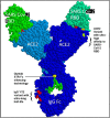
Critical features of Paradigm’s chimeric ACE-2/Fc-silent antibody technology. These include: the use of a full length human ACE-2 domain (AAs 18–740, blue), which when modified as described next, substantially increases binding affinity for SARS CoV-2 RBD/spike protein (green); the choice of specific mutations in ACE-2 (top right red orange, light green AAs) that impart picomolar affinity for the RBD and ~ femtomolar affinity for the full-length spike protein; linkage of the ACE-2 construct to a completely Fc-silent “STR” mutated monoclonal antibody (yellow, Mike Clark PhD, used with permission). The use of STR technology in an ACE2 chimeric prophylactic for SARS CoV-2 is patent pending, see (Fig. 15). In addition, one of the two mAb chimeras (“LiVE-Longer” vs. “LiVE”) utilizes a Y-T-E variant (red AAs, bottom) for increased half-life by binding to FcRn, which recycles IgG and is thereby predicted to increase its biological half-life by 3–4-fold (see Fig. 16 and text for details). The “LiVE” mAB does not contain the YTE sequence; both LiVE and LiVE-Longer are tested in (Figs 11–13 and Table 1). This figure was generated by Protean 3D, Version 17.3 (DNASTAR. Madison, WI).
Figure 2
Figure 2
Three-dimensional model of the SARS-CoV-2/ACE-2 molecular interface. Multiple In Silico modeling programs were used to predict optimal AA substitutions in ACE-2 (green) that would retain high binding affinity to the widest possible range of AA variations in the SARS-CoV-2 spike protein RBD (RBD, red). Two of the three optimal substitutions in ACE-2 depicted here are: T27L (ACE2 L27, blue lower) and H34V (ACE2 V34, blue upper right), shown here in close proximity to RBD AAs Y473 and F456 versus L455 and Y453, respectively. The third substitution N90E (ACE2 E90, blue upper left) is discussed in (Fig. 3). See text for details.
Figure 3
Figure 3
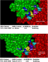
The effect of eliminating the glycosylation site at AA 90 of ACE-2. The asparagine at AA 90 of w.t. ACE-2 (N90) is a site for N-linked glycosylation (left panel, ACE-2 N90 CHO) which, when the sugar is present, causes steric hindrance of ACE-2/RBD interaction. Multiple modeling platforms predicted that elimination of the glycosylation site by the mutation N90E (asparagine to glutamate) would relieve steric hindrance and allow closer ACE-2/RBD interaction, particularly in combination with the ACE-2 H34V substitution (right panel, top). Subsequent figures below show data consistent with this prediction. See text for details.
Figure 4
Figure 4
The designed ACE-2 variant LVE/STR chimera scores higher than other ACE-2 Fusion protein competitors by the DFIRE scoring method. Molecular modeling was conducted with Protean 3D Version 17.3 (DNASTAR. Madison, WI) software, aimed at testing Paradigms’ proprietary LiVE variant (ACE-2 mutations T27L, H34V, and N90E) binding to w.t. SARS-2 RBD (DFIRE = −6.67), as it compares to the ACE-2 variant YTY (ACE-2 mutations T27Y, L79T and N330Y) binding to the w.t. SARS-2 RBD (DFIRE = −4.53). A lower DFIRE score predicts tighter (stabilizing) protein–protein interactions. See Methods for details.
Figure 5
Figure 5
In silico testing of SARS CoV-2 Delta variant predicts Paradigm’s ACE-2 variant LVE/STR chimera out-scores competing ACE-2 chimeric technologies. Top panel: molecular modeling with Protean 3D Version 17.3 (DNASTAR. Madison, WI) software of ACE-2 LVE construct (green) binding to w.t. SARS-CoV-2 RBD (red) yields DFIRE score = −6.67. Bottom panel: identical modeling of ACE-2 LiVE construct (green) binding to SARS-CoV-2 Delta variant yields DFIRE score = −6.80, slightly tighter (stabilizing) than that for the w.t. RBD. Note rotation of RBD F456 (yellow) in Delta variant toward ACE-2 AA 27, made permissive by the T27L mutation.
Figure 6
Figure 6
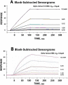
High binding affinity of Paradigm’s ACE-2 variant LVE/STR chimera to the RBD of Alpha and Delta variants of SARS-CoV-2. SPR assays were performed to determine binding affinities between the synthesized, purified LVE/STR chimera and synthesized RBDs of SARS-CoV-2 Alpha or Delta variants. Panel A: the S1 subunit with RBD of SARS-CoV-2 Alpha variant B.1.1.7 was synthesized, purified and subjected to SPR assay against the purified LVE/STR chimera. Determined binding affinity was 0.378 nM (Acro Biosystems). Note slow “off rate” compared with Panel B. Panel B: the RBD of Delta variant B.1.617.2 was synthesized, purified and subjected to SPR assay against the purified LiVE/STR chimera. Determined binding affinity was 0.554 nM (Acro Biosystems). See (Table 1) for more complete listing of binding affinities.
Figure 7
Figure 7
Paradigm’s ACE-2 variant LVE/STR chimera neutralizes Delta Variant B.1.617.2 RBD or w.t. SARS-CoV-2 RBD (Wuhan strain) with nearly equal potency. sVNTs (see Methods) were performed with SARS-CoV-2 RBD proteins expressing the Delta Variant B.1.617.2 sequence (T478K and L452R mutants, red bars) or the w.t. (original Wuhan strain) sequence (blue bars) incubated with the ACE-2 LiVE/STR construct. Dilutions shown are 0.05–2.4 ng/ml, left to right. Note similar sVNT titers of ~ 4.9 ng/ml.
Figure 8
Figure 8
Paradigm’s ACE-2 variant LVE/STR chimera neutralizes Beta Variant B.1.351 RBD significantly better than w.t. SARS-CoV-2 RBD. sVNT assays were performed with SARS-CoV-2 RBD proteins expressing the Beta Variant B.1.351 sequence (K417N, E484K and N501Y mutants, red bars) or the w.t. (original Wuhan strain) sequence (blue bars) incubated with the ACE-2 LiVE/STR construct. Dilutions shown are 0.05–2.4 ng/ml, left to right. Note sVNT titers of ~ 2.4 ng/ml for neutralization of the SARS-2 Beta variant.
Figure 9
Figure 9
Paradigm’s ACE-2 variant LVE/STR chimera neutralizes Alpha Variant B.1.1.7 RBD significantly better than GenScript IgG FL18–740 w.t. ACE-2 mAB. sVNT assays were performed with SARS-CoV-2 RBD proteins expressing the Alpha Variant B.1.1.7 sequence (N501Y mutant) challenged with either Paradigm’s ACE-2 variant LVE/STR chimera (dark blue bars) or with the GenScript IgG FL18–740 w.t. ACE-2 mAB (light blue bars, binding affinity 3 nM). Dilutions shown are 0.05–2.4 ng/ml, left to right. Note sVNT titers of ~ 6.3 μg/ml for the Genscript mAB for neutralization of the Alpha variant, in contrast to ~ 4.9 ng/ml for neutralization of the Alpha variant by the Paradigm construct LiVE-STR.
Figure 10
Figure 10
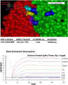
Paradigms’ ACE-2 variant LVE/STR chimera binds to SARS-2 Omicron variant B.1.1.529 spike protein trimer with high affinity. Top panel: molecular modeling of Paradigms’ ACE-2 variant LVE/STR chimera binding to the Omicron variant sequence as of 1 November 2022. Omicron RBD (red) mutations Y473, F456, L455 and N417 are shown at upper left, and ACE-2 (green) mutations LVE are shown in blue. The ACE-2 H34V makes contact with the aliphatic straight chain of Omicron mutation Q493R and K417N. Note the vertical orientation of N417 in close contact with V34, which enabled higher affinity binding compared to the w.t. K417, which assumed a more horizontal orientation in earlier SARS-2 variants. Molecular modeling performed identically to that in (Figs 4 and 5) yields superior DFIRE score of −3.38 (stabilizing). See Discussion for details. Bottom panel: the S1 subunit trimer of SARS-CoV-Omicron variant described above was synthesized, purified and subjected to SPR assay against the purified LVE/STR chimera. Determined binding affinity was 0.144 nM (Acro Biosystems); see (Table 1) for more complete listing of binding affinities.
Figure 11
Figure 11
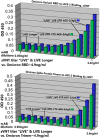
Paradigm’s ACE-2 variant LVE/STR chimeras potently neutralize the SARS-CoV-2 Omicron Variant B.1.1.529. sVNT assays were performed with purified recombinant SARS-CoV-2 RBD (top panel) or spike protein trimers (bottom panel) expressing the Omicron variant sequences described in (Fig. 10). The bargraphs show inhibition of the binding of Omicron RBD (top) or Omicron spike protein trimer (bottom) to purified recombinant ACE-2 by either Paradigm’s “LiVE” ACE-2 variant LVE/STR STR Fusion protein chimera or by the “LiVE Longer” LVE/STR STR–YTE IgG chimera. Dilutions shown are 0.05–2.4 ng/ml, left to right. Note similar sVNT titers of ~ 4.9 ng/ml for neutralization of Omicron RBD or Omicron spike trimers, and slightly better neutralization by the “LiVE Longer” chimera (green). See (Figs 1 and 13) for additional details about the ACE-2/Fusion protein chimeras.
Figure 12
Figure 12
Antiviral effects of “LiVE” or “LiVE Longer” fusion proteins against SARS-CoV-2 B.1.1.214 or BA.1 infection of human AO cultures. After 1 day in culture, organoid cultures (1.0 × 104 cells/well) were infected with 0.1 MOI SARS-CoV-2 B.1.1.214 (A) or BA.1 (B) and then cultured with the medium containing a serially diluted antibody for 2 days. The viral RNA copy number in the cell culture supernatant was measured by qPCR. Data are represented as means ± SD (n = 3). See Methods for details.
Figure 13
Figure 13
Antiviral effects of the “LiVE”, “LiVE Longer” or related chimeric fusion proteins against SARS-CoV-2 Omicron BA.2 or BA.5 infection of human AO cultures. Organoid cultures were exposed to Omicron variants BA.2 (top panels) or BA.5 (bottom panels) as described in Materials and Methods and were challenged with the fusion proteins “LiVE” (leftmost pair), “LiVE Longer” (3rd pair from left), modified LiVE without the STR motif (2nd from left) or a modified LiVE LONGER with tyrosine substituted for leucine at position 27 (Y-V-E*, rightmost panels). Note the most potent inhibition of Omicron BA.5 replication by the LiVE (IC50 = 29.9 ng/ml) and LiVE LONGER fusion proteins (IC50 = 26.9 ng/ml). See text for details.
Figure 14
Figure 14
ACE-2 enzymatic activity of the chimeric fusion proteins. Equal amounts of rhACE-2 were subjected to measurements of ACE-2 enzyme activity as described in Materials and Methods, using standard ACE-2 enzyme assay methods based on fluorogenic substrate conversion. Results are the mean ± S.E.M. of eight replicates per group. Note inhibition of ACE-2 activity by the competitive inhibitor peptide DX600 (at 1 μM) and the extremely low to negligible activity in any of the chimeric fusion proteins tested. See text for details.
Figure 15
Figure 15
Paradigm's Fc fusion protein including the STR Fc silencing mutations licensed by MAbsolve is Superior to LALA and other Reduced Fc Effector Function Technologies. A comparison of Fc-silencing technologies shows the relative binding of purified IgG1 (control) versus a variety of Fc-mutant antibody technologies to immobilized human FcγRI (CD64), as determined by SPR assay (data courtesy of MAbsolve, https://mabsolve.com/science/#linkone). Note extremely low binding of either the YTE variant used by Paradigm (far right bar) or MAbsolve’s STR variant. Inset: The binding affinities of the non-YTE LiVE or YTE-variant LiVE-Longer ACE-2/Fusion protein chimeras to purified FcRn receptor [24] were determined by SPR assay (Acro Biosystems). Note high affinity binding of the YTE chimera (27 nM) to FcRn. See text for details.
Figure 16
Figure 16
Longer half-life of nasally applied chimeric ACE-2/Fc-silent Fusion protein is due to kinetics of FcRn-Dependent IgG Uptake into Olfactory Mucosa. Nasally applied IgG is slowly transcytosed through binding to FcRn receptor expressed in porcine or human olfactory epithelia. In this figure, Ladel et al. [24] used ex vivo porcine olfactory mucosa to show to track the uptake of allogenic IgG (red) into porcine olfactory epithelia. (A) Basal levels of endogenous pIgG (porcine IgG) were detected with a low signal at the apical side, in the basal cell layer, glands, cavernous bodies and blood vessels. This signal served as a blank and was subtracted from the photos showing the penetration of exogenous pIgG. (B) After 30 min, only the areas close to the apical side show immunoreactivity for pIgG, but some signal was detected in the lamina propria. (C) After 4 h, the pIgGs obviously distributed into the lamina propria. *Indicate round structure filled with cells and mostly spared from IgG (D) After 8 h, pIgG were detected throughout the whole lamina propria. Nuclei stained with DAPI; epithelial control: quality control for tissue integrity, stained with HE. Scale bar: 100 μm. On this basis, the YTE variant of Paradigm’s ACE-2 LVE/IgG YTE chimera (which binds longer to FcRn (see Zhu et al. [25]) is predicted to have 3–4-fold longer biological half-life when administered nasally. Reprinted with publisher’s permission from Ladel et al., Allogenic Fc Domain-Facilitated Uptake of IgG in Nasal Lamina Propria. Pharmaceutics, 10:107, 2018. See text for details.

References

    1. Samavati, L, Uhal, B. ACE-2, much more than just a receptor for SARS-CoV-2. Front Cell Infect Microbiol 2020; 10: 317.eCollection 2020PMID: 32582574. - PMC - PubMed
    1. Heurich, A, Hofmann-Winkler, H, Gierer, Set al. . TMPRSS2 and ADAM17 cleave ACE2 differentially and only proteolysis by TMPRSS2 augments entry driven by the severe acute respiratory syndrome coronavirus spike protein. J Virol 2014; 88: 1293–307Epub 2013 Nov 13. PMID: 24227843; PMCID: PMC3911672. - PMC - PubMed
    1. Seyran, M, Takayama, K, Uversky, VNet al. . The structural basis of accelerated host cell entry by SARS-CoV-2†. FEBS J 2021; 288: 5010–20PMID: 33264497. - PMC - PubMed
    1. Celik, I, Khan, A, Dwivany, FMet al. . Computational prediction of the effect of mutations in the receptor-binding domain on the interaction between SARS-CoV-2 and human ACE2. Mol Divers 2022; 26: 3309–24PMID: 35138508. - PubMed
    1. Oh, SJ, Shin, OS. SARS-CoV-2-mediated evasion strategies for antiviral interferon pathways. J Microbiol 2022; 5: 1–10PMID: 35122601; PMCID: PMC8817151. - PMC - PubMed