Review
. 2023 Jan 17:13:1085551.
doi: 10.3389/fimmu.2022.1085551. eCollection 2022.

Airway microbiome-immune crosstalk in chronic obstructive pulmonary disease

Affiliations
Review

Airway microbiome-immune crosstalk in chronic obstructive pulmonary disease

Alex Kayongo et al. Front Immunol. .

Abstract

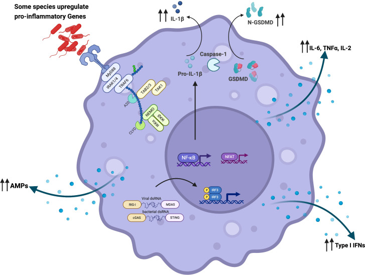
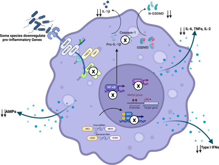
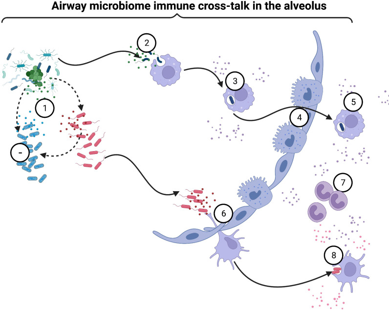
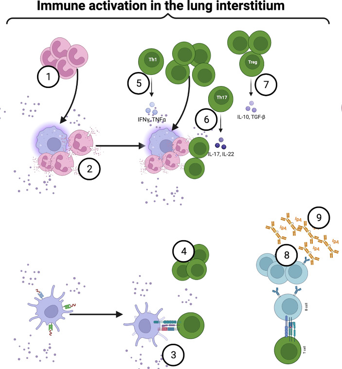
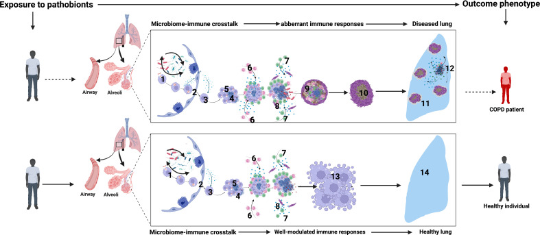
Chronic Obstructive Pulmonary Disease (COPD) has significantly contributed to global mortality, with three million deaths reported annually. This impact is expected to increase over the next 40 years, with approximately 5 million people predicted to succumb to COPD-related deaths annually. Immune mechanisms driving disease progression have not been fully elucidated. Airway microbiota have been implicated. However, it is still unclear how changes in the airway microbiome drive persistent immune activation and consequent lung damage. Mechanisms mediating microbiome-immune crosstalk in the airways remain unclear. In this review, we examine how dysbiosis mediates airway inflammation in COPD. We give a detailed account of how airway commensal bacteria interact with the mucosal innate and adaptive immune system to regulate immune responses in healthy or diseased airways. Immune-phenotyping airway microbiota could advance COPD immunotherapeutics and identify key open questions that future research must address to further such translation.

Keywords: COPD; adaptive immunity; inflammation; innate immunity; lung microbiome; mucosal immunity.


Conflict of interest statement

The authors declare that the research was conducted in the absence of any commercial or financial relationships that could be construed as a potential conflict of interest.

Figures

Figure 1
Figure 1
Microbial interaction with the innate immune system activates and reprograms immune cells: Airway microbiome-derived products such as lipopolysaccharide are detected by surface and intracellular immune sensors such as Toll-like receptors (TLRs). In a MyD88-dependent manner, airway epithelial cells, alveolar macrophages, and dendritic cells become activated and upregulate the expression of pro-inflammatory cytokines such as IL-1β, IL-18, TNFα, IL-6, and IL-2. These activated cells orchestrate immune inflammation. Microbicidal activity of alveolar macrophages is also potentiated through increased gene expression of antimicrobial peptides and other lytic proteins. The microbiome has also been shown to potentiate macrophage-bacterial killing and clearance via GM-CSF signaling. Furthermore, depletion of commensal bacteria has been shown to reprogram alveolar macrophages from classical (M1) to alternative (M2) phenotype characterized by the expression of higher levels of Arg1, CCL24, IL-13, IL-10, IL-6, and IL-1β. In addition to MyD88-dependent activation, intracellular sensing of microbial-derived products by NOD1, NOD2, and NLRP6 in the epithelial cells set the pace for a pro-inflammatory signal along the mucosa. This restricts mucosal colonization by pathogenic bacteria via the secretion of IL-1β and IL-18. Microbiome-driven-type-I-IFN responses dependent on cGAS-STING activation have also been described. Created with BioRender.com.
Figure 2
Figure 2
Airway microbiome-driven immunomodulation: Airway microbiome downregulates pro-inflammatory signals via interference with immune signaling machinery. For instance, R. mucilaginosa has been shown to inhibit immune activation via NF-κB dependent mechanisms. As an immune evasion strategy, pathobionts have been described to inhibit TRIF and NFAT signaling, phagolysosomal fusion and acidification, as well as inflammasome activation. In other scenarios, gene methylation may promote transcriptional repression of pro-inflammatory genes. Created with BioRender.com.
Figure 3
Figure 3
Airway microbiome immune crosstalk in alveoli: Following successful colonization of the respiratory tract, bacterial species replicate rapidly and establish dense biological communities (1). Some bacteria inhibit other species via the secretion of antimicrobial peptides and lytic enzymes(1). Alveolar macrophages engulf bacteria(2) and become activated via MyD88-dependent and -independent mechanisms(3). Airway microbiome sensing potentiates bacterial killing(3). Some intracellular pathobionts infect alveolar macrophages and access the lung interstitium (4). Activated macrophages secrete chemoattractants and promote the migration of other cellular players, such as monocytes, neutrophils, and adaptive immune response cells, into the airways to clear pathobionts (5). Dendritic cells (DCs) continuously sample airway bacteria that attach and colonize the mucosa via protruding dendrites(6). Airway microbiome sensing by bronchial epithelial cells, alveolar macrophages, and DCs activate innate lymphoid cells (ILCs) (7), which modulate other immune cells’ activity. Depending on the type and degree of microbial exposure, DCs induce a wide range of immune responses, from immune tolerance induced by plasmacytoid DCs (pDCs) to inflammation induced by conventional DCs (cDCs)(8). Continuous microbial sampling and trafficking by activated DCs, and alveolar macrophages deliver processed microbial antigens to naïve CD4+T and CD8+T cells within mucosa-associated lymphoid tissue (MALT) and draining lymph nodes (8). Created with BioRender.com.
Figure 4
Figure 4
Airway microbiome immune crosstalk in the lung interstitium: Some intracellular pathobionts infect alveolar macrophages and access the interstitium. Activated macrophages secrete chemoattractants and promote neutrophilic infiltration into the airways to clear pathobionts (1). Upon bacterial sensing, neutrophils become activated, degranulate and release NETs (2). Continuous sampling and trafficking by activated DCs, and alveolar macrophages deliver processed microbial antigens to naïve CD4+T within mucosa-associated lymphoid tissue (MALT) and draining lymph nodes (4). Under a steady state, mature lung cDCs are preferentially programmed to induce a Th2 immune response. However, following immune sensing and activation, the production of IL-12, IL-23, IL-27, and notch ligand by airway DCs, alveolar macrophages, and epithelial cells induce a Th1 response (5). Among CD4+T cell phenotypes, microbiome-mediated mucosal inflammation has been strongly linked to aberrant Th17 (6). Besides the Th17 phenotype, microbial interaction with mucosal CD4+T cells induces immune tolerance (7). MyD88-dependent TLR2 activation by capsular polysaccharide A induces the expansion of Foxp3+T cells within the mucosa (7). Foxp3+T cells drive IL-10 production, facilitating mucosal immune tolerance (7). In another mechanism, microbial-induced Tregs promote mucosal memory B or plasma cells’ IgA secretion (8). Created with BioRender.com.
Figure 5
Figure 5
Airway microbiome-immune crosstalk in healthy versus diseased lungs (COPD): 1. Initial crosstalk between airway microbial communities and the mucosal innate and adaptive immune cell activation sets the tone of airway mucosal immune responses. 2. Infected macrophages from the distal airways and alveolar spaces migrate into the interstitium in an IL-1R-dependent manner. 3. Intracellular bacteria replicate within macrophages. 4. Some intracellular pathobionts induce infected macrophage apoptosis and expression of host lytic proteins in an ESX-1-dependent manner. 5. Newly recruited macrophages engulf infected cell debris. 6. Neutrophils infiltrate the airways to orchestrate inflammation, engulf dying infected cells, and kill bacteria through NETosis and the release of lytic enzymes. 7. Microbial-specific T cells arrive in airways and produce cytokines such as IFN-γ, which enhance the microbicidal activity of alveolar macrophages, monocytes, and DCs 8. Macrophage and neutrophil necroptosis lead to the release of lytic proteins such as MMPs, defensins, and cathepsin G into the extracellular space. Subsequent induction of lytic proteins causes extensive tissue damage. 9. Over time, with attempted healing and subsequent inflammation, extensive lung damage, fibrosis, and reduced lung function ensue. 10-11. Extensive fibrosis further reduces lung compliance and worsens lung function. 12. Sustained dysbiosis driven by periodic insults such as bacterial, viral, and fungal infections and air pollution or smoking orchestrate more damage and susceptibility. 13. In contrast, in healthy airways, robust innate and adaptive immune reprogramming promotes a highly modulated immune microenvironment with balanced lethal and resolving inflammatory states. This is optimal to contain airway commensals and pathobionts hence maintaining healthy lungs seen in 14. Created with BioRender.com.

References

    1. Venkatesan P. GOLD report: 2022 update. Lancet Respir Med (2022) 10(2):e20. doi: 10.1016/S2213-2600(21)00561-0 - DOI - PubMed
    1. Chung K, Adcock I. Multifaceted mechanisms in COPD: inflammation, immunity, and tissue repair and destruction. Eur Respir J (2008) 31(6):1334–56. doi: 10.1183/09031936.00018908 - DOI - PubMed
    1. Barbu C, Iordache M, Man M. Inflammation in COPD: pathogenesis, local and systemic effects. Rom J Morphol Embryol (2011) 52(1):21–7. - PubMed
    1. Barnes P, Burney P, Silverman EK, Celi BR, Vestbo J, Wedzicha JA, et al. . Chronic obstructive pulmonary disease. Nat Rev Dis Primers (2015) 1:15076. doi: 10.1038/nrdp.2015.76 - DOI - PubMed
    1. Fabbri L, Luppi F, Beghé B, Rabe K. Complex chronic comorbidities of COPD. Eur Respir J (2008) 31(1):204–12. doi: 10.1183/09031936.00114307 - DOI - PubMed

Publication types