. 2022 May 20;1(3):pgac063.
doi: 10.1093/pnasnexus/pgac063. eCollection 2022 Jul.

SLC46A3 is a lysosomal proton-coupled steroid conjugate and bile acid transporter involved in transport of active catabolites of T-DM1

Affiliations

SLC46A3 is a lysosomal proton-coupled steroid conjugate and bile acid transporter involved in transport of active catabolites of T-DM1

Ryuto Tomabechi et al. PNAS Nexus. .

Abstract

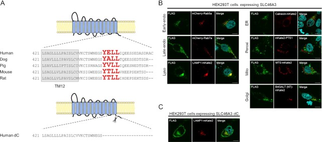
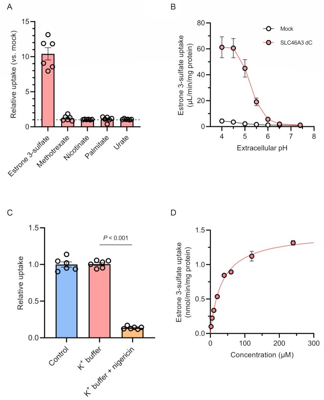
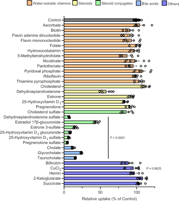
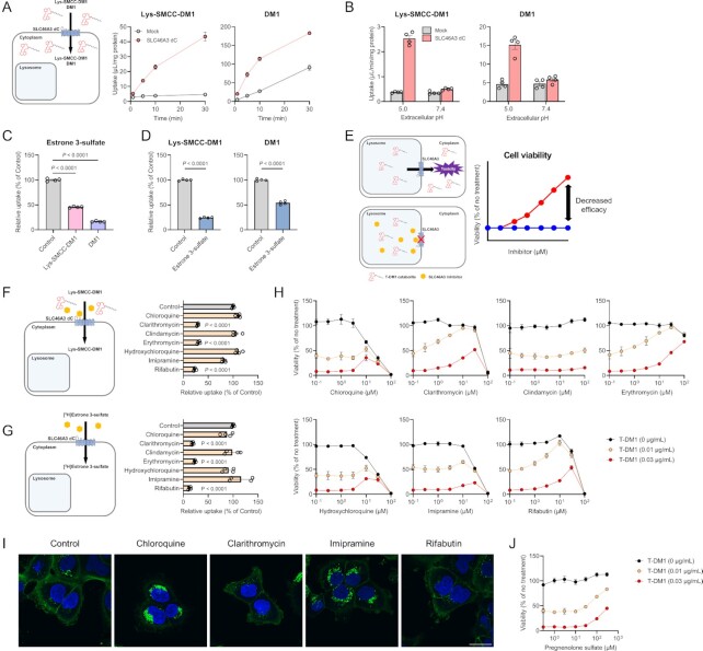
Antibody-drug conjugates (ADCs) represent a new class of cancer therapeutics that enable targeted delivery of cytotoxic drugs to cancer cells. Although clinical efficacy has been demonstrated for ADC therapies, resistance to these conjugates may occur. Recently, SLC46A3, a lysosomal membrane protein, was revealed to regulate the efficacy of trastuzumab emtansine (T-DM1), a noncleavable ADC that has been widely used for treating breast cancer. However, the role of SLC46A3 in mediating T-DM1 cytotoxicity remains unclear. In this study, we discovered the function of SLC46A3 as a novel proton-coupled steroid conjugate and bile acid transporter. SLC46A3 preferentially recognized lipophilic steroid conjugates and bile acids as endogenous substrates. In addition, we found that SLC46A3 directly transports Lys-SMCC-DM1, a major catabolite of T-DM1, and potent SLC46A3 inhibitors attenuate the cytotoxic effects of T-DM1, suggesting a role in the escape of Lys-SMCC-DM1 from the lysosome into the cytoplasm. Our findings reveal the molecular mechanism by which T-DM1 kills cancer cells and may contribute to the rational development of ADCs that target SLC46A3.

Keywords: SLC46A3; and trastuzumab emtansine; antibody–drug conjugate; bile acid; steroid conjugate.


Figures

Fig. 1.
Fig. 1.
Mammalian SLC46A3 shares a lysosomal-sorting motif in its C-terminal region. (A) Predicted membrane topology of SLC46A3 by TMHMM (http://www.cbs.dtu.dk/services/TMHMM/) and sequence alignment of its C-terminal region between mammalian species. The sequences predicted as tyrosine-based lysosomal-sorting motifs are shown with red color characters. Human dC is a C-terminal amino acid sequence deleted in SLC46A3 mutants in this study. (B) and (C) Immunofluorescence staining of FLAG-tagged SLC46A3 (green) in transfected cells by anti-FLAG antibody. HEK293T cells were transfected with 3 × FLAG-tagged wild-type human SLC46A3 (B) or C-terminal deleted mutant human SLC46A3 (SLC46A3 dC) (C) and mCherry or mKate2-tagged organelle marker protein (red). The results were replicated in three independent experiments. Blue color, nucleus. Scale bar, 10 μm. PTS1, peroxisomal targeting signal 1; MTS, mitochondrial targeting sequence; and B4GALT (NT), N-terminal 81-amino acid human beta-1,4-galactosyltransferase.
Fig. 2.
Fig. 2.
SLC46A3 mediates proton-coupled estrone 3-sulfate uptake. (A) Uptake of [3H]estrone 3-sulfate (10 nM), methotrexate (10 μM), [3H]nicotinate (200 nM), [3H]palmitate (20 nM), and [14C]urate (1.8 μM) with 50 μM urate in MDCKII/SLC46A3 dC cells. Uptake for 5 min was measured in HBSS (pH 5.5). Relative uptake was calculated by dividing the uptake in MDCKII/SLC46A3 dC cells by that in mock cells. (B) pH dependence of [3H]estrone 3-sulfate (10 nM) uptake. Uptake for 1 min was measured in HBSS (pH 7.4) or modified-HBSS-containing citrate for pH 4.0 to 4.5, or MES for pH 5.0 to 6.5. (C) Effect of a proton gradient on [3H]estrone 3-sulfate uptake. The uptake was measured in HBSS (pH 5.0) or modified-HBSS in which NaCl was replaced with KCl with or without 10 μg/mL nigericin. (D) Concentration-dependent estrone 3-sulfate uptake. Uptake for 1 min was measured in HBSS (pH 5.0). SLC46A3 dC-specific uptake was calculated by subtracting the uptake in mock cells from that in MDCKII/SLC46A3 dC cells. The solid line represents the computer-fitted profile. Data are represented as the mean ± SEM obtained from two biologically independent experiments. P-values are indicated in the different panels (one-way ANOVA followed by Tukey's multiple comparisons test).
Fig. 3.
Fig. 3.
Steroid conjugates and bile acids strongly suppress SLC46A3 dC-mediated estrone 3-sulfate transport. Effect of various compounds on the uptake of [3H]estrone 3-sulfate (10 nM) in MDCKII/SLC46A3 dC cells. Uptake was performed at 37°C, pH 5.0, for 1 min in the absence or presence of inhibitors (20 μM for cholesterol, dehydroepiandrosterone, estrone, pregnenolone, 25-hydroxyvitamin D3, 25-hydroxyvitamin D3 glucuronide, 25-hydroxyvitamin D3 sulfate, hemin, and bilirubin or 200 μM for the other compounds). Data are presented as the mean ± SEM from two biologically independent experiments. P-values are indicated in the different panels (one-way ANOVA followed by Dunnett's multiple comparison).
Fig. 4.
Fig. 4.
SLC46A3 mediates T-DM1 cytotoxicity by directly transporting Lys-SMCC-DM1. (A) Time-dependent uptake of Lys-SMCC-DM1 (5 μM) and DM1 (1 μM) in the MDCKII/SLC46A3 dC cells. Uptake was performed at pH 5.0. Data are represented as the mean ± SEM obtained from two biologically independent experiments. (B) pH-dependent uptake of Lys-SMCC-DM1 (5 μM) and DM1 (1 μM) in the MDCKII/SLC46A3 dC cells. Data are represented as the mean ± SEM obtained from two biologically independent experiments. (C) Inhibitory effect of Lys-SMCC-DM1 and DM1 on the uptake of [3H]estrone 3-sulfate (10 nM) in MDCKII/SLC46A3 dC cells. Uptake was performed at pH 5.0 in the absence or presence of Lys-SMCC-DM1 and DM1 (200 μM). Data are represented as the mean ± SEM obtained from two biologically independent experiments. P-values are indicated in the different panels (one-way ANOVA followed by Dunnett's multiple comparison). (D) Inhibitory effect of estrone 3-sulfate on the uptake of Lys-SMCC-DM1 (10 µM) and DM1 (1 µM). Uptake was performed at pH 5.0 in the absence or presence of estrone 3-sulfate (200 µM). Data are presented as the mean ± SEM from two biologically independent experiments. P-values are indicated in the different panels (two-tailed unpaired t test). (E) Predicted schematic and results of Lys-SMCC-DM1–drug interaction through SLC46A3 in lysosomes. Impairment of lysosomal SLC46A3 function by a potent SLC46A3 inhibitor cause the inhibition of Lys-SMCC-DM1 escape from lysosomes, thereby reducing T-DM1 cytotoxicity. The predicted profiles of cell viability treated with T-DM1 in the presence (a red line) or absence (a blue line) of a SLC46A3 inhibitor are shown. (F) and (G) Inhibitory effect of lysosomotropic drugs on the uptake of Lys-SMCC-DM1 (5 μM) (F) or [3H]estrone 3-sulfate (10 nM) (G) in MDCKII/SLC46A3 dC cells. Uptake was performed at pH 5.0 in the absence or presence of lysosomotropic drugs (200 μM). Data are represented as the mean ± SEM obtained from two biologically independent experiments. P-values are indicated in the different panels (one-way ANOVA followed by Dunnett's multiple comparison). (H) Effect of lysosomotropic drugs on T-DM1 cytotoxicity. SK-BR-3 cells were exposed to T-DM1 (0 μg/mL; black, 0.01 μg/mL; yellow, and 0.03 μg/mL; red) for 4 days in the presence of lysosomotropic drugs as indicated. Data are represented as the mean ± SEM obtained from two biologically independent experiments. (I) Immunofluorescence staining of T-DM1(green). HCC1954 cells were pulsed with 1.5 μg/mL T-DM1 for 15 min at 37°C, and chased for 1 day in the presence or absence of chloroquine (10 μM), clarithromycin (30 μM), imipramine (30 μM), and rifabutin (30 μM). The results were replicated in two biologically independent experiments. Blue color, nucleus. Scale bar, 30 μm. (J) Effect of pregnenolone sulfate on T-DM1 cytotoxicity. SK-BR-3 cells were exposed to T-DM1 (0 μg/mL; black, 0.01 μg/mL; yellow, and 0.03 μg/mL; red) for 4 days with pregnenolone sulfate as indicated concentration. Data are presented as the mean ± SEM obtained from two biologically independent experiments.

References

    1. Chau CH, Steeg PS, Figg WD. 2019. Antibody-drug conjugates for cancer. Lancet. 394:793–804. - PubMed
    1. Khongorzul P, Ling CJ, Khan FU, Ihsan AU, Zhang J. 2020. Antibody-drug conjugates: a comprehensive review. Mol Cancer Res. 18:3–19. - PubMed
    1. Tsao L-C, Force J, Hartman ZC. 2021. Mechanisms of therapeutic antitumor monoclonal antibodies. Cancer Res. 81:4641–4651. - PMC - PubMed
    1. Coats S, et al. . 2019. Antibody-drug conjugates: future directions in clinical and translational strategies to improve the therapeutic index. Clin Cancer Res. 25:5441–5448. - PubMed
    1. Tsui CK, et al. . 2019. CRISPR-Cas9 screens identify regulators of antibody-drug conjugate toxicity. Nat Chem Biol. 15:949–958. - PMC - PubMed