Review
. 2023 Jan 19:14:1002960.
doi: 10.3389/fnsyn.2022.1002960. eCollection 2022.

Segregation of D1 and D2 dopamine receptors in the striatal direct and indirect pathways: An historical perspective

Affiliations
Review

Segregation of D1 and D2 dopamine receptors in the striatal direct and indirect pathways: An historical perspective

Charles R Gerfen. Front Synaptic Neurosci. .

Abstract

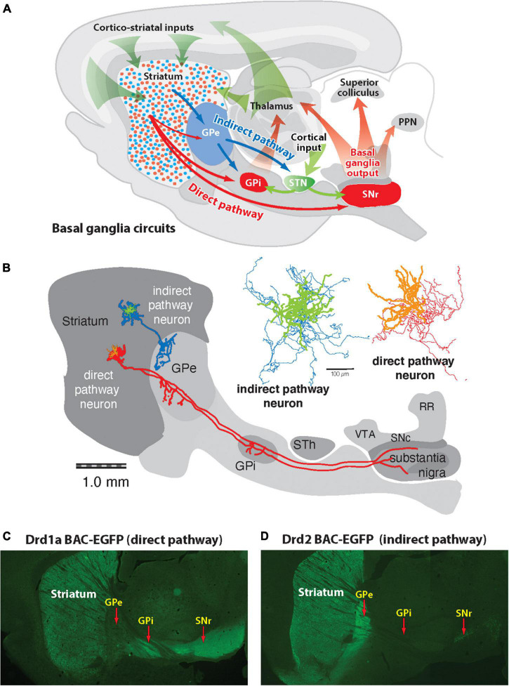
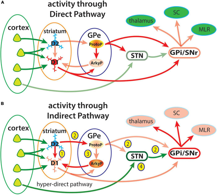
The direct and indirect striatal pathways form a cornerstone of the circuits of the basal ganglia. Dopamine has opponent affects on the function of these pathways due to the segregation of the D1- and D2-dopamine receptors in the spiny projection neurons giving rise to the direct and indirect pathways. An historical perspective is provided on the discovery of dopamine receptor segregation leading to models of how the direct and indirect affect motor behavior.

Keywords: Parkinson’s disease; basal ganglia; dopamine; motor function; striatum.


Conflict of interest statement

The author declares that the research was conducted in the absence of any commercial or financial relationships that could be construed as a potential conflict of interest.

Figures

FIGURE 1
FIGURE 1
Diagram of basic basal ganglia circuits. (A) The striatum receives excitatory corticostriatal and thalamic inputs. Outputs of the basal ganglia arise from the internal segment of the globus pallidus (GPi) and substantia nigra pars reticulata (SNr), which are directed to the thalamus, superior colliculus, and pedunculopontine nucleus (PPN). The direct pathway originates from Drd1a-expressing SPNs that project to the GPi and SNr output nuclei. The indirect pathway originates from Drd2-expressing SPNs that project only to the external segment of the globus pallidus (GPe), which together with the subthalamic nucleus (STN) contain transynaptic circuits connecting to the basal output nuclei. (B) Tracings of individual indirect and direct striatal projection neurons drawn in place on a sagittal brain diagram (Kawaguchi et al., 1989). The indirect striatal pathway neuron (green) has axon collaterals (blue) that spreads locally within the striatum and one that projects into the GPe, where it arborizes, and does not extend beyond this nucleus. The direct striatal pathway neuron (orange) has a local collateral within the striatum and projection axons (red) that extend some collaterals into the globus pallidus (GPe), and others to the globus pallidus internal segment (GPi) and substantia nigra. (C) Fluorescent imaging of a brain section from a mouse expressing enhanced green fluorescent protein (eGFP) under regulation of the Drd1a promoter shows Drd1a-expressing SPNs in the striatum that project axons through the GPe, which terminate in the GPi and GPe. (D) Fluorescent imaging of a Drd2-eGFP mouse shows that labeled SPNs provide axonal projections that terminate in the GPe but do not extend to the GPi or SNr.
FIGURE 2
FIGURE 2
(I) Expression of SP and D1 in striatonigral and ENK and D2r in striatopallidal neurons. Striatal neurons retrogradely labeled with the fluorescent dye fluorogold after injection into the substantia nigra combined with darkfield illumination of silver grains produced by ISHH labeling with 35S labeled oligonucleotide probes for (A) substance P (SP), (B) the DI dopamine receptor (DI), (C) enkephalin (ENK), and (D) the D2 dopamine receptor (D2). Striatonigral neurons show ISHH labeling for both substance P [(A), solid arrows] and the D1 dopamine receptor [(B), solid arrows]. Striatal neurons that are unlabeled by fluorogold, and presumably project to the globus pallidus, show ISHH labeling for both enkephalin [(C), open arrows] and the D2 dopamine receptor [(D), open arrows]. (II) In situ hybridization in the striatum from brain sections apposed to autoradiographic film labeled with 35S-labeled oligonucleotide probes complementary to (A–D) enkephalin (ENK), (E–H) substance P (SP), and (I–L) dynorphin (DYN). Sections in the first two columns are from the same saline-treated animal showing ISHH labeling on the unlesioned control side (A,E,I) and lesioned 6-OHDA-injected side (B,F,J). Sections in the third column are from the lesioned, 6-OHDA-injected side of an animal that received intermittent treatment with the D1 receptor selective agonist SKF38393 (C,G,K). Sections in the fourth column are from the lesioned, 6-OHDA-injected lesioned side of animal that received continuous treatment with the D2 agonist quinpirole (D,H,L). The increase in enkephalin ISHH labeling caused by 6-OHDA lesions (B) is not affected by D1 agonist treatment (C) but is reversed by continuous D2 agonist treatment (D). The decrease in substance P ISHH labeling caused by 6-OHDA lesions (F) is reversed by intermittent D1 agonist treatment (G) but is unaffected by D2 agonist treatment (H). Dynorphin ISHH labeling is not significantly altered by 6-OHDA lesions (J) but is elevated by D1 agonist treatment (K) but not affected by D2 agonist treatment (L) (Gerfen et al., 1990).
FIGURE 3
FIGURE 3
Expression of the IEG, zif268 (egr2) in D1r- and D2r-neurons in the control dopamine intact striatum (A), in the dopamine-depleted 6-OHDA lesioned striatum (B), in the lesioned striatum of animals treated with a D1r-selective agonist (C) and in the lesioned striatum of animals treated with both a D1r- and D2r-selective agonists (D). Expression of zif268 is determined by the number of silver grains (white) generated with 35S-oligonucleotide labeling of zif268 mRNA concentrated over striatal neurons labeled with alkaline-phosphatase labeled ribonucleotide probes for ENK (blue arrows) or over putative D1r expressing neurons that do not express ENK (red arrows). Histogram distribution of the average number of zif268_ISHH generated silver grains per ENK+ (D2) and ENK– (D1) cells from five animals are plotted for each condition. In the unlesioned striatum there is no significant difference in zif268 levels between D1 and D2 cells (A’). In the 6-OHDA lesioned striatum there is a significant increase in D2 cells and decrease in D1 cells (B’) compared to levels in the dopamine intact striatum (distributions from panel A’ are shaded in panel B’). In animals with 6-OHDA lesions to deplete striatal dopamine that were treated with a D1r selective agonist (SKF38393, 1.0 mg/kg i.p.), there is no significant change in zif268 levels in D2 cells but a significant increase in D1 cells (C’) compared to levels in the dopamine depleted striatum without agonist treatment (distributions from panel B’ are shown shaded in panel C’). Treatment with combined D1r-agonist (SKF38393, 1.0 mg/kg i.p.) and D2r-agonist (quinpirole, 1.0 mg/kg) resulted in a reduction in zif268 levels in D2-cells and a significant increase in D1-cells compared to levels compared with levels in the lesioned striatum of animals treated with the D1r-agoinist alone (distribution levels from panel C’ are shaded in panel D’). These data demonstrate that D1r- and D2r-selective agonists have opposite acute effects on dSPN and iSPNs (Gerfen et al., 1995).
FIGURE 4
FIGURE 4
(I) D1 dopamine receptor-mediated phosphorylation of ERK1/2 (p-ERK1/2) in the dopamine-depleted striatum. Unilateral lesion of the nigrostriatal dopamine systems is demonstrated by the loss of tyrosine hydroxylase immunoreactivity in the right lesioned striatum (A). After treatment (15 min) with the partial D1 dopamine agonist SKF38393 (2 mg/kg, i.p.), p-ERK1/2 is not evident in the dopamine-intact striatum (B) but is present in numerous neurons in the dopamine-depleted striatum (C). To determine the type of striatal neuron in which p-ERK1/2 is present, sections were processed to display both p-ERK1/2 with a green fluorescent label and enkephalin mRNA with a red fluorescent label (D”). Nearly all p-ERK1/2-immunoreactive neurons (blue arrows) are enkephalin negative. Only a small number enkephalin-positive neurons display p-ERK1/2 immunoreactivity (yellow arrow), whereas the vast majority are p-ERK1/2 negative (orange arrows). (II) Demonstration of distinct mechanisms of D1 dopamine receptor-mediated gene regulation in the dopamine (DA)-intact and -depleted striatum, using the full D1 agonist SKF81297 alone or combined with other drugs. (A–D) In situ hybridization histochemical localization of mRNA encoding c-fos 45 min after different drug combinations: (A) SKF81297 (0.5 mg/kg); (B) SKF81297 (2.0 mg/kg); (C) SKF81297 (2.0 mg/kg) combined with the muscarinic receptor antagonist scopolamine (5 mg/kg); or (D) SKF81297 (2.0 mg/kg) combined with the D2 dopamine receptor agonist (1 mg/kg) and scopolamine. The low dose of agonist alone (A) demonstrates the supersensitive response by the selective induction of c-fos in the dopamine depleted striatum. Bilateral induction of c-fos IEG in both the dopamine-intact and -depleted striatum follows treatment with high dose of the full D1 agonist alone (B) or in combination with other drugs (C,D). However, when animals receiving any of these treatments (15 min survival) p-ERK1/2-immunoreactive neurons are evident only in the dopamine-depleted striatum and not in the dopamine-intact striatum (data not shown). The treatment combining full D1 agonist with both the D2 agonist and scopolamine produces the most robust c-fos IEG response in both the DA-intact (E) and DA-depleted (F) striatum at 45 min. This treatment also results in persistent p-ERK1/2 (H) and phosphorylated c-jun (J) in the dopamine-depleted striatum but does not activate p-ERK1/2 (G) or phosphorylated c-jun (I) in neurons in the dopamine-intact striatum. These results demonstrate that, although D1 dopamine receptor-mediated induction of the IEG c-fos occurs in both the dopamine intact and -depleted striatum, activation of ERK1/2 occurs only in the dopamine-depleted striatum (Gerfen et al., 2002).
FIGURE 5
FIGURE 5
Electrical stimulation of the nigrostriatal pathway results in the induction of the IEG c-fos throughout the striatum and nucleus accumbens, but activation of ERK1/2 is restricted principally to the nucleus accumbent. Electrodes were placed in the junction between dopamine (DA) neurons in the ventral tegmental area (VTA) and substantia nigra pars compacta (SNc) and stimulated (A,E). In animals killed 45 min after stimulation onset (A–D), c-fos is induced throughout the dorsal striatum and nucleus accumbens (B). Higher-power photomicrographs reveal c-fos-immunoreactive nuclei in the nucleus accumbens (C) and in the dorsal striatum (D). In animals killed 15 min after stimulation onset (E–H), the time point that is optimal for detecting phosphorylated ERK1/2, immunoreactive neurons are observed only in the nucleus accumbens (F). Higher power photomicrographs reveal numerous rons in the nucleus accumbens (G), whereas in the dorsal striatum, only scattered large immunoreactive neurons are observed (H) and not in SPNs (Gerfen et al., 2002).
FIGURE 6
FIGURE 6
Proposed model for direct dopamine-dependent ERK1/2 activation in dSPNs (Jiang et al., 2021). Cocaine acts by increasing synaptic dopamine leading to ERK activation in NAc, required for locomotor sensitization and CPP. It has previously been proposed that D1 receptor activation affects ERK activity only indirectly, via PKA- and DARPP-32/STEP-mediated inhibition of ERK dephosphorylation (Svenningsson et al., 2004; Valjent et al., 2005), whereas direct ERK activation itself occurs in the D1 MSNs only via NMDAR-dependent glutamatergic signaling (Pascoli et al., 2011; Cahill et al., 2014) through calcium-sensitive Ras-guanine nucleotide releasing factor (Ras-GRF1) (Farnsworth et al., 1995; Fasano et al., 2009). We propose here a more parsimonious mechanism for ERK-dependent cocaine-induced dopaminergic signaling, in which cAMP elevation by dopamine in D1-MSNs results in parcelation of signaling between ERK and CREB with separate cellular consequences under the control of each pathway. An indirect modulatory role for PKA-dependent ERK phosphatase inhibition after psychomotor stimulant administration (Valjent et al., 2005; Sun et al., 2007), and additional ERK regulation by glutamatergic input to D1 dopaminoceptive neurons in the context of cellular plasticity underlying cocaine addiction (Valjent et al., 2000; Park et al., 2013; Cahill et al., 2014; Pascoli et al., 2014) is not contradicted by this model. We posit that D1 receptor activation, and cAMP elevation, in D1 MSNs likely results in parallel effects on ERK, both directly via NCS-Rapgef2 and indirectly via PKA with the effects of cocaine requiring multiple necessary, but perhaps individually insufficient inputs activated by dopamine, that converge on D1-MSN ERK phosphorylation. These include PKA/DARPP/STEP/PP1 (Svenningsson et al., 2004), PKA/RasGRP2/Rap1 (Nagai et al., 2016a,b), a NMDAR-dependent Ras activation (Fasano et al., 2009; Pascoli et al., 2011), and NCS-Rapgef2/Rap1/B-Raf/MEK.
FIGURE 7
FIGURE 7
Diagram of simplified view of activity through the direct and indirect pathways highlighting some connections of the GPe. Cortical neurons provide excitatory inputs to D1r- or D2r-expressing SPNs that give rise to the direct and indirect striatal pathways to the output pathways of the basal arising from GABAergic neurons in the GPi and SNr. These output neurons are tonically active, providing inhibitory input to circuits engaged in movement including the thalamus, superior colliculus (SC) and midbrain locomotor region (MLS). (A) Activity through the direct pathway: Cortical excitatory input to dSPNs (D1) provides direct inhibition of GPi/SNr neurons to disinhibit their targets in the thalamus, superior colliculus (SC) and midbrain locomotor region (MLR) (Chevalier et al., 1985). Prototypical GPe neurons are tonically active and provide inhibitory inputs to the subthalamic nucleus (STN) and GPi/SNr, which also contribute to disinhibition of basal ganglia targets. (B) Activity in the indirect pathway may have opponent effects on the direct pathway at multiple sites. (1) iSPNs may directly inhibit activity of neighboring dSPNs through collateral axons (Czubayko and Plenz, 2002; Taverna et al., 2008; Matamales et al., 2020). (2) The original indirect pathway circuit involves activity of iSPNs provide inhibition of prototypical GPe (ProtoP) neurons, which are tonically active, resulting in disinhibition of the STN and increased inhibition of basal ganglia targets (Pamukcu et al., 2020). (3) iSPN inhibition of ProtoGPe neurons also disinhibits Arkypallidal (ArkyP) neurons, which provide inhibitory inputs selectively to dSPNs resulting in suppression of movement (Aristieta et al., 2021; Cui et al., 2021a,b). (4) The hyper-direct pathway from the cerebral cortex directly to the STN provides an additional mechanism to increase the inhibitory output of the basal ganglia.

Similar articles

Cited by

References

    1. Albin R., Young A., Penney J. (1989). The functional anatomy of basal ganglia disorders. Trends Neurosci. 12 366–375. 10.1016/0166-2236(89)90074-X - DOI - PubMed
    1. Alexander G., DeLong M., Strick P. (1986). Parallel organization of functionally segregated circuits linking basal ganglia and cortex. Annu. Rev. Neurosci. 9 357–381. 10.1146/annurev.ne.09.030186.002041 - DOI - PubMed
    1. Anderson M. E., Horak F. B. (1985). Influence of the globus pallidus on arm movements in monkeys III. Timing of movement-related information. J. Neurophysiol. 54 433–448. 10.1152/jn.1985.54.2.433 - DOI - PubMed
    1. Anderson M. E., Inase M., Buford J., Turner R. S., Mano N., Hamada I., et al. (1993). “Movement and preparatory activity of neurons in pallidal-receiv ing areas of the monkey thalamus,” in Role of the cerebellum and basal ganglia in voluntary movement, (Amsterdam: Elsevier Science Publishers; ).
    1. Arenkiel B. R., Peca J., Davison I. G., Feliciano C., Deisseroth K., Augustine G. J., et al. (2007). In vivo light-induced activation of neural circuitry in transgenic mice expressing channelrhodopsin-2. Neuron 54, 205–218. 10.1016/j.neuron.2007.03.005 - DOI - PMC - PubMed

LinkOut - more resources