Review
. 2022 Dec 2;14(3):409-442.
doi: 10.1039/d2sc05672k. eCollection 2023 Jan 18.

Metal complexes for catalytic and photocatalytic reactions in living cells and organisms

Affiliations
Review

Metal complexes for catalytic and photocatalytic reactions in living cells and organisms

Hugo Madec et al. Chem Sci. .

Abstract

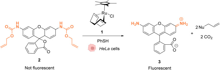
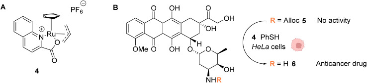
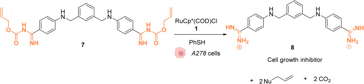
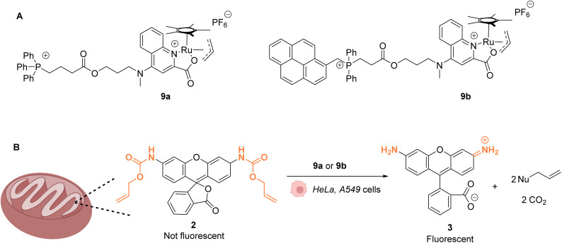
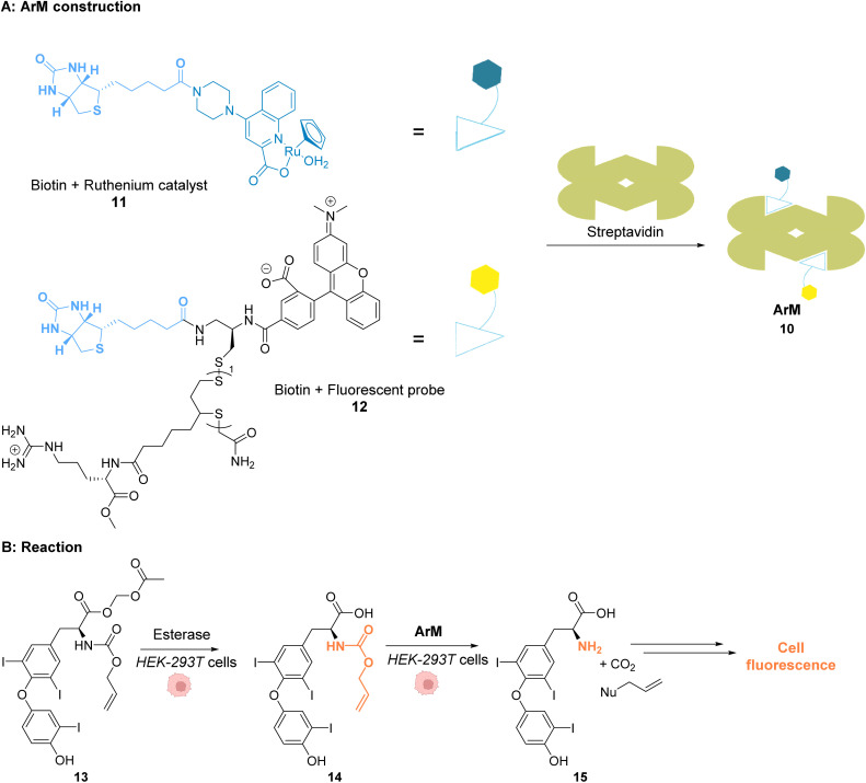
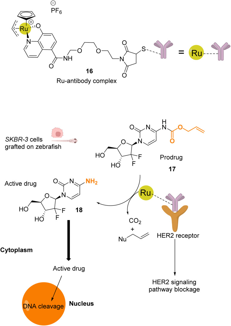
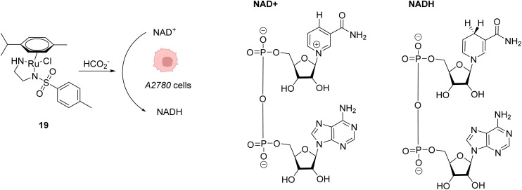
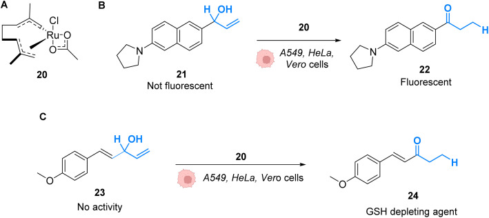
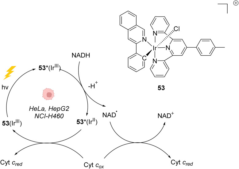
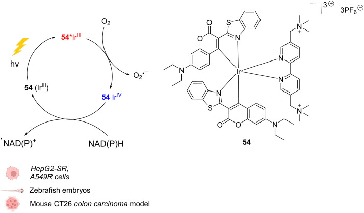
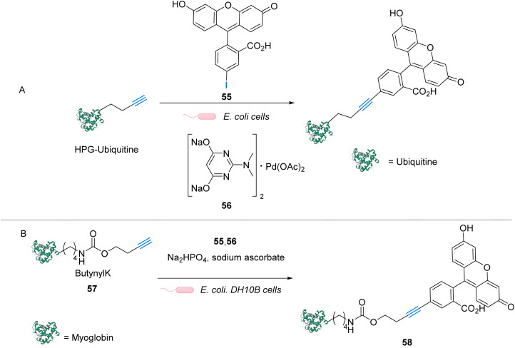
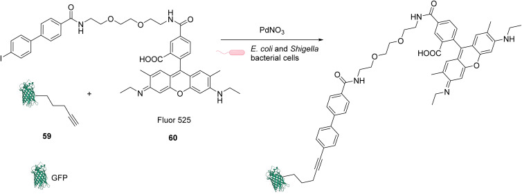
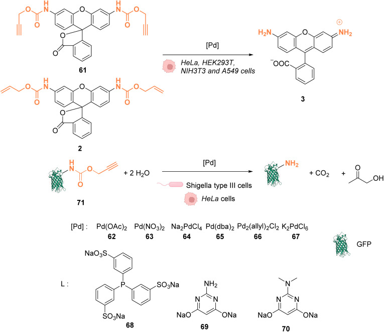
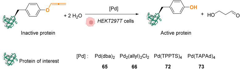
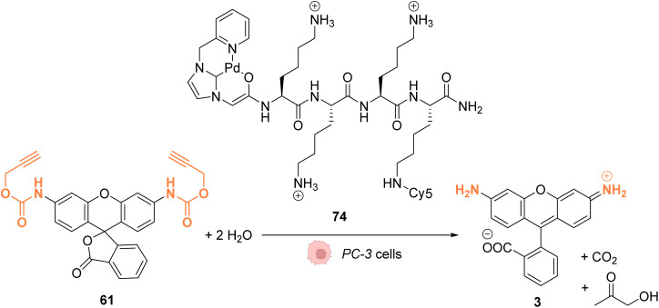
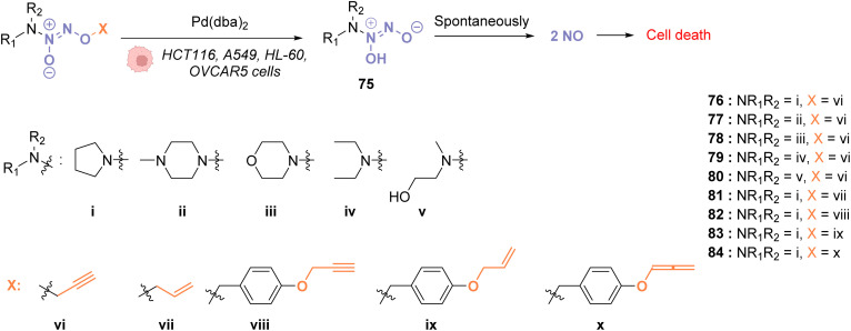
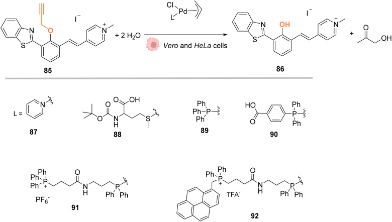
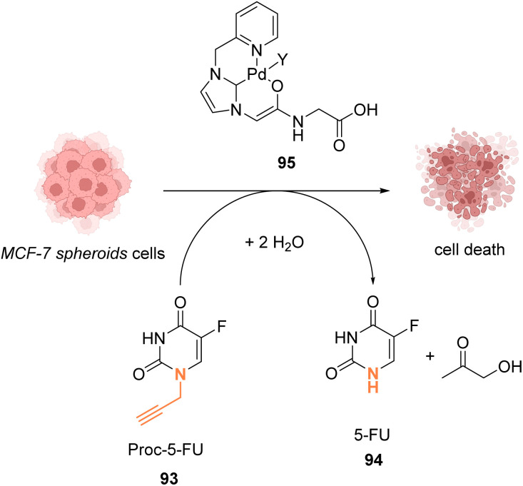
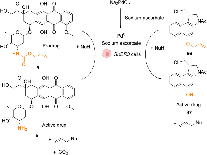
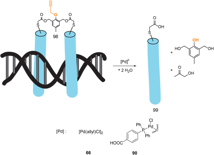
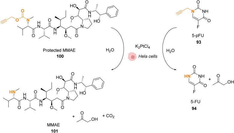
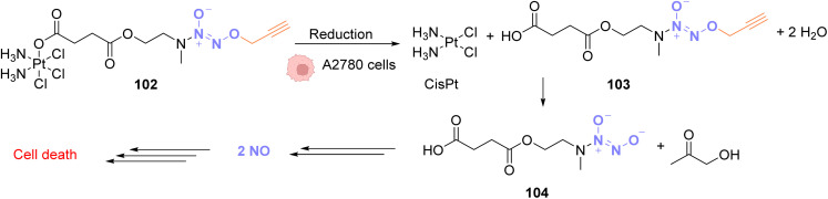
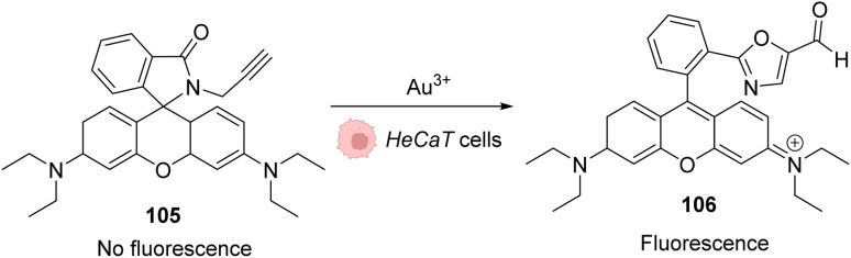
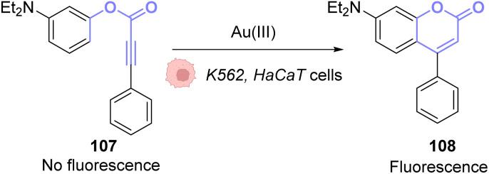
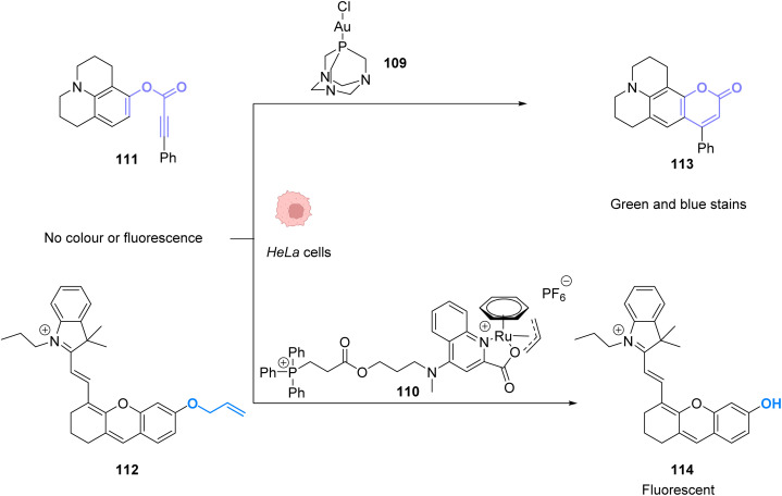
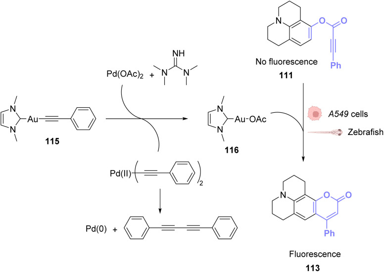
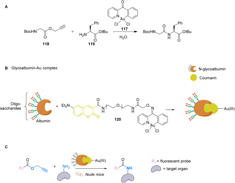
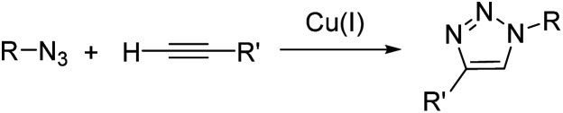
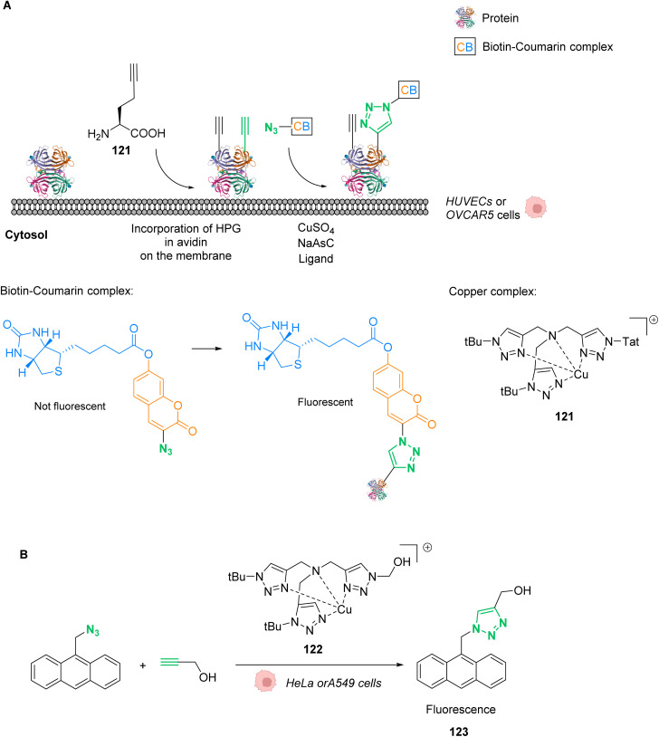
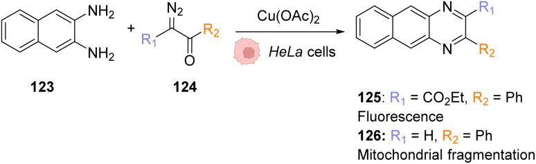
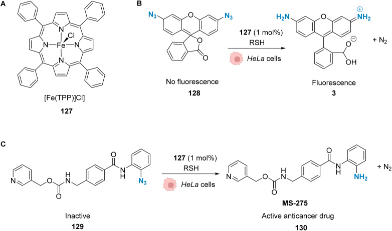
The development of organometallic catalysis has greatly expanded the synthetic chemist toolbox compared to only exploiting "classical" organic chemistry. Although more widely used in organic solvents, metal-based catalysts have also emerged as efficient tools for developing organic transformations in water, thus paving the way for further development of bio-compatible reactions. However, performing metal-catalysed reactions within living cells or organisms induces additional constraints to the design of reactions and catalysts. In particular, metal complexes must exhibit good efficiency in complex aqueous media at low concentrations, good cell specificity, good cellular uptake and low toxicity. In this review, we focus on the presentation of discrete metal complexes that catalyse or photocatalyse reactions within living cells or living organisms. We describe the different reaction designs that have proved to be successful under these conditions, which involve very few metals (Ir, Pd, Ru, Pt, Cu, Au, and Fe) and range from in cellulo deprotection/decaging/activation of fluorophores, drugs, proteins and DNA to in cellulo synthesis of active molecules, and protein and organelle labelling. We also present developments in bio-compatible photo-activatable catalysts, which represent a very recent emerging area of research and some prospects in the field.


Conflict of interest statement

There are no conflicts of interest to declare.

Figures

Fig. 1
Fig. 1. Ruthenium(ii)-catalysed Alloc deprotection of the caged fluorophore to rhodamine 110 in HeLa cells in the presence of thiols.
Fig. 2
Fig. 2. (A) Structure of the cationic ruthenium(iv) precatalyst 4; (B) Ru-induced uncaging of the anticancer drug doxorubicin inside HeLa cells.
Fig. 3
Fig. 3. Ru-catalysed decaging of double stranded DNA binding agents by [RuCp*(COD)Cl] (1) inside A278 cancer cells.
Fig. 4
Fig. 4. (A) Structure of mitochondria targeting RuIV complexes 9a and 9b; (B) catalysis of the uncaging of fluorophore rhodamine 110 inside HeLa and A549 cells, preferentially occurring inside mitochondria.
Fig. 5
Fig. 5. (A) Assembly of a cell-penetrating Ru-based artificial metalloenzyme (ArM). The ruthenium catalyst and the fluorescent probe/cell penetrating peptide are both attached to biotin units interacting with streptavidin to create the artificial metalloprotein; (B) the ArM catalyses the Alloc deprotection of compound 14, generating the hormone 15. The HEK-293T cells have been modified to become fluorescent once 15 is liberated.
Fig. 6
Fig. 6. Schematic illustration of HER2-targeted chemotherapy using a gemcitabine-based prodrug and the RuIV complex 16 as the catalyst. The prodrug is activated by the catalyst near the membrane and is taken up by the cell as an active anticancer drug. The reaction was performed in vivo inside zebrafish larvae grafted with SKBR-3 cells.
Fig. 7
Fig. 7. Induction of reductive stress in cancer cells by the RuII arene complex 19 in the presence of formate ions, leading to selective cancer cell death.
Fig. 8
Fig. 8. (A) Ruthenium(iv) complex 20 inducing intracellular isomerisation of allylic alcohols. (B) and (C) Generation of functional unsaturated ketones inside A549, HeLa and Vero cells.
Fig. 9
Fig. 9. (A) Ru complexes 1 and 25–26 used for intracellular [2 + 2 + 2] cycloaddition reactions; (B) intramolecular and (C) intermolecular cycloaddition reactions promoted by ruthenium complexes inside HeLa cells for the generation of a fluorescent probe (28) or an anthraquinone (31), respectively.
Fig. 10
Fig. 10. ROS generation by a ruthenium(ii) complex inside bacterial cells: (A) hydride transfer mechanism in formate abundant Gram+ strains and (B) SET mechanism in formate deficient strains.
Fig. 11
Fig. 11. Allylcarbamate (Alloc) cleavage by photo-activatable ruthenium(ii) precatalyst 34 inside HeLa cells allowing spatial and temporal control of the reaction.
Fig. 12
Fig. 12. (A) Mechanism of the photoreduction of the azide. (B) [RuII(bipy)3]-type complexes and azide/rhodamine substrates linked to protein ligands for an intracellular protein templated reaction induced by visible light irradiation. Photoreduction of the azide to the aniline leads to immolative linker decomposition and uncaging of rhodamine.
Fig. 13
Fig. 13. Nucleic acid templated reaction catalysed by [RuII(bipy)3]-type complexes under visible light irradiation for imaging of miRNAs in BT474 and HeLa cancer cells.
Fig. 14
Fig. 14. Azide reduction of a pro-fluorescent probe inside different cell lines by the ruthenium(ii) complex 36, which is conjugated with a protein ligand R, allowing specific localisation of the reaction in targeted parts of the cell by two-photon excitation at 730 nm.
Fig. 15
Fig. 15. Intracellular visible light-induced targeted protein modification by Single Electron Transfer (SET) catalysed by [RuII(bpy)3]2+ complex 39 in mouse erythrocytes. Labelling of carbonic anhydrase (CA) by using a benzene sulfonamide-conjugated Ru photocatalyst and a biotin N-modified tyrosyl radical trapping agent.
Fig. 16
Fig. 16. Ru-gefitinib photocatalyst 41 reported by Nakamura et al. for targeted knockdown of the epidermal growth factor receptor (EGFR) protein within A431 cells.
Fig. 17
Fig. 17. Photo-induced azide-thioalkyne cycloaddition promoted by ruthenium(ii) complexes 34 and 42–43 in HeLa cells.
Fig. 18
Fig. 18. Structures of IrIII complexes 44–45 inducing ROS production inside cancer cells and proposed reaction pathways for ROS generation and activation/deactivation of Ir complexes 44–45 inside mammalian cells.
Fig. 19
Fig. 19. Hydride transfer reaction catalysed by IrIII complex 46 inside NIH-3T3 mouse embryo fibroblast cells inducing intracellular fluorescence enhancement.
Fig. 20
Fig. 20. (A) Allylcarbamate (Alloc) cleavage reactions by IrI complexes 49–51 used in PBS buffer and (B) within HeLa cells for decaging of protected rhodamine (R = Alloc).
Fig. 21
Fig. 21. Photoredox IrIII-based catalytic cycle proposed by Sadler et al. for in cellulo light-induced NADH oxidation under hypoxic conditions.
Fig. 22
Fig. 22. Biocompatible IrIII complex 54 developed by Huang et al. and proposed photoredox IrIII/IrIV-based catalytic cycle for in cellulo light-induced ROS generation and NAD(P)H reduction in the presence of O2.
Fig. 23
Fig. 23. (A) Copper-free Sonogashira cross-coupling reaction inside E. coli for fluorescent labelling of HPG-Ub protein by modified fluorescein using a discrete PdII precatalyst. (B) Sonogashira cross-coupling labeling of Myoglobin inside E. coli.
Fig. 24
Fig. 24. Sonogashira cross-coupling inside E. coli or Shigella cells with Pd(NO3)2 as a precatalyst for coupling of fluorescent protein GFP with Fluor 525 and bioorthogonal labeling of toxin proteins in living pathogenic Shigella cells.
Fig. 25
Fig. 25. Alloc and propargyloxycarbonyl deprotection of profluorophores by palladium complexes inside cells.
Fig. 26
Fig. 26. Allenyl group cleavage on tyrosine residue inside HEKT297T cells to restore the activity of Tyr-dependent enzymes.
Fig. 27
Fig. 27. Propargyl deprotection of a profluorescent probe by a discrete palladium complex inside PC-3 cancer cells. The ligand is linked to a polycationic cell penetrating peptide to favor cellular uptake.
Fig. 28
Fig. 28. Cleavage of different protecting groups by Pd(dba)2 inducing NO release in living cancer cells.
Fig. 29
Fig. 29. Propargyl cleavage on a profluorophore catalysed by well-defined palladium(allyl) complexes in Vero and HeLa cells.
Fig. 30
Fig. 30. Propargyl cleavage of a prodrug inside MCF-7 cells and in a tumor model using a well-defined NHC-PdII precatalyst.
Fig. 31
Fig. 31. Allyl ether or allyl carbamate cleavage by a palladium complex inside SKBR3 cells.
Fig. 32
Fig. 32. Control of DNA binding based on a palladium-mediated uncaging process under mild and physiological conditions reported by Mascareñas et al.
Fig. 33
Fig. 33. Pentynoyl and propargyl amine deprotection of prodrugs by platinum(ii) complexes inside HeLa cells.
Fig. 34
Fig. 34. O 2-propargyl cleavage and NO release inside A2780 cells using a platinum(iv) precatalyst of low toxicity.
Fig. 35
Fig. 35. Generation of fluorescence inside HeCaT cells by the generation of 106 after incubation with AuIII.
Fig. 36
Fig. 36. Gold(iii)-catalysed hydroarylation of non-fluorescent 107 into a fluorescent coumarin.
Fig. 37
Fig. 37. Simultaneous AuI and RuII catalysis inside HeLa cells. Gold-catalysed hydroarylation and allyl-deprotection catalysed by Ru.
Fig. 38
Fig. 38. PdII-triggered transmetalation activates AuI compound 115 and induces hydroarylation of the coumarin precursor to form a fluorescent coumarin in A549 cancer cells and zebrafish.
Fig. 39
Fig. 39. (A) Cyclometalated AuIII complex 117 structure and reactivity. (B) Preparation of glycoalbumins as “transition-metal carriers” to produce glyco-Au complexes. (C) In vivo fluorescence labelling by 120-catalysed amide bond formation between propargyl ester-based imaging probes and surface.
Fig. 40
Fig. 40. CuAAC reaction.
Fig. 41
Fig. 41. (A) In cellulo CuAAC reaction between coumarin/biotin azides and HPG-incorporating proteins. (B) In cellulo CuAAC reaction that produces a fluorescent probe without the use of sodium ascorbate.
Fig. 42
Fig. 42. Copper-catalysed metal carbene transfer reaction in HeLa cells for the assembly of quinoxalines such as the fluorescent compound 125 and the toxic compound 126, capable of inducing mitochondrial fragmentation.
Fig. 43
Fig. 43. (A) Structure of a 5,10,15,20-tetraphenyl-21H,23H-porphine (TPP)-containing complex, [Fe(TPP)Cl]. (B) Iron-mediated reduction of a rhodamine 110 derivative to form fluorescent rhodamine 110. (C) Reduction of the azide molecule 129 to the anticancer drug candidate MS-275.
None
Hugo Madec
None
Francisca Figueiredo
None
Kevin Cariou
None
Sylvain Roland
None
Matthieu Sollogoub
None
Gilles Gasser

Similar articles

Cited by

References

    1. Chen J. Wang J. Li K. Wang Y. Gruebele M. Ferguson A. L. Zimmerman S. C. Polymeric “Clickase” Accelerates the Copper Click Reaction of Small Molecules, Proteins, and Cells. J. Am. Chem. Soc. 2019;141(24):9693–9700. doi: 10.1021/jacs.9b04181. doi: 10.1021/jacs.9b04181. - DOI - DOI - PubMed
    1. Luther D. C. Huang R. Jeon T. Zhang X. Lee Y.-W. Nagaraj H. Rotello V. M. Delivery of Drugs, Proteins, and Nucleic Acids Using Inorganic Nanoparticles. Adv. Drug Deliv. Rev. 2020;156:188–213. doi: 10.1016/j.addr.2020.06.020. doi: 10.1016/j.addr.2020.06.020. - DOI - DOI - PMC - PubMed
    1. Liu Y. Bai Y. Design and Engineering of Metal Catalysts for Bio-Orthogonal Catalysis in Living Systems. ACS Appl. Bio Mater. 2020;3(8):4717–4746. doi: 10.1021/acsabm.0c00581. doi: 10.1021/acsabm.0c00581. - DOI - DOI - PubMed
    1. Hambley T. W. Developing New Metal-Based Therapeutics: Challenges and Opportunities. Dalton Trans. 2007;43:4929. doi: 10.1039/b706075k. doi: 10.1039/B706075K. - DOI - DOI - PubMed
    1. Patra M. Gasser G. Organometallic Compounds: An Opportunity for Chemical Biology? ChemBioChem. 2012;13(9):1232–1252. doi: 10.1002/cbic.201200159. doi: 10.1002/cbic.201200159. - DOI - DOI - PubMed