

Case report: Large-size intramuscular nodular fasciitis, a challenging histopathologic diagnosis confirmed by molecular detection of USP6 gene rearrangement: Case report and literature review
- PMID: 36741963
- PMCID: PMC9894875
- DOI: 10.3389/pore.2023.1610785
Case report: Large-size intramuscular nodular fasciitis, a challenging histopathologic diagnosis confirmed by molecular detection of USP6 gene rearrangement: Case report and literature review
Abstract
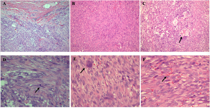
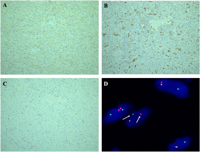
The intramuscular subtype of nodular fasciitis (NF) is rare with lesions normally not more than 2 cm in size and characterized by pseudosarcomatous morphology. We report a case of a 27-year-old man with a large-size intramuscular NF. The patient came for treatment complaining of an increasingly enlarged mass in the left upper arm for 4 months. Magnetic resonance imaging (MRI) confirmed the presence of a well-defined tumor measuring 5 cm within the outer edge of the middle humerus. Microscopically, the neoplasm was rich in fibroblasts and myofibroblasts in an interlaced pattern with high mitotic index and evident multinuclear giant cells. Erythrocyte extravasation was easily seen in the stroma. The tumor border was infiltrative. Immunohistochemically, the tumor cells were positive for smooth muscle actin (SMA) and negative for cytokeratin, desmin, H-Caldesmon, CD34, S100, ALK, and β-catenin. Fibrosarcoma was highly suspected by histopathological and immunohistochemical examination. Molecular detection demonstrated evidence of ubiquitin-specific peptidase 6 (USP6) gene rearrangement in this tumor. Based on the findings, the tumor was diagnosed as intramuscular NF. At 56 months after the initial surgery, the patient had recovered with no evidence of recurrence or metastasis. Large-size intramuscular NF is very rare and easily overdiagnosed as malignant tumor due to its obvious pseudosarcomatoid pathological features. USP6 gene rearrangement detection can effectively avoid this major misdiagnosis.
Keywords: USP6; gene rearrangement; intramuscular; large-size; nodular fasciitis.
Copyright © 2023 Wang, Wang, Xu and Xiang.
Conflict of interest statement
The authors declare that the research was conducted in the absence of any commercial or financial relationships that could be construed as a potential conflict of interest.
Figures

References
-
- Chen J, Ye X, Li Y, Wei C, Zheng Q, Zhong P, et al. Chromosomal translocation involving USP6 gene in nodular fasciitis. Zhonghua Bing Li Xue Za Zhi (2014) 43:533–6. - PubMed
-
- Erber R, Agaimy A. Misses and near misses in diagnosing nodular fasciitis and morphologically related reactive myofibroblastic proliferations: Experience of a referral center with emphasis on frequency of USP6 gene rearrangements. Virchows Arch (2018) 473:351–60. 10.1007/s00428-018-2350-0 - DOI - PubMed
Publication types
MeSH terms
Substances
LinkOut - more resources
Full Text Sources