. 2023 Jan 20:13:882635.
doi: 10.3389/fneur.2022.882635. eCollection 2022.

Genetic removal of synaptic Zn2+ impairs cognition, alters neurotrophic signaling and induces neuronal hyperactivity

Affiliations

Genetic removal of synaptic Zn2+ impairs cognition, alters neurotrophic signaling and induces neuronal hyperactivity

Emily C Vogler et al. Front Neurol. .

Abstract

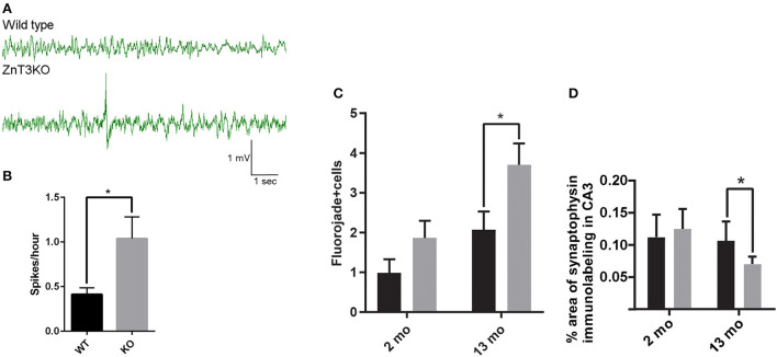
Vesicular Zn2+ (zinc) is released at synapses and has been demonstrated to modulate neuronal responses. However, mechanisms through which dysregulation of zinc homeostasis may potentiate neuronal dysfunction and neurodegeneration are not well-understood. We previously reported that accumulation of soluble amyloid beta oligomers (AβO) at synapses correlates with synaptic loss and that AβO localization at synapses is regulated by synaptic activity and enhanced by the release of vesicular Zn2+ in the hippocampus, a brain region that deteriorates early in Alzheimer's disease (AD). Significantly, drugs regulating zinc homeostasis inhibit AβO accumulation and improve cognition in mouse models of AD. We used both sexes of a transgenic mouse model lacking synaptic Zn2+ (ZnT3KO) that develops AD-like cognitive impairment and neurodegeneration to study the effects of disruption of Zn2+ modulation of neurotransmission in cognition, protein expression and activation, and neuronal excitability. Here we report that the genetic removal of synaptic Zn2+ results in progressive impairment of hippocampal-dependent memory, reduces activity-dependent increase in Erk phosphorylation and BDNF mRNA, alters regulation of Erk activation by NMDAR subunits, increases neuronal spiking, and induces biochemical and morphological alterations consistent with increasing epileptiform activity and neurodegeneration as ZnT3KO mice age. Our study shows that disruption of synaptic Zn2+ triggers neurodegenerative processes and is a potential pathway through which AβO trigger altered expression of neurotrophic proteins, along with reduced hippocampal synaptic density and degenerating neurons, neuronal spiking activity, and cognitive impairment and supports efforts to develop therapeutics to preserve synaptic zinc homeostasis in the brain as potential treatments for AD.

Keywords: Alzheimer's disease (AD); ZnT3; hippocampus; neurodegeneration; neuronal hyperactivity; neurotrophic signaling; synaptic zinc; zinc transporter.


Conflict of interest statement

The authors declare that the research was conducted in the absence of any commercial or financial relationships that could be construed as a potential conflict of interest.

Figures

Figure 1
Figure 1
Age-dependent deficits in hippocampal-dependent memory in ZnT3KO mice. Object location memory (OLM) task discrimination index shows age-dependent memory impairment in ZnT3KO mice, becoming significant at 3 months of age and profound by 6 months. WT, wild type, n = 8–13; KO, ZnT3KO, n = 14–16; DI, discrimination index [(novel-familiar)/(novel+familiar)*100]; *p < 0.05, **p < 0.01. Data were analyzed by two-tailed unpaired t-test. Error bars indicate the mean ± SEM.
Figure 2
Figure 2
Protein level and activity-dependent phosphorylation of AKT and Erk1/2 are reduced in ZnT3KO hippocampus. (A) Basal levels of AKT and Erk1/2 proteins are reduced in ZnT3KO. (B) Incubation with 20 mM KCl results in a greater increase in p-Erk1/2 in WT than in ZnT3KO hippocampus. WT, wild type; KO, ZnT3KO; AKT, Erk1/2, n = 6; p-Erk1/2, WT, n = 4, KO, n = 5; NT, no treatment (vehicle); KCl, 10-min incubation in 20 mM KCl; *p < 0.05, **p < 0.01; values expressed as normalized densitometry relative to GAPDH. Data were analyzed by two-tailed unpaired t-test. Error bars indicate the mean ± SEM.
Figure 3
Figure 3
Activity-dependent phosphorylation of Erk1/2 is differentially regulated by NMDAR subunits in ZnT3KO hippocampus. (A) Treatment with ifenprodil, an antagonist of the NMDAR NR2B subunit, showed a decrease in activity-dependent Erk1/2 phosphorylation with a similar percent decrease in both WT and ZnT3KO mice. (B) The activity-dependent phosphorylation of Erk1/2 is increased by incubation with the NR2A antagonist PEAQX, with a significant percent increase in ZnT3KO. WT, wild type; KO, ZnT3KO; ifenprodil, WT, n = 4 KO, n = 6; PEAQX, WT and KO, n = 5; K, 10-min incubation in 20 mM KCl; K/I, 10-min co-incubation with 20 mM KCl and 50 μM ifenprodil: K/P, 30-min incubation with 0.5 μM PEAQX followed by 10-min co-incubation with 0.5 μM PEAQX/20 mM KCl; *p < 0.05, ***p < 0.0005; values expressed as normalized densitometry relative to GAPDH. Data were analyzed by two-tailed unpaired t-test. Error bars indicate the mean ± SEM.
Figure 4
Figure 4
Activity and age-dependent alterations in BDNF expression in ZnT3KO hippocampus. (A) Age-dependent alterations in BDNF protein levels were assessed in hippocampus tissue from age-matched WT and ZnT3KO mice by Western blot analysis, finding that ZnT3KO hippocampus has an age-dependent decrease in BDNF protein levels, becoming significant by 15 months of age. (B) Activity-dependent increase in BDNF mRNA was assessed through RT-PCR and qPCR analysis in acute hippocampal slices from 3-4-week-old WT and ZnT3KO after 3.5-h incubation in 20 mM KCl. WT, wild type; KO, ZnT3KO; BDNF immunoblots, 6 mo, n = 6; 12 mo, n = 5; 15 mo, n = 6; BDNF mRNA, n = 4; NT, no treatment (vehicle); KCl, 3.5 h incubation in 20 mM KCl, n = 6; *p < 0.05, ****p < 0.0001; immunoblot values expressed as normalized densitometry relative to GAPDH; BDNF mRNA values expressed as fold change ratio to actin. Data were analyzed by two-tailed unpaired t-test. Error bars indicate the mean ± SEM.
Figure 5
Figure 5
ZnT3KO hippocampus exhibit age-dependent alterations in calbindin and NPY expression that are consistent with alterations resulting from epileptiform activity. (A) Western blot analysis of hippocampal tissue shows an age-dependent decrease of calbindin protein levels in ZnT3KO mice. (B) Quantification of calbindin immunolabeling shows reduced calbindin in the dentate gyrus, but not in the CA1, region of ZnT3KO hippocampus. (C) Representative images of 12-month-old ZnT3KO and WT mice quantified in (B). (D) Dot blot analysis of hippocampal tissue shows an age-dependent increase in NPY protein levels in ZnT3KO mice. (E) Quantification of NPY immunolabeling shows NPY expression is increased in the CA3 region of ZnT3KO hippocampus. (F) Representative images of 12-month old ZnT3KO and WT hippocampus quantified in (E). WT, wild type; KO, ZnT3KO; calbindin immunoblots, 3 and 6 mos. n = 5, 12 mo. n = 8, 15 mo. n = 7; NPY dot blots, 3 and 15 mos. n = 4, 6 mo. n = 3, 12 mo. n = 5; scale bars a and d, 100 μm; b, c, e, and f, 10 μm; *p < 0.05, **p < 0.001; two fields from each subject was imaged at 63x magnification, quantified using the Axiovision (Zeiss) automated measurement program and the percent area calculated. Immunoblot values expressed as normalized densitometry relative to GAPDH or MAP2. Data were analyzed by two-tailed unpaired t-test. Error bars indicate the mean ± SEM.
Figure 6
Figure 6
Aberrant, age-dependent mossy fiber sprouting in ZnT3KO hippocampus. (A) Quantification of synaptoporin puncta shows an age-dependent increase in mossy sprouting in the molecular layer of the dentate gyrus in ZnT3KO hippocampus. (B) Representative image of synaptoporin immunolabeling quantified in A and magnification of KO inset box. (C) Quantification of the percent area of GAP43 immunolabeling in the molecular layer of the dentate gyrus shows an age-dependent increase in ZnT3KO hippocampus. (D) Representative image of GAP43 immunolabeling quantified in C and magnification of KO inset box. WT, wild type; KO, ZnT3KO; synaptoporin, 6 mo. n = 10, 13 mo. WT n = 8, KO n = 11; scale bars 100 μm; *p < 0.05, ***p < 0.0002; three fields from each subject were imaged at 63X, puncta counted by an experimenter blinded to subject conditions and values expressed as fold change relative to WT puncta for each subject; GAP43, n = 5; ten fields from each subject were imaged at 63X, quantified using the Axiovision (Zeiss) automated measurement program and the average percent area calculated. Data were analyzed by two-tailed unpaired t-test. Error bars indicate the mean ± SEM.
Figure 7
Figure 7
ZnT3KO mice exhibit elevated epileptiform spiking activity, fluorojade+ cells, and reduced synaptophysin expression. (A) Representative EEG of 2-month-old WT and ZnT3KO shows spiking epileptiform activity in ZnT3KO. (B) 2-month-old ZnT3KO have an elevated rate of spikes per hour. n = 3. EEG data during movement detected by implanted Neurologger recording device was manually removed and raw EEG data was bandpass filtered from 1 to 80 Hz. Spike threshold was a peak amplitude 2.5X greater than the average baseline. (C) Image analysis of immunolabeling in hippocampus CA3 region shows an age-dependent decrease in synaptophysin protein levels in ZnT3KO. 2 mo: WT, n = 8, KO, n = 9; 13 mo: n = 6; scale bars 20 μm; one field from each subject was imaged at 63X, quantified using the Axiovision (Zeiss) automated measurement program and the average percent area calculated. (D) Quantification of Fluorojade+ cells counted in the CA3 region of the hippocampus show that there is an increased number in 13-month-old KO. 2 mo: WT, n = 11, KO, n = 9; 13 mo. WT, n = = 9, ZnT3KO n = 11; the CA3 region was imaged at 20X magnification using the Mosaic tool in Axiovision to image and stitch together adjacent fields and the number of Fluorojade positive cells were counted by an experimenter blinded to subject conditions. *p < 0.05. Data were analyzed by two-tailed unpaired t-test. Error bars indicate the mean ± SEM.

References

    1. Frederickson CJ. Neurobiology of zinc and zinc-containing neurons. Int Rev Neurobiol. (1989) 31:145–238. 10.1016/s0074-7742(08)60279-2 - DOI - PubMed
    1. Palmiter RD, Cole TB, Quaife CJ, Findley SD. ZnT-3, a putative transporter of zinc into synaptic vesicles. Proc Natl Acad Sci U S A. (1996) 93:14934–9. 10.1073/pnas.93.25.14934 - DOI - PMC - PubMed
    1. Hwang JJ, Park MH, Choi SY, Koh JY. Activation of the Trk signaling pathway by extracellular zinc. Role of metalloproteinases J Biol Chem. (2005) 280:11995–2001. 10.1074/jbc.M403172200 - DOI - PubMed
    1. Besser L, Chorin E, Sekler I, Silverman WF, Atkin S, Russell JT, et al. . Synaptically released zinc triggers metabotropic signaling via a zinc-sensing receptor in the hippocampus. J Neurosci. (2009) 29:2890–901. 10.1523/jneurosci.5093-08.2009 - DOI - PMC - PubMed
    1. Paoletti P, Vergnano AM, Barbour B, Casado M. Zinc at glutamatergic synapses Neuroscience. (2008) 158:126–136. 10.1016/jneuroscience01 - DOI - PubMed