. 2022 Nov 22;15(3):e23.
doi: 10.12786/bn.2022.15.e23. eCollection 2022 Nov.

Classification and Diagnosis of Adult Glioma: A Scoping Review

Affiliations

Classification and Diagnosis of Adult Glioma: A Scoping Review

Yoon Hwan Byun et al. Brain Neurorehabil. .

Abstract

Gliomas are primary central nervous system tumors that arise from glial progenitor cells. Gliomas have been classically classified morphologically based on their histopathological characteristics. However, with recent advances in cancer genomics, molecular profiles have now been integrated into the classification and diagnosis of gliomas. In this review article, we discuss the clinical features, imaging findings, and molecular profiles of adult-type diffuse gliomas based on the new 2021 World Health Organization Classifications of Tumors of the central nervous system.

Keywords: Brain Neoplasms; Classification; Diagnosis; Glioma.


Conflict of interest statement

Conflict of Interest: The authors have no potential conflicts of interest to disclose.

Figures

Fig. 1
Fig. 1. The 2021 WHO classification of adult-type diffuse gliomas.
Adult-type diffuse gliomas are classified as: 1) astrocytoma, IDH-mutant, 2) oligodendroglioma, IDH-mutant and 1p/19q-codeleted, and 3) glioblastoma, IDH-wildtype. WHO, World Health Organization; IDH, isocitrate dehydrogenase; ATRX, alpha-thalassemia/mental retardation, X-linked; CDKN2A/B, cyclin-dependent kinase inhibitor 2A/B; EGFR, epidermal growth factor receptor; TERT, telomerase reverse transcriptase; MGMT, O-5-methylguanine-DNA methyltransferase. *IDH-wildtype glioma without high-grade features (WHO grade 2/3) should be investigated further and classified into other categories.
Fig. 2
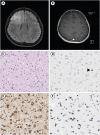
Fig. 2. Diffuse astrocytoma, IDH-mutant, grade 3.
(A) A T2-FLAIR image and (B) a T1 contrast-enhanced image show an ill-defined T2 high SI lesion without significant contrast enhancement in the right frontal cortex. The tumor shows (C) increased cellularity and nuclear polymorphism, (D) an intermediate mitotic count (3/10 HPF) on PHH3 immunohistochemistry (arrowhead), (E) IDH1 mutation positivity, and (F) ATRX mutation positivity (ATRX loss). FLAIR, fluid-attenuated inversion recovery; SI, signal intensity; PHH3, phosphohistone H3; HPF, high-power field; IDH, isocitrate dehydrogenase, ATRX, alpha-thalassemia/mental retardation, X-linked.
Fig. 3
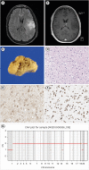
Fig. 3. Oligodendroglioma, IDH-mutant, 1p/19q-codeleted.
(A) A T2-FLAIR image and (B) a T1 contrast-enhanced image show an ill-defined T2 high-SI lesion without significant contrast enhancement in the left frontoparietal lobe. (C) Gross photograph of the resected tumor. The tumor shows (D) a “fried egg” appearance with uniformly rounded nuclei and clear halos, (E) IDH1 mutation positivity, (F) ATRX mutation negativity, and (G) 1p/19q co-deletion. IDH, isocitrate dehydrogenase; FLAIR, fluid-attenuated inversion recovery; SI, signal intensity; ATRX, alpha-thalassemia/mental retardation, X-linked; CNV, copy number variation.
Fig. 4
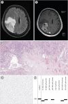
Fig. 4. Glioblastoma, IDH-wild type.
(A) A T2-FLAIR image and (B) a T1 contrast-enhanced image show an enhancing mass in the left parietal lobe with multiple necrosis and perilesional T2 high-SI infiltrations. The tumor shows (C) necrosis and microvascular proliferation, (D) IDH1 mutation negativity, and (E) positive results for MGMT methylation. IDH, isocitrate dehydrogenase; FLAIR, fluid-attenuated inversion recovery; SI, signal intensity; MGMT, O-5-methylguanine-DNA methyltransferase.

References

    1. Sanai N, Alvarez-Buylla A, Berger MS. Neural stem cells and the origin of gliomas. N Engl J Med. 2005;353:811–822. - PubMed
    1. Weller M, van den Bent M, Preusser M, Le Rhun E, Tonn JC, Minniti G, Bendszus M, Balana C, Chinot O, Dirven L, French P, Hegi ME, Jakola AS, Platten M, Roth P, Rudà R, Short S, Smits M, Taphoorn MJ, von Deimling A, Westphal M, Soffietti R, Reifenberger G, Wick W. EANO guidelines on the diagnosis and treatment of diffuse gliomas of adulthood. Nat Rev Clin Oncol. 2021;18:170–186. - PMC - PubMed
    1. Kang H, Song SW, Ha J, Won YJ, Park CK, Yoo H, Jung KW. A nationwide, population-based epidemiology study of primary central nervous system tumors in Korea, 2007-2016: a comparison with United States data. Cancer Res Treat. 2021;53:355–366. - PMC - PubMed
    1. Cancer Genome Atlas Research Network. Comprehensive genomic characterization defines human glioblastoma genes and core pathways. Nature. 2008;455:1061–1068. - PMC - PubMed
    1. Louis DN, Perry A, Wesseling P, Brat DJ, Cree IA, Figarella-Branger D, Hawkins C, Ng HK, Pfister SM, Reifenberger G, Soffietti R, von Deimling A, Ellison DW. The 2021 WHO classification of tumors of the central nervous system: a summary. Neuro-oncol. 2021;23:1231–1251. - PMC - PubMed

Publication types