

Robot-Assisted Removal of a Partially Intravesical Intrauterine Device (IUD) and Large Bladder Stone
- PMID: 36742352
- PMCID: PMC9897916
- DOI: 10.1155/2023/8074689
Robot-Assisted Removal of a Partially Intravesical Intrauterine Device (IUD) and Large Bladder Stone
Abstract
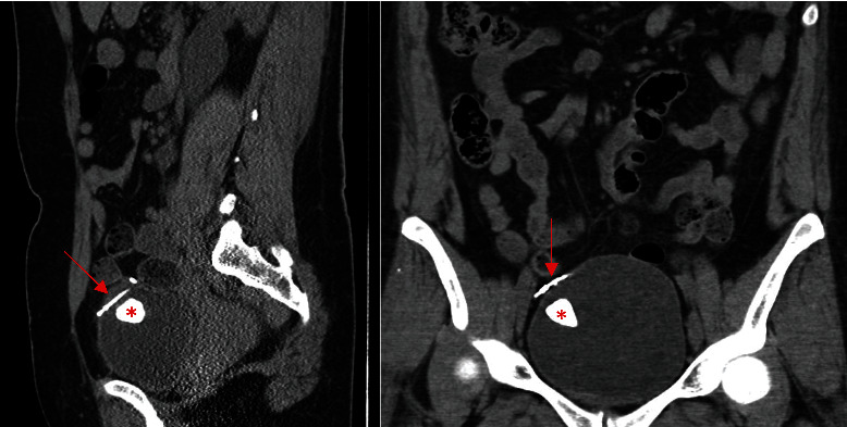
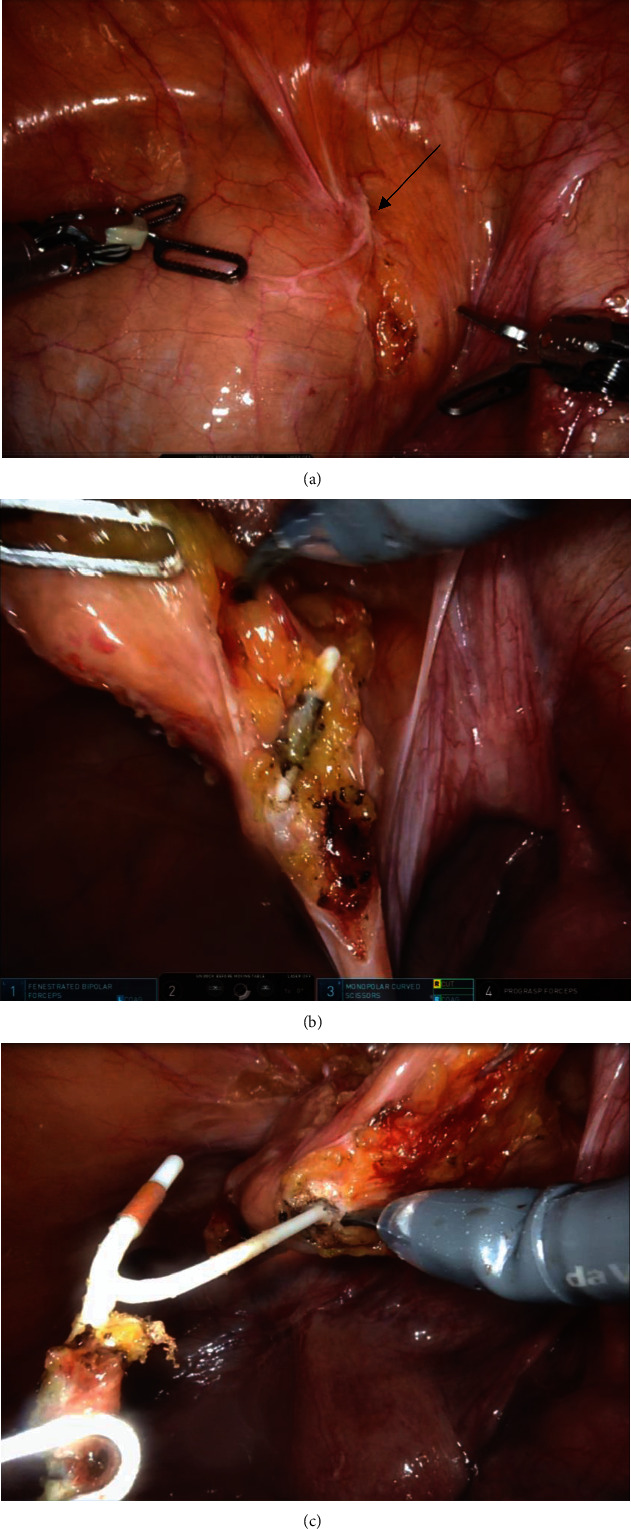
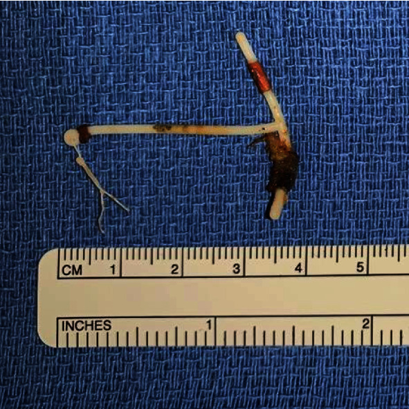
An intrauterine device (IUD) is a highly effective and widely utilized option for long-acting reversible contraception. IUDs are generally well-tolerated with a low rate of serious complications. Perforation of an IUD through the uterine wall and into the urinary bladder is a rare event that may be asymptomatic. The approach for surgical removal primarily depends on the location of the device. We present a case report of a 41-year-old woman who was found to have a partially intravesical IUD and associated 2.4 cm bladder calculus. Removal of the intravesical IUD and stone was achieved with cystoscopy, cystolitholapaxy, and robot-assisted laparoscopic cystotomy.
Copyright © 2023 Cassandra Heaney et al.
Conflict of interest statement
The authors declare that here is no conflict of interest.
Figures
Similar articles
-
Perforation of the bladder by the intrauterine device.Obstet Gynecol Surv. 1984 Feb;39(2):59-66. doi: 10.1097/00006254-198402000-00001. Obstet Gynecol Surv. 1984. PMID: 6229704 Review.
-
Case report: Bladder stone caused by dislocated Cu-IUD.Int J Surg Case Rep. 2024 Dec;125:110562. doi: 10.1016/j.ijscr.2024.110562. Epub 2024 Nov 7. Int J Surg Case Rep. 2024. PMID: 39522410 Free PMC article.
-
Using laparoscope to remove an ectopic intrauterine device in the anterior wall of urinary bladder: A case report.World J Clin Cases. 2024 Jun 16;12(17):3221-3225. doi: 10.12998/wjcc.v12.i17.3221. World J Clin Cases. 2024. PMID: 38898866 Free PMC article.
-
A Case Report of Intrauterine Device Migration: Uterine Penetration and Bladder Involvement with Secondary Stones 3 Years Post-Insertion.Int J Womens Health. 2024 Nov 9;16:1903-1907. doi: 10.2147/IJWH.S492865. eCollection 2024. Int J Womens Health. 2024. PMID: 39539644 Free PMC article.
-
Intrauterine devices migrated into the bladder: two case reports and literature review.BMC Womens Health. 2021 Aug 16;21(1):301. doi: 10.1186/s12905-021-01443-w. BMC Womens Health. 2021. PMID: 34399735 Free PMC article. Review.
Cited by
-
Migrated intra-uterine device to infra-umbilical skin: a rare case report.BMC Womens Health. 2024 Dec 30;24(1):672. doi: 10.1186/s12905-024-03522-0. BMC Womens Health. 2024. PMID: 39736668 Free PMC article.
-
Intrauterine device migration into the bladder with urolithiasis: A case report.Int J Surg Case Rep. 2025 Jun;131:111409. doi: 10.1016/j.ijscr.2025.111409. Epub 2025 May 10. Int J Surg Case Rep. 2025. PMID: 40349496 Free PMC article.
References
-
- United Nations. Contraceptive Use by Method 2019: Data Booklet . UN iLibrary; 2019. Department of Economic and Social Affairs, population division. - DOI
Publication types
LinkOut - more resources
Full Text Sources
