. 2022 Dec;74(Suppl 3):5782-5785.
doi: 10.1007/s12070-020-02308-8. Epub 2021 Jan 23.

Hyoid Chondroma

Affiliations

Hyoid Chondroma

Satish Chandra Tripuraneni et al. Indian J Otolaryngol Head Neck Surg. 2022 Dec.

Abstract

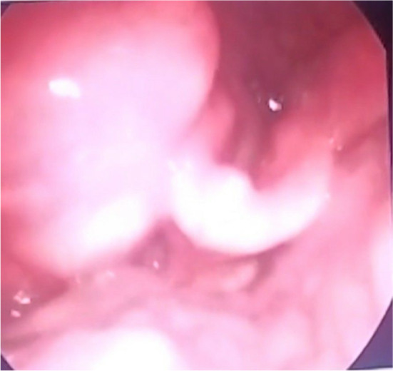
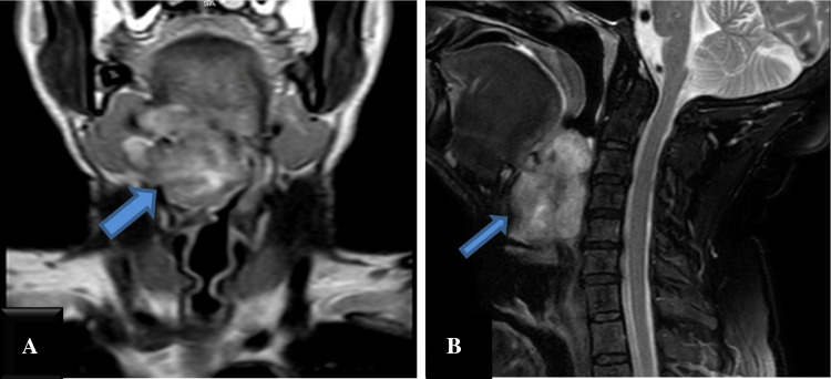
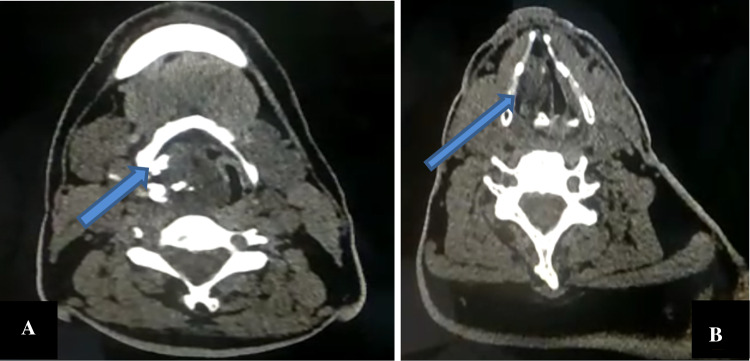
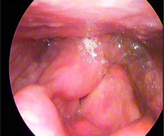
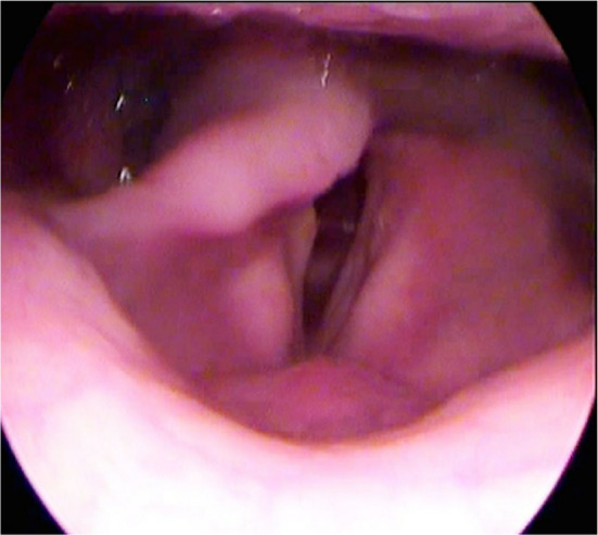
Mass lesions of the larynx are one of the most common clinical entity which we come across in routine otorhinolaryngology and head neck practice with varied symptomatology. Among all the mass lesions of the larynx, Epithelial neoplasms constitute up to 97%. Mesenchymal tumours of the larynx constitute only 0.3-1.0% of all the laryngeal tumors. Abundance of cartilage structures in the larynx made it a spot for mesenchymal tumors [chondromas and chandrosacrcomas]. The spectrum of mesenchymal neoplasms can vary from chondromas, chondroblastoma to chondrosarcoma. Here we want to share our experience of a mesenchymal tumour of the larynx. This case is reported for the rarity and ambiguity in diagnosis. Though these are slow-growing tumours with an early presentation, in our case, the patient had a supportive tracheostomy without definitive treatment for more than 2 years. We managed this patient by excising the mass by lateral pharyngotomy with the preservation of larynx followed by successful Decannulation in 20 days.

Keywords: Cartilaginous tumours; Hyoid chondroma; Larynx; Lateral pharyngotomy.


Figures

Fig. 1
Fig. 1
VLS examination showing a Supraglottic mass on right side extending from the right pyriform fossa, compressing the right aryepiglottic fold and narrowing the laryngeal inlet
Fig. 2
Fig. 2
T2-weighted MRI of neck a coronal section b sagittal section showing broad right sided neck mass lesion (arrow), spanning over right parapharyngeal space, pharyngeal mucosa, deep parotid space, and also extending to the left side infiltrating oropharynx, the upper part of the hypopharynx and supraglottic larynx reaching up to glottic area causing airway obstruction
Fig. 3
Fig. 3
Non contrast CT Axial section a at the level of oropharynx and hyoid bone showing well defined lobulated hyperdense mass lesion with multiple calcifications extending across Rt parapharyngeal space, oropharynx with erosion of adjacent hyoid bone b at the level of vocal cord showing narrowing of the airway
Fig. 4
Fig. 4
a, b Intraoperative images showing subplatysmal plane c excised specimen of chondroma d image showing mass attached to greater cornu of the hyoid bone(arrow)
Fig. 5
Fig. 5
Videolaryngoscopy on postoperative day 7 showing the larynx free of mass
Fig. 6
Fig. 6
Videolaryngoscopy during follow up after 2 months showing the adequate airway

References

    1. Thomé R, Thomé DC, De La Cortina RAC. Long-term follow-up of cartilaginous tumors of the larynx. Otolaryngol Head Neck Surg. 2001;124(6):634–640. - PubMed
    1. Travers F. A case of ossification and bony growth of the cartilages of the larynx. Med Chir Trans. 1816;7:150. doi: 10.1177/095952871600700109. - DOI - PMC - PubMed
    1. Dalla Palma P, Piazza M. Peripheral chondrosarcoma of the hyoid bone: report of a case. Appl Pathol. 1983;1:333–338. - PubMed
    1. Grayson A, Bain M. Juxtacortical chondroma of the hyoid bone: report of a case. Arch Otolaryngol. 1967;86:679–680. doi: 10.1001/archotol.1967.00760050681015. - DOI - PubMed
    1. Umekita Y, Yoshida A, Yoshida H. Parosteal chondrosarcoma attached to the hyoid bone. Int J Oral Maxillofac Surg. 1998;27:215–216. doi: 10.1016/S0901-5027(98)80014-3. - DOI - PubMed

LinkOut - more resources