

Giant Osteoma of the Mandible: Report of a Rare Case with Review of Literature
- PMID: 36742645
- PMCID: PMC9895695
- DOI: 10.1007/s12070-021-02565-1
Giant Osteoma of the Mandible: Report of a Rare Case with Review of Literature
Abstract
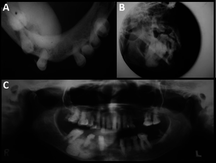
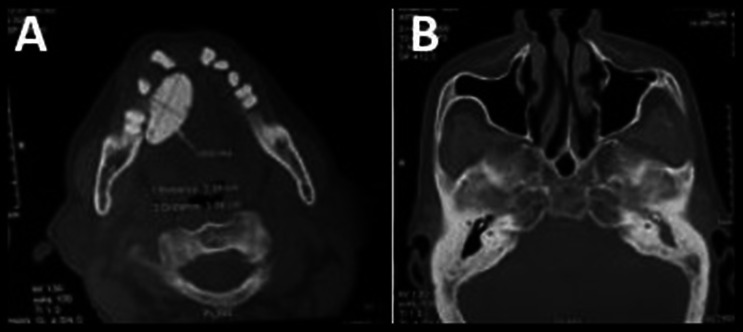
Osteoma is a slow growing, asymptomatic, benign bony tumor composed of compact and cancellous bones. Central, peripheral, and extra skeletal osteomas are the three types based on the site of origin. They are mostly observed on routine radiographic screening, mostly in the paranasal sinuses. Gnathic involvement is an uncommon occurrence, and if present, mandibular involvement is more frequently seen. Mostly, osteomas are small asymptomatic lesions and very rarely they become symptomatic and acquire larger size. Multiple osteomas are a feature of Gardner's syndrome; however, solitary osteomas are non-syndromic. Oral health professional may be the first to diagnose Gardner's syndrome as the osteomas may be initial manifestation of the disorder. Treatment protocol of osteomas varies based on the associated signs and symptoms. Small, asymptomatic cases are treated conservatively by periodic clinical and radiographic evaluation. However, larger, symptomatic lesions require surgical intervention. Herby, reporting an unusual case of Giant peripheral osteoma of the mandible. Our case is unique in few aspects because of its unusually large size (5 × 4 cm) and involvement of lingual aspect of the mandible in the region of sublingual fossa, with compression of the floor of mouth.
Keywords: Gardner’s syndrome; Giant osteoma; Mandible; Osteoma.
© Association of Otolaryngologists of India 2021.
Conflict of interest statement
Conflict of interestThe author declares that they have no conflict of interest.
Figures


References
-
- Fourcade A, Salmon B, Pelletier FL, Ejeil AL. Peripheral osteoma of the mandibular crest: a short case study. J Oral Med Oral Surg. 2018;24:29–32. doi: 10.1051/mbcb/2017020. - DOI
-
- Kamimura R, Fukumoto C, Hasegawa T, Komiyama Y, Fujita A, Kawamata H. A case of mandibular peripheral osteoma on the inferior border of the mandible. Oral Sci Int. 2020;00:1–5.
-
- Figueiredo NR, Meena M, Dinkar AD, Malik S. Peripheral osteoma of the angle of mandible. Int J Oral Health Sci. 2014;4:33–36. doi: 10.4103/2231-6027.151620. - DOI
LinkOut - more resources
Full Text Sources