

The C-S Approach for The Management of Median or Paramedian Frontal Sinus Lesion
- PMID: 36742757
- PMCID: PMC9895234
- DOI: 10.1007/s12070-021-02896-z
The C-S Approach for The Management of Median or Paramedian Frontal Sinus Lesion
Abstract
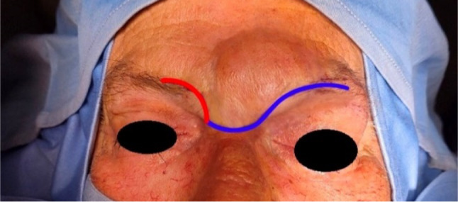
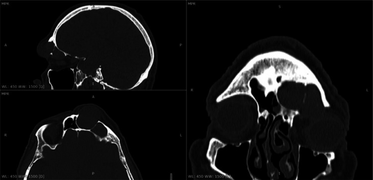
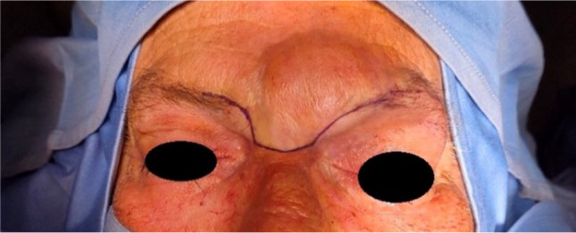
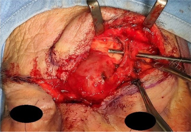
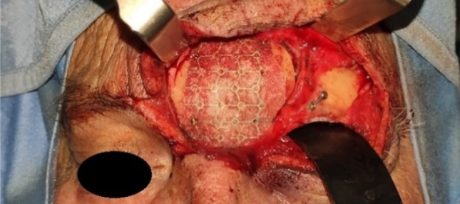
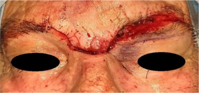
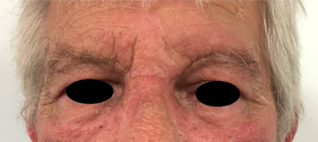
The frontal sinus is one the most complex of the paranasal sinuses, its proximity to the cranial vault and the orbit cause that frontal sinus pathologies can progress to involve these structures and lead to significant morbidity, or even death. Surgical management of the frontal sinus is technically challenging, the most commonly used surgical approaches are coronal, butterfly, gullwing and suprabrow. The purpose of this article is to propose the C-S approach, an interesting alternative to the gullwing approach for the managing of median and paramedian frontal sinus lesions or isolated displaced fractures of the anterior wall. The main advantage of this technique is represented by the fact that it follows the new tension lines described in the literature, a curved vertical line that follows the glabellar frown.
Keywords: Fractures; Frontal sinus; Incision; Mucocele; Tumors.
© Association of Otolaryngologists of India 2021.
Conflict of interest statement
Conflicts of interestThe author(s) declared no potential conflicts of interest with respect to the research, authorship, and/or publication of this article.
Figures
Similar articles
-
Overview of Frontal Sinus Pathology and Management.Otolaryngol Clin North Am. 2016 Aug;49(4):899-910. doi: 10.1016/j.otc.2016.03.014. Epub 2016 Jun 18. Otolaryngol Clin North Am. 2016. PMID: 27329984 Review.
-
Reduction of Closed Frontal Sinus Fractures through Suprabrow Approach.Arch Craniofac Surg. 2017 Dec;18(4):230-237. doi: 10.7181/acfs.2017.18.4.230. Epub 2017 Dec 23. Arch Craniofac Surg. 2017. PMID: 29349046 Free PMC article.
-
Surgical approach of isolated fracture of the anterior wall of the frontal sinus: The upper eyelid incision.J Stomatol Oral Maxillofac Surg. 2019 Jun;120(3):240-243. doi: 10.1016/j.jormas.2018.11.010. Epub 2018 Nov 26. J Stomatol Oral Maxillofac Surg. 2019. PMID: 30496847
-
[Bicoronal approach in the management of frontal sinus fractures].Minerva Stomatol. 1995 Nov;44(11):507-14. Minerva Stomatol. 1995. PMID: 8868584 Italian.
-
Bilateral Transorbital and Transnasal Endoscopic Resection of a Frontal Sinus Osteoblastoma and Orbital Mucocele: A Case Report and Review of the Literature.Ann Otol Rhinol Laryngol. 2018 Nov;127(11):864-869. doi: 10.1177/0003489418798388. Epub 2018 Sep 6. Ann Otol Rhinol Laryngol. 2018. PMID: 30187776 Review.
Cited by
-
Maxillofacial Injuries in Padel Game.J Maxillofac Oral Surg. 2022 Dec;21(4):1393-1396. doi: 10.1007/s12663-022-01725-2. Epub 2022 Jul 26. J Maxillofac Oral Surg. 2022. PMID: 36896067 Free PMC article.
-
New 3-D Technologies in Salivary Gland Surgery: The Exoscope-Assisted Surgery for Treatment of Benign Tumors of Parotid Gland.Indian J Otolaryngol Head Neck Surg. 2024 Oct;76(5):4506-4515. doi: 10.1007/s12070-024-04898-z. Epub 2024 Jul 20. Indian J Otolaryngol Head Neck Surg. 2024. PMID: 39376411
References
-
- Brown TM, Drake TM, Krishnamurthy K (2020) Anatomy, head and neck, procerus muscle. In: StatPearl. StatPearls Publishing, Treasure Island (FL) - PubMed
LinkOut - more resources
Full Text Sources