. 2022 Dec;74(Suppl 3):3733-3737.
doi: 10.1007/s12070-021-02519-7. Epub 2021 Mar 31.

Management of Meningitis Due to Cystic Cochleovestibular Malformation: a Stitch in Time

Affiliations

Management of Meningitis Due to Cystic Cochleovestibular Malformation: a Stitch in Time

Prajakta Golhar et al. Indian J Otolaryngol Head Neck Surg. 2022 Dec.

Abstract

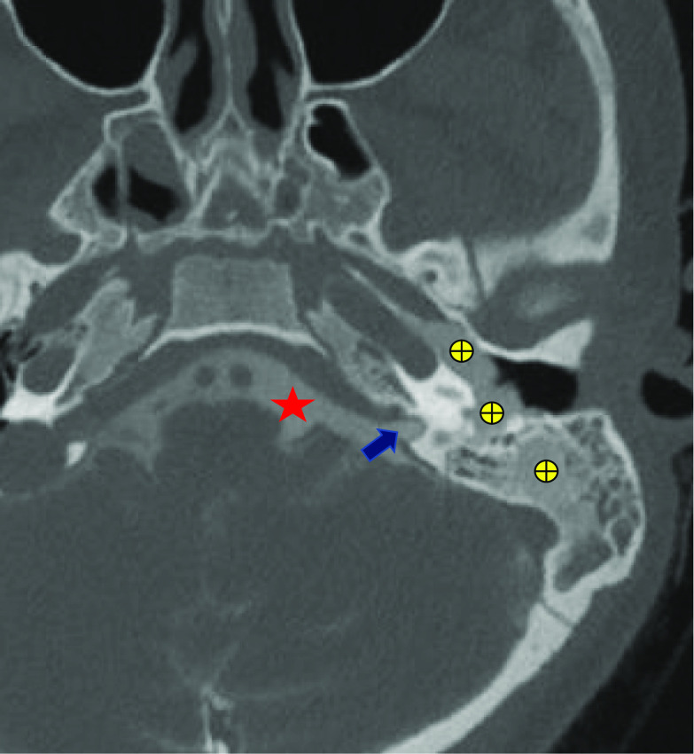
Spontaneous CSF otorrhea alludes to those cases which have no known etiology like traumatic, iatrogenic, neoplastic or infectious. The presentation of the patient depends on the anatomical integrity of the eustachian tube as well as the tympanic membrane. Children with certain congenital inner ear malformations, including incomplete partition deformity, show a higher incidence of spontaneous CSF leaks and the resultant meningitis. Cystic vestibulocochlear deformity accounts for approximately 20% of inner ear malformations. In this case report, we discuss a child who presented with first episode of meningitis and with the help of thorough clinical otorhinolaryngological examination and radiology, was diagnosed with a congenital ear anomaly. With this paper, we stress upon the importance of keeping an open mind regarding the differential diagnosis of any condition, and the value of timely intervention.

Keywords: Cerebrospinal fluid (CSF) otorrhea; Computed tomography (CT); Congenital ear malformation; Cystic cochleovestibular anomaly; Meningitis.


Conflict of interest statement

Conflict of interestThe authors declare that they have no conflict of interest.

Figures

Fig. 1
Fig. 1
Axial MRI suggestive of meningitis. Yellow star: white lines depict interfolial leptomeningeal enhancement-suggestive of meningitis. Blue circle: fluid in mastoid air cells
Fig. 2
Fig. 2
T2-weighted FLAIR images of Axial MRI suspicious of CSF leak. Red star: cystic cochleovestibular malformation with focal enhancement. Blue circle: fluid in mastoid air cells
Fig. 3
Fig. 3
Axial HRCT of the Temporal Bone showing Left-sided Cystic cochleovestibular anomaly. Red star: cystic empty cochlea and vestibule (figure of eight appearance). Strike: soft tissue/fluid in middle ear, surrounding ossicles and in mastoid air cells
Fig. 4
Fig. 4
CT Cisternography. Red star: contrast in basal cisterns. Arrow: Internal audiotry canal. Encircled plus: contrast opacification in middle ear and mastoid cells

Similar articles

References

    1. Goddard JC, Meyer T, Nguyen S, Lambert PR. New considerations in the cause of spontaneous cerebrospinal fluid otorrhea. Otol Neurotol. 2010;31(6):940–945. doi: 10.1097/MAO.0b013e3181e8f36c. - DOI - PubMed
    1. Pachauri R, Nagaonkar S, Kirtane MV. Spontaneous Csf otorrhoea—presenting as conductive deafness. Indian J Otolaryngol Head Neck Surg. 2002;54(1):51–54. doi: 10.1007/BF02911008. - DOI - PMC - PubMed
    1. Tyagi I, Syal R, Goyal A. Cerebrospinal fluid otorhinorrhoea due to inner-ear malformations: clinical presentation and new perspectives in management. J Laryngol Otol. 2005;119(9):714–718. doi: 10.1258/0022215054797934. - DOI - PubMed
    1. Boo SH, Goh YB, Han CS. Repair of spontaneous cerebrospinal fluid otorrhea from defect of middle cranial fossa. Korean J Audiol. 2013;17(3):148–151. doi: 10.7874/kja.2013.17.3.148. - DOI - PMC - PubMed
    1. Sennaroğlu L, Bajin MD. Classification and current management of inner ear malformations. Balkan Med J. 2017;34(5):397–411. doi: 10.4274/balkanmedj.2017.0367. - DOI - PMC - PubMed

LinkOut - more resources