Review
. 2023 Jan 17;8(4):3667-3683.
doi: 10.1021/acsomega.2c06098. eCollection 2023 Jan 31.

Natural Bioactive Molecules as Neuromedicines for the Treatment/Prevention of Neurodegenerative Diseases

Affiliations
Review

Natural Bioactive Molecules as Neuromedicines for the Treatment/Prevention of Neurodegenerative Diseases

Afshin Babazadeh et al. ACS Omega. .

Abstract

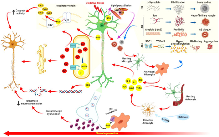
The brain is vulnerable to different types of stresses, particularly oxidative stress as a result of oxygen requirements/utilization in the body. Large amounts of unsaturated fatty acids present in the brain increase this vulnerability. Neurodegenerative diseases (NDDs) are brain disorders that are characterized by the gradual loss of specific neurons and are attributed to broad evidence of cell-level oxidative stress. The accurate characterization of neurological disorders relies on several parameters along with genetics and environmental risk factors, making therapies less efficient to fight NDDs. On the way to tackle oxidative damage and discover efficient and safe therapies, bioactives are at the edge of NDD science. Naturally occurring bioactive compounds such as polyphenols, carotenoids, essential fatty acids, phytosterols, essential oils, etc. are particularly of interest owing to their potent antioxidant and anti-inflammatory activities, and they offer lots of brain-health-promoting features. This Review focuses on probing the neuroefficacy and bioefficacy of bioactives and their role in supporting relatively low antioxidative and low regenerative capacities of the brain, neurogenesis, neuroprotection, and ameliorating/treating NDDs.


Conflict of interest statement

The authors declare no competing financial interest.

Figures

Figure 1
Figure 1
Oxidative stress-induced neurodegeneration. The oxidative stress state can induce neurodegeneration and neuroinflammation. In an OS state, levels of ROS/RNS are increased, which can lead the activation of signaling pathways responsible for major glial inflammatory actions. The glial cells (astrocytes and microglia) secrete proinflammatory agents (TNF-α, tumor necrosis factor alpha; iNOS, inducible nitric oxide synthase; NOX, NADPH oxidase; COX-2, cyclooxigenase-2; RNS, reactive nitrogen species; IL, interleukin; ROS, reactive oxygen species) which leads to neurodegeneration, resulting in neuron injury and death. Myelin is damaged by the increased ROS leading to an increase in Ca2+ influx, which further contributes to the rise in NOX1 and Calpain1 (C1), causing OS damage and myelin degradation. Glutamate (Glu) is normally cleared by the glial cell transporters and returns to neurons via repackaging into synaptic vesicles. An inadequate clearance of Glu could lead to excessive entry of Ca2+ into the presynaptic neuron, which can cause excitotoxicity that is as a result of damaged synaptic mitochondria, and consequently resulting in ROS formation, altered synaptic transmission, and neuron dysfunction. Moreover, OS leads to lipid peroxidation through an attack of ROS and triggering a chain process, whereby products such as 4-hydroxy-2-nonenal, malondialdehyde, acrolein, isoprostanes, or neuroprostanes (iso/Neuro Ps) are formed, which, in turn, changes protein functions causing neuron dysfunctions.
Figure 2
Figure 2
Possible effects of bioactive agents in the clearance of protein aggregates in neurodegenerative disease. Bioactive agents are potent agents to enhance neuroprotection by enhancing protein clearance pathways and clearing aggregated proteins which are one of the main causative factors in neurodegenerative diseases. TAR DNA/RNA binding protein (TDP), tau, and α-synuclein are the main protein aggregations in ALS, Azheimer’s, and Parkinson’s diseases. The enhancement may occur through two main protein clearance pathways: the ubiquitin–proteasome pathway and autophagy–lysosome pathway.
Figure 3
Figure 3
Neuron health-promoting effects of bioactives in neurodegeneration. Bioactives can show their antioxidant roles in neurons by modulating damage caused by ROS and/or RNS using several pathways. These are enhancing glutathione peroxidase (GPx) activity directly, ROS RNS scavenging capacity, activating nuclear factor erythroid 2-related factor 2 (Nrf-2) transcription of antioxidant enzymes, and promoting functionality and health of mitochondria. Genes that can be activated by Nrf2 include gamma-glutamylcysteine ligase (γ-GCL), glutathione (GSH), and Cu,Zn-superoxide dismutase (SOD1) which can then be used in conjunction with GPx and glutathione reductase, detoxifying ROS/RNS to avoid apoptosis. Another neuron health-promoting effect of bioactive compounds is based on calcium homeostasis and excitotoxicity which can be achieved through binding to the excitatory compounds kainite (Kai) or glutamate (Glu). The massive calcium influx caused by their interfering cognate receptors can interfere with the endoplasmic reticulum (ER) protein folding functioning, causing ER stress, activating proapoptotic signaling cascades, depolarizing the mitochondria, and uncoupling the electron transport chain, resulting in mitochondrial dysfunction, OS, permeability transition pore openings (releasing apoptogenic substances into the cytosol), and activating proapoptotic factors (calpains) directly, which can cause neuron death. Bioactive compounds are potent agents protecting neurons from excitotoxicity by avoiding increments of intracellular Ca2+ through Glu and Kai signaling pathways. Effects of bioactive compounds on neuroinflammation can be achieved via inflammatory stimuli, such as depositing aggregated proteins, activating toll-like receptor-4 (TLR4), and downstream induction of Akt, extracellular regulating signal kinase 1/2 (ERK1/2), and p38-mitogen-activated protein kinase (p38-MAPK), which subsequently activate nuclear factor-kappa B (NF-κB) in astrocytes and microglia followed by translocation of NF-κB to the nucleus and initiating transcription of proinflammatory genes such as cyclooxygenase-2 (COX-2), NADPH-oxidase-2 (NOX-2), inducible nitric oxide synthase (iNOS), and tumor necrosis factor-α (TNF-α). It is thought that bioactive agents inhibit this pathway through blocking activation of TLR4, Akt, ERK1/2, and p38-MAPK. Effects of bioactive agents on prosurvival/apoptotic signaling pathways can be achieved by modulating some signaling pathways that play roles in neuron death or survival. Bioactive agents can inhibit the c-Jun N-terminal kinase (JNK) and p53 activities, which are responsible for activation of proapoptotic proteins like B-cell lymphoma-2 (Bcl-2), Bad, Puma, Bim, and Noxa. Puma, Bim, and Noxa are identified for inhibiting the prosurvival functionalities of Bcl-2, resulting in activating the Bax (a proapoptotic protein). Then, Bax can form mitochondrial membrane pores, causing the Cyt C release. Cyt C then can interact with caspase-9 and apoptosis protease activating factor-1 (APAF-1) and form the apoptosome complex. Then, apoptosome can cleave procaspase-3 to form active caspase-3, stimulating apoptosis. In addition to inhibiting p53 activity, bioactive agents can also enhance the phosphoinositide-3-kinase (PI3K)/Akt prosurvival signaling pathway activity, which inhibits Bcl-2, Bim, Bad, and Bax activities. This neuron health-promoting activity of bioactive agents can prevent neurons from going to caspase-dependent apoptosis. Moreover, bioactive agents might also prohibit caspase-independent apoptosis via blocking the translocation of apoptosis inducing factor (AIF) from mitochondria to the nucleus.

Similar articles

Cited by

References

    1. Babazadeh A.; Mohammadi Vahed F.; Jafari S. M. Nanocarrier-Mediated Brain Delivery of Bioactives for Treatment/Prevention of Neurodegenerative Diseases. J. Controlled Release 2020, 321, 211–221. 10.1016/j.jconrel.2020.02.015. - DOI - PubMed
    1. Wang K.; Wang Y.; Li K.; Wan Y.; Wang Q.; Zhuang Z.; Guo Y.; Li H. Uptake, Translocation and Biotransformation of Selenium Nanoparticles in Rice Seedlings (Oryza Sativa L.). J. Nanobiotechnology 2020, 18 (1), 103.10.1186/s12951-020-00659-6. - DOI - PMC - PubMed
    1. Singh A.; Kukreti R.; Saso L.; Kukreti S. Oxidative Stress: A Key Modulator in Neurodegenerative Diseases. Molecules 2019, 24 (8), 1583.10.3390/molecules24081583. - DOI - PMC - PubMed
    1. Testa G.; Staurenghi E.; Zerbinati C.; Gargiulo S.; Iuliano L.; Giaccone G.; Fantò F.; Poli G.; Leonarduzzi G.; Gamba P. Changes in Brain Oxysterols at Different Stages of Alzheimer’s Disease: Their Involvement in Neuroinflammation. Redox Biol. 2016, 10, 24–33. 10.1016/j.redox.2016.09.001. - DOI - PMC - PubMed
    1. Mahalakshmi K.; Parimalanandhini D.; Sangeetha R.; Livya Catherene M.; Beulaja M.; Thiagarajan R.; Arumugam M.; Janarthanan S.; Manikandan R. Influential Role of 7-Ketocholesterol in the Progression of Alzheimer’s Disease. Prostaglandins Other Lipid Mediat. 2021, 156, 106582.10.1016/j.prostaglandins.2021.106582. - DOI - PubMed