

Spheroid Engineering in Microfluidic Devices
- PMID: 36743071
- PMCID: PMC9893254
- DOI: 10.1021/acsomega.2c06052
Spheroid Engineering in Microfluidic Devices
Abstract
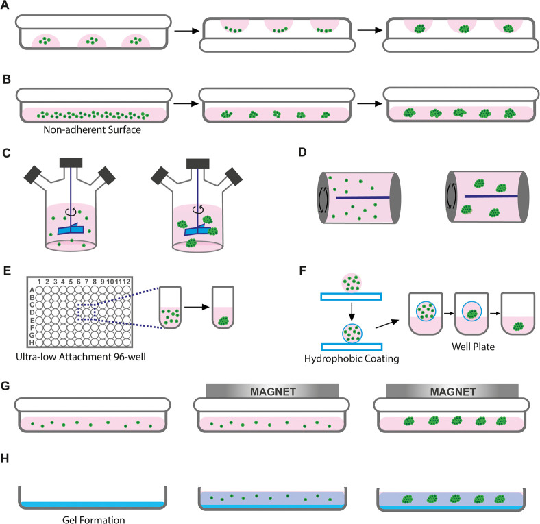
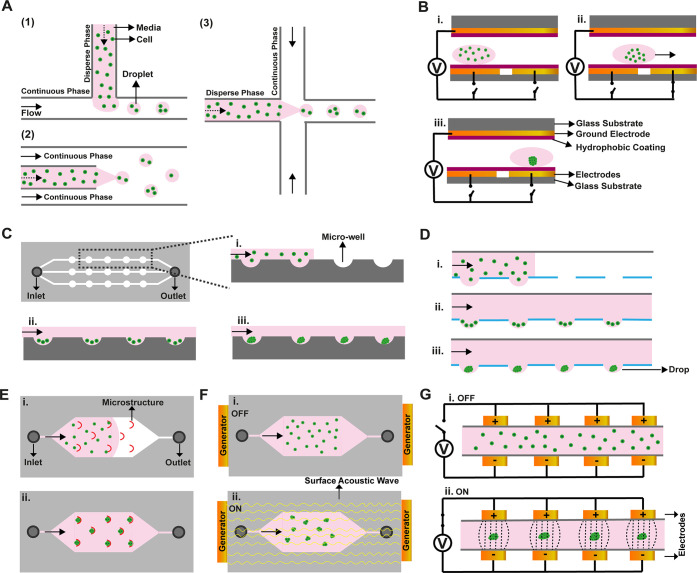
Two-dimensional (2D) cell culture techniques are commonly employed to investigate biophysical and biochemical cellular responses. However, these culture methods, having monolayer cells, lack cell-cell and cell-extracellular matrix interactions, mimicking the cell microenvironment and multicellular organization. Three-dimensional (3D) cell culture methods enable equal transportation of nutrients, gas, and growth factors among cells and their microenvironment. Therefore, 3D cultures show similar cell proliferation, apoptosis, and differentiation properties to in vivo. A spheroid is defined as self-assembled 3D cell aggregates, and it closely mimics a cell microenvironment in vitro thanks to cell-cell/matrix interactions, which enables its use in several important applications in medical and clinical research. To fabricate a spheroid, conventional methods such as liquid overlay, hanging drop, and so forth are available. However, these labor-intensive methods result in low-throughput fabrication and uncontrollable spheroid sizes. On the other hand, microfluidic methods enable inexpensive and rapid fabrication of spheroids with high precision. Furthermore, fabricated spheroids can also be cultured in microfluidic devices for controllable cell perfusion, simulation of fluid shear effects, and mimicking of the microenvironment-like in vivo conditions. This review focuses on recent microfluidic spheroid fabrication techniques and also organ-on-a-chip applications of spheroids, which are used in different disease modeling and drug development studies.
© 2023 The Authors. Published by American Chemical Society.
Conflict of interest statement
The authors declare no competing financial interest.
Figures
References
Publication types
LinkOut - more resources
Full Text Sources
