

Gold nanoparticles capped with L-glycine, L-cystine, and L-tyrosine: toxicity profiling and antioxidant potential
- PMID: 36743265
- PMCID: PMC9888356
- DOI: 10.1007/s10853-023-08209-9
Gold nanoparticles capped with L-glycine, L-cystine, and L-tyrosine: toxicity profiling and antioxidant potential
Abstract
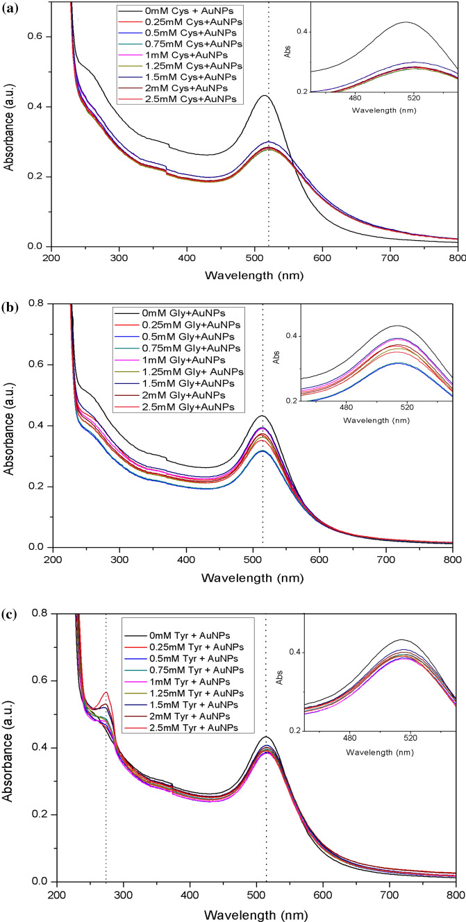
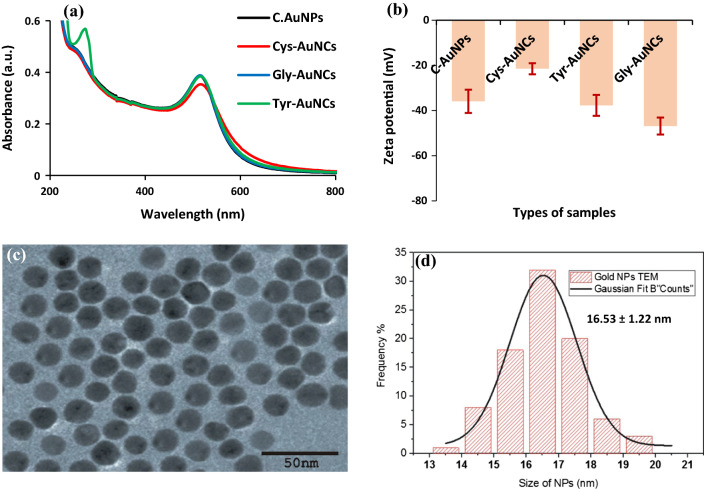
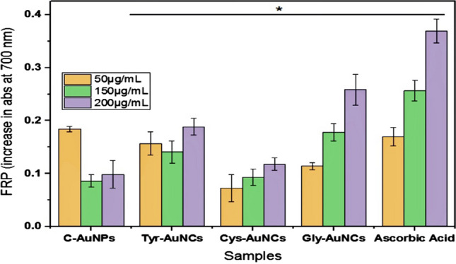
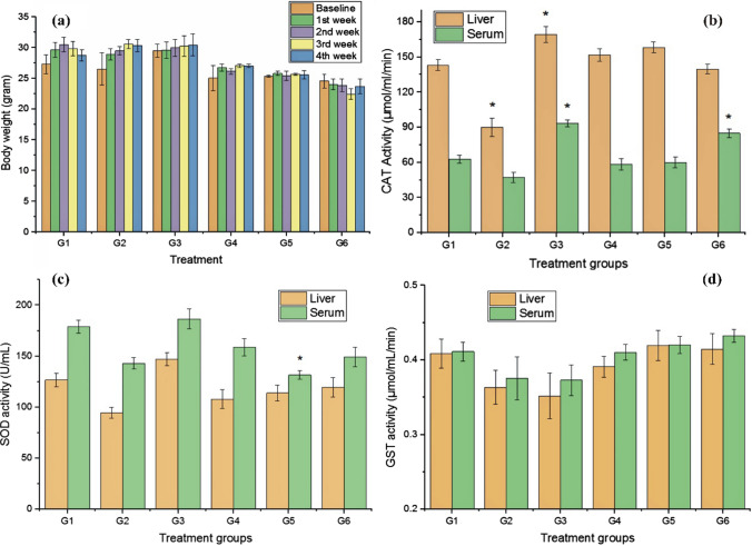
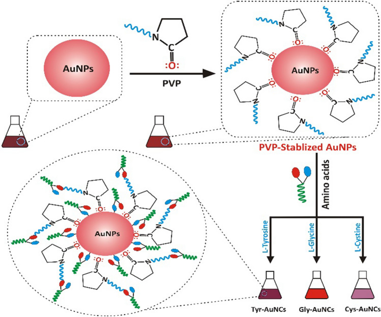
Biomolecules-based surface modifications of nanomaterials may yield effective and biocompatible nanoconjugates. This study was designed to evaluate gold nanoconjugates (AuNCs) for their altered antioxidant potential. Gold nanoparticles (AuNPs) and their conjugates gave SPR peaks in the ranges of 512-525 nm, with red or blueshift for different conjugates. Cys-AuNCs demonstrated enhanced (p < 0.05) and Gly-AuNCs (p > 0.05) displayed reduced DPPH activity. Gly-AuNCs and Tyr-AuNCs displayed enhanced ferric-reducing power and hydrogen peroxide scavenging activity, respectively. Cadmium-intoxicated mice were exposed to gold nanomaterials, and the level of various endogenous parameters, i.e., CAT, GST, SOD, GSH, and MTs, was evaluated. GSH and MTs in liver tissues of the cadmium-exposed group (G2) were elevated (p < 0.05), while other groups showed nonsignificance deviations than the control group. It is concluded that these nanoconjugates might provide effective nanomaterials for biomedical applications. However, more detailed studies for their safety profiling are needed before their practical applications.
© The Author(s), under exclusive licence to Springer Science+Business Media, LLC, part of Springer Nature 2023, Springer Nature or its licensor (e.g. a society or other partner) holds exclusive rights to this article under a publishing agreement with the author(s) or other rightsholder(s); author self-archiving of the accepted manuscript version of this article is solely governed by the terms of such publishing agreement and applicable law.
Conflict of interest statement
Conflict of interestThe authors declare that they have no conflict of interest.
Figures


Similar articles
-
Cellular Uptake and Tissue Biodistribution of Functionalized Gold Nanoparticles and Nanoclusters.J Biomed Nanotechnol. 2017 Feb;13(2):167-79. J Biomed Nanotechnol. 2017. PMID: 29377647
-
Peptide-induced aggregation of glutathione-capped gold nanoclusters: A new strategy for designing aggregation-induced enhanced emission probes.Anal Chim Acta. 2019 Oct 31;1078:101-111. doi: 10.1016/j.aca.2019.05.069. Epub 2019 May 31. Anal Chim Acta. 2019. PMID: 31358207
-
Regulation of the Enzymatic Activities of Lysozyme by the Surface Ligands of Ultrasmall Gold Nanoclusters: The Role of Hydrophobic Interactions.Langmuir. 2021 Nov 23;37(46):13787-13797. doi: 10.1021/acs.langmuir.1c02719. Epub 2021 Nov 15. Langmuir. 2021. PMID: 34779209
-
Peroxyl radical scavenging activity measurement of antioxidants using histidine-stabilized and glutathione-capped fluorescent gold nanoclusters.Spectrochim Acta A Mol Biomol Spectrosc. 2023 Jan 15;285:121876. doi: 10.1016/j.saa.2022.121876. Epub 2022 Sep 15. Spectrochim Acta A Mol Biomol Spectrosc. 2023. PMID: 36122463
-
Design and applications of gold nanoparticle conjugates by exploiting biomolecule-gold nanoparticle interactions.Nanoscale. 2013 Apr 7;5(7):2589-99. doi: 10.1039/c3nr33870c. Nanoscale. 2013. PMID: 23423633 Review.
Cited by
-
Development and characterization of gold nanoparticle-based colorimetric sensing assay functionalized with L-cysteine for the detection of calcium carbide in ripened mango and banana fruits.Heliyon. 2025 Feb 15;11(4):e42614. doi: 10.1016/j.heliyon.2025.e42614. eCollection 2025 Feb 28. Heliyon. 2025. PMID: 40066027 Free PMC article.
-
Design Principles for Engineering Ionic Liquid-Gold Nanoparticles for Therapeutic Delivery to the Brain.ACS Nano. 2025 Jul 15;19(27):24806-24816. doi: 10.1021/acsnano.5c02375. Epub 2025 Jul 3. ACS Nano. 2025. PMID: 40605559 Free PMC article.
References
-
- Zhu Q-L, Xu Q. Immobilization of ultrafine metal nanoparticles to high-surface-area materials and their catalytic applications. Chem. 2016;1(2):220–245. doi: 10.1016/j.chempr.2016.07.005. - DOI
-
- Soto-Alvaredo J, López-Chaves C, Sánchez-González C, Montes-Bayón M, Llopis J, Bettmer J. Speciation of gold nanoparticles and low-molecular gold species in Wistar rat tissues by HPLC coupled to ICP-MS. J Anal At Spectrom. 2017;32(1):193–199. doi: 10.1039/C6JA00248J. - DOI
-
- Csapó E, Sebők D, Makrai Babić J, Šupljika F, Bohus G, Dékány I, Kallay N, Preočanin T. Surface and structural properties of gold nanoparticles and their biofunctionalized derivatives in aqueous electrolytes solution. J Dispersion Sci Technol. 2014;35(6):815–825. doi: 10.1080/01932691.2013.817314. - DOI
-
- Jazayeri MH, Amani H, Pourfatollah AA, Pazoki-Toroudi H, Sedighimoghaddam B. Various methods of gold nanoparticles (GNPs) conjugation to antibodies. Sens Bio-sensing Res. 2016;9:17–22. doi: 10.1016/j.sbsr.2016.04.002. - DOI
LinkOut - more resources
Full Text Sources
Research Materials
Miscellaneous