. 2023 Jan 19:11:1116128.
doi: 10.3389/fcell.2023.1116128. eCollection 2023.

Brief research report: Effects of Pinch deficiency on cartilage homeostasis in adult mice

Affiliations

Brief research report: Effects of Pinch deficiency on cartilage homeostasis in adult mice

Xiaohao Wu et al. Front Cell Dev Biol. .

Abstract

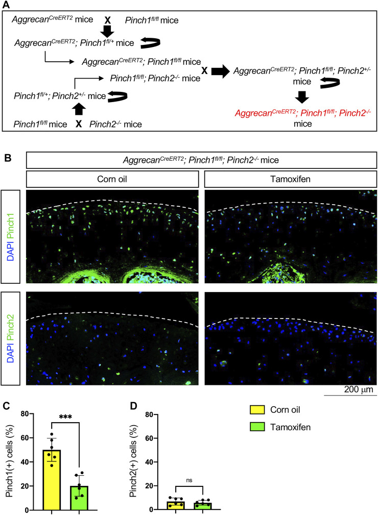
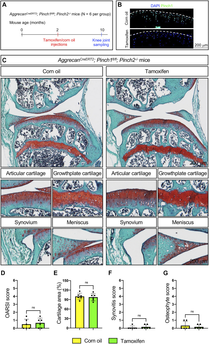
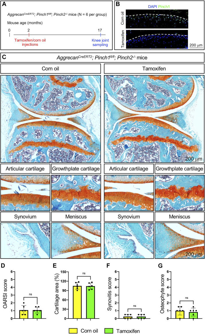
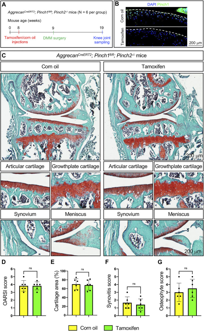
Pinch1 and Pinch2 are LIM domain-containing proteins with crucial functions in mediating focal adhesion formation. Our previous studies have demonstrated that Pinch1/2 expression is essential for cartilage and bone formation during skeletal development in mice. Loss of Pinch expression (Prx1Cre; Pinch1flox/flox; Pinch2-/-) inhibits chondrocyte proliferation and promotes chondrocyte apoptosis, resulting in severe chondrodysplasia and limb shortening. Based on these observations, we wonder if Pinch proteins have a role in adult cartilage and whether Pinch deficiency will compromise cartilage homeostasis and promote osteoarthritis (OA)-related defects in adult mice. To this end, we generated the AggrecanCreERT2; Pinch1flox/flox; Pinch2-/- mice, in which the Pinch1 gene can be inducibly deleted in aggrecan-expressing chondrocytes by tamoxifen and the Pinch2 gene is globally inactivated. Immunofluorescent staining confirmed that the expression of Pinch proteins was significantly decreased in articular cartilage in tamoxifen-treated adult AggrecanCreERT2; Pinch1flox/flox; Pinch2-/- mice. Unexpectedly, our results showed that Pinch loss did not induce marked abnormalities in articular cartilage and other joint tissues in the knee joints of either adult (10-month-old) mice or aged (17-month-old) mice. In a destabilization of the medial meniscus (DMM)-induced OA model, the surgically-induced OA lesions were comparable between Pinch-deficient mice and control mice. Given the fact that Pinch proteins are essential for chondrogenesis and cartilage formation during skeletal development, these findings suggest that Pinch expression is seemingly not indispensable for adult cartilage homeostasis in mice.

Keywords: Pinch; articular cartilage; focal adhesion; homeostasis; osteoarthritis.


Conflict of interest statement

The authors declare that the research was conducted in the absence of any commercial or financial relationships that could be construed as a potential conflict of interest. The handling editor DX declared a shared parent affiliation with the author(s) RL, LL at the time of review. The reviewer GH declared a shared parent affiliation with the author(s) RL, LL to the handling editor at the time of review. The reviewer FW declared a shared parent affiliation with the author(s) SL, XZ to the handling editor at the time of review.

Figures

FIGURE 1
FIGURE 1
Generation of AggrecanCreERT2; Pinch1flox/flox; Pinch2−/− mice. (A) A schema showing the breeding of AggrecanCreERT2; Pinch1flox/flox; Pinch2−/− mice. Tamoxifen was used to induce Pinch1 deletion in aggrecan-positive chondrocytes in 2-month-old AggrecanCreERT2; Pinch1flox/flox; Pinch2−/− mice. Corn oil was used as the control treatment of tamoxifen. (B) Immunofluorescence staining images showing the expression of Pinch1 and Pinch2 proteins in articular cartilage of knee joint. Scale bar: 200 μm. (C–D) Percentages of Pinch1- and Pinch2-positive chondrocytes in tamoxifen- and corn oil-treated AggrecanCreERT2; Pinch1flox/flox; Pinch2−/− mice, respectively. Note: N = 6 mice for each group. Results are expressed as mean ± standard deviation (s.d.). ***p < 0.001; ****p < 0.0001.
FIGURE 2
FIGURE 2
Pinch1/2 loss does not induce marked abnormalities in knee joints in adult mice. (A) A schema illustrating the experimental design. (B) Immunofluorescence staining of Pinch1 in articular cartilage. Scale bar: 200 μm. (C) Representative safranin O and fast green-stained images of knee joint sections from tamoxifen- or corn oil-treated AggrecanCreERT2; Pinch1flox/flox; Pinch2−/− mice at 10 months of age. Higher magnification images in lower panels are showing the areas of articular cartilage, growth-plate cartilage, synovium, and meniscus. Scale bar: 200 μm. (D–G) Quantitative histological analyses of OARSI (osteoarthritis research society international) score, cartilage areas, synovitis score, and osteophyte score using knee joint sections. Note: N = 6 mice for each group. Results are expressed as mean ± standard deviation (s.d.). ns: not significant.
FIGURE 3
FIGURE 3
Pinch1/2 loss does not promote OA defects in aged mice. (A) A schema illustrating the experimental design. (B) Immunofluorescence staining of Pinch1 in articular cartilage. Scale bar: 200 μm. (C) Representative safranin O and fast green-stained images of knee joint sections from tamoxifen- or corn oil-treated AggrecanCreERT2; Pinch1flox/flox; Pinch2−/− mice at 17 months of age. Higher magnification images in lower panels are showing the areas of articular cartilage, growth-plate cartilage, synovium, and meniscus. Scale bar: 200 μm. (D–G) Quantitative histological analyses of OARSI score, cartilage areas, synovitis score, and osteophyte score using knee joint sections. Note: N = 6 mice for each group. Results are expressed as mean ± standard deviation (s.d.). ns: not significant.
FIGURE 4
FIGURE 4
Pinch1/2 loss does not exacerbate surgically induced OA lesions in mice. (A) A schema illustrating the experimental design. (B) Immunofluorescence staining of Pinch1 in articular cartilage. Scale bar: 200 μm. (C) Representative safranin O and fast green-stained images of knee joint sections from tamoxifen- or corn oil-treated AggrecanCreERT2; Pinch1flox/flox; Pinch2−/− mice at 8 weeks after DMM surgery. Higher magnification images in lower panels are showing the areas of articular cartilage, growth-plate cartilage, synovium, and meniscus. Scale bar: 200 μm (D–G) Quantitative histological analyses of OARSI score, cartilage areas, synovitis score, and osteophyte score using knee joint sections. Note: N = 6 mice for each group. Results are expressed as mean ± standard deviation (s.d.). ns: not significant.

Similar articles

Cited by

References

    1. Chen K., Tu Y., Zhang Y., Blair H. C., Zhang L., Wu C. (2008). PINCH-1 regulates the ERK-Bim pathway and contributes to apoptosis resistance in cancer cells. J. Biol. Chem. 283, 2508–2517. 10.1074/jbc.M707307200 - DOI - PubMed
    1. Chen S., Wu X., Lai Y., Chen D., Bai X., Liu S., et al. (2022). Kindlin-2 inhibits Nlrp3 inflammasome activation in nucleus pulposus to maintain homeostasis of the intervertebral disc. Bone Res. 10, 5. 10.1038/s41413-021-00179-5 - DOI - PMC - PubMed
    1. Fukuda T., Chen K., Shi X., Wu C. (2003). PINCH-1 is an obligate partner of integrin-linked kinase (ILK) functioning in cell shape modulation, motility, and survival. J. Biol. Chem. 278, 51324–51333. 10.1074/jbc.M309122200 - DOI - PubMed
    1. Gao H., Zhong Y., Ding Z., Lin S., Hou X., Tang W., et al. (2021). Pinch loss ameliorates obesity, glucose intolerance, and fatty liver by modulating adipocyte apoptosis in mice. Diabetes 70, 2492–2505. 10.2337/db21-0392 - DOI - PubMed
    1. Goldring M. B. (2012). Chondrogenesis, chondrocyte differentiation, and articular cartilage metabolism in health and osteoarthritis. Ther. Adv. Musculoskelet. Dis. 4, 269–285. 10.1177/1759720X12448454 - DOI - PMC - PubMed

LinkOut - more resources