. 2023:3:1102467.
doi: 10.3389/fsysb.2023.1102467. Epub 2023 Jan 18.

Computational design of custom therapeutic cells to correct failing human cardiomyocytes

Affiliations

Computational design of custom therapeutic cells to correct failing human cardiomyocytes

Andrew Tieu et al. Front Syst Biol. 2023.

Abstract

Background: Myocardial delivery of non-excitable cells-namely human mesenchymal stem cells (hMSCs) and c-kit+ cardiac interstitial cells (hCICs)-remains a promising approach for treating the failing heart. Recent empirical studies attempt to improve such therapies by genetically engineering cells to express specific ion channels, or by creating hybrid cells with combined channel expression. This study uses a computational modeling approach to test the hypothesis that custom hypothetical cells can be rationally designed to restore a healthy phenotype when coupled to human heart failure (HF) cardiomyocytes.

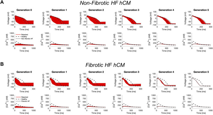
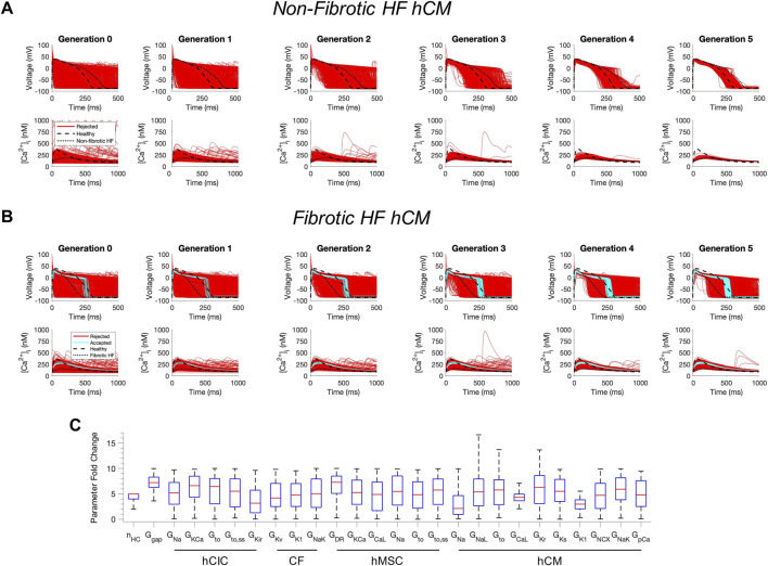
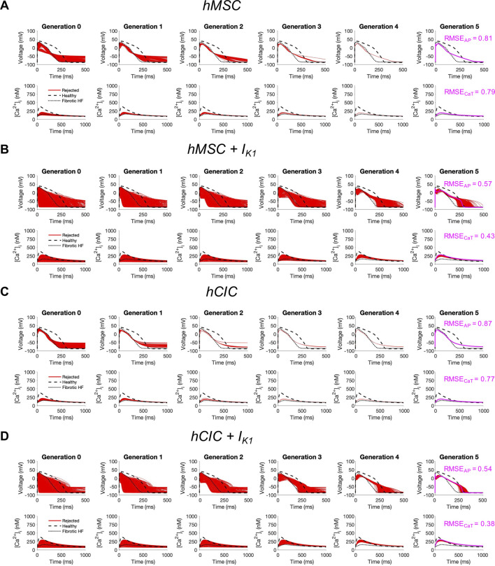
Methods: Candidate custom cells were simulated with a combination of ion channels from non-excitable cells and healthy human cardiomyocytes (hCMs). Using a genetic algorithm-based optimization approach, candidate cells were accepted if a root mean square error (RMSE) of less than 50% relative to healthy hCM was achieved for both action potential and calcium transient waveforms for the cell-treated HF cardiomyocyte, normalized to the untreated HF cardiomyocyte.

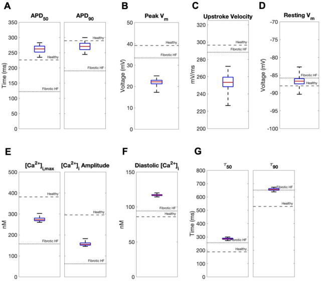
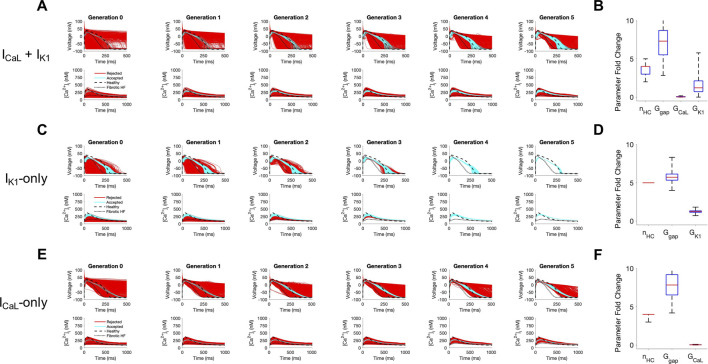
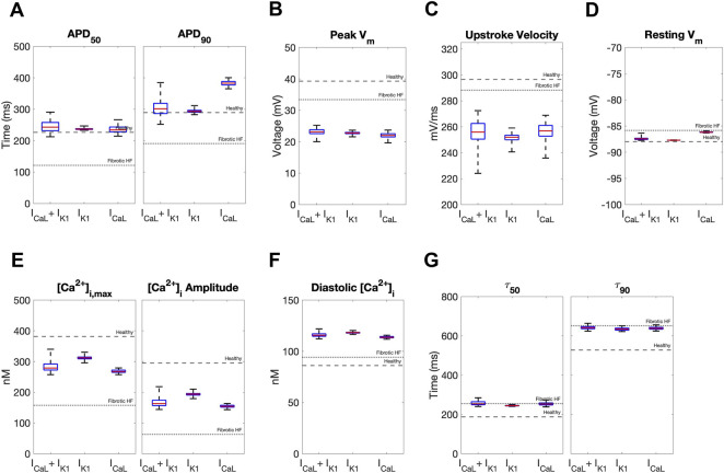
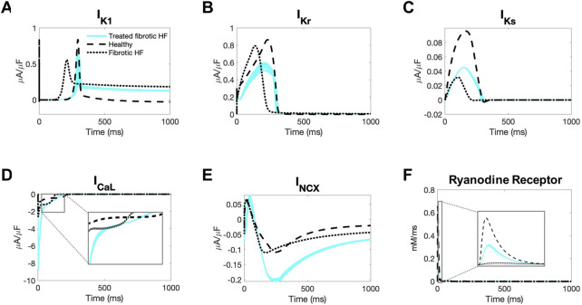
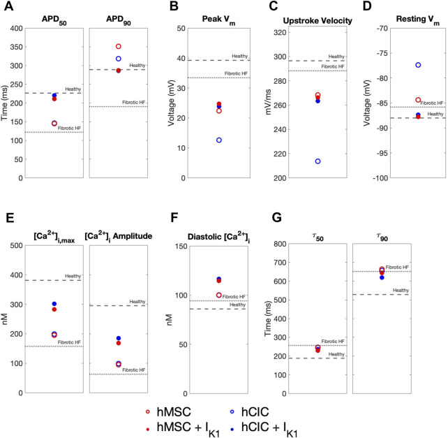
Results: Custom cells expressing only non-excitable ion channels were inadequate to restore a healthy cardiac phenotype when coupled to either fibrotic or non-fibrotic HF cardiomyocytes. In contrast, custom cells also expressing cardiac ion channels led to acceptable restoration of a healthy cardiomyocyte phenotype when coupled to fibrotic, but not non-fibrotic, HF cardiomyocytes. Incorporating the cardiomyocyte inward rectifier K+ channel was critical to accomplishing this phenotypic rescue while also improving single-cell action potential metrics associated with arrhythmias, namely resting membrane potential and action potential duration. The computational approach also provided insight into the rescue mechanisms, whereby heterocellular coupling enhanced cardiomyocyte L-type calcium current and promoted calcium-induced calcium release. Finally, as a therapeutically translatable strategy, we simulated delivery of hMSCs and hCICs genetically engineered to express the cardiomyocyte inward rectifier K+ channel, which decreased action potential and calcium transient RMSEs by at least 24% relative to control hMSCs and hCICs, with more favorable single-cell arrhythmia metrics.

Conclusion: Computational modeling facilitates exploration of customizable engineered cell therapies. Optimized cells expressing cardiac ion channels restored healthy action potential and calcium handling phenotypes in fibrotic HF cardiomyocytes and improved single-cell arrhythmia metrics, warranting further experimental validation studies of the proposed custom therapeutic cells.

Keywords: action potential; calcium transient; cardiac electrophysiology; cell therapy; computational modeling; heart failure; heterocellular coupling.


Conflict of interest statement

Conflict of interest KDC discloses his role as scientific co-founder and Chief Scientific Officer of Novoheart International Ltd. Novoheart did not play any role in the design or conduct of this study. The remaining authors declare that the research was conducted in the absence of any commercial or financial relationships that could be construed as a potential conflict of interest.

Figures

FIGURE 1
FIGURE 1
Genetic algorithm-based approach to identify custom cells expressing non-excitable cell channels are unable to restore failing cardiomyocyte phenotype. Initial populations of 2500 custom cell models were generated for heterocellular coupling with non-fibrotic (A) and fibrotic (B) human heart failure (HF) cardiomyocytes with pseudo-random perturbations of non-excitable cell ion channel maximal conductivities, gap junction conductances, and number of coupled cells. Custom cell populations underwent 5 generations of genetic algorithm evolution. Custom cells were accepted if root mean square error was less than 50% relative to untreated HF cardiomyocytes for both action potential and calcium transient waveforms; in this set of simulations, all models were outside this range and were therefore rejected (red). Waveforms for healthy human cardiomyocytes (hCMs) and untreated HF cardiomyocytes are indicated by dashed and dotted lines, respectively.
FIGURE 2
FIGURE 2
Custom cells expressing excitable cell channels restore a healthy phenotype in fibrotic failing cardiomyocytes. Initial populations of 2500 custom cell models were generated for heterocellular coupling with non-fibrotic (A) and fibrotic (B) heart failure (HF) human cardiomyocytes (hCMs) with pseudo-random perturbations of gap junction conductances, number of coupled cells, and both cardiomyocyte and non-excitable cell ion channel maximal conductivities. Custom cell populations underwent 5 generations of genetic algorithm evolution, whereby custom cells were accepted if root mean square error was less than 50% for both action potential and calcium transient waveforms (blue); models outside this range were rejected (red). Waveforms for healthy and untreated HF cardiomyocytes are indicated by dashed and dotted lines, respectively. (C) Distribution of accepted custom cell model parameter fold changes (relative to baseline values) in the fibrotic HF condition shown as box and whisker plots. Note for certain parameters, a fold-change >10 is achieved due to the genetic algorithm mutation step. Abbreviations: hMSCs, human mesenchymal stem cells; hCIC, c-kit+ cardiac interstitial cells; CF, cardiac fibroblast; hCM, human cardiomyocyte; nHC, Number of heterocellularly coupled cells; Ggap, Gap junction conductance; GNa, Na+ channel conductance; GKCa, Large-conductance Ca2+-activated K+ channel conductance; Gto, Transient outward K+ channel conductance; Gto,ss, Transient outward K+ channel steady-state conductance; GKir, Inward rectifying K+ channel conductance; GKv, Time- and voltage-dependent K+ channel conductance; GK1, Inward rectifier K+ channel conductance; GNaK, Na+/K+ pump activity; GDR, Delayed rectifier K+ channel conductance; GCaL, L-type Ca2+ channel conductance; GNaL, Na+ channel conductance, late component; GKr, Rapid delayed rectifier K+ channel conductance; GKs, Slow delayed rectifier K+ channel conductance; GNCX, Na+/Ca2+ exchanger conductance; GpCa Sarcolemmal Ca2+ pump activity.
FIGURE 3
FIGURE 3
Effects of accepted custom cell heterocellular coupling on fibrotic heart failure cardiomyocyte electrophysiology and calcium handling. Action potential and calcium transient metrics of fibrotic heart failure (HF) cardiomyocytes when coupled to accepted custom cells are shown as box and whisker plots. Action potential metrics include: (A) Action potential duration (APD) at 50% repolarization (APD50) and 90% repolarization (APD90); (B) peak voltage; (C) upstroke velocity; and (D) resting membrane potential. Calcium transient metrics include: (E) peak intracellular calcium and calcium transient amplitude; (F) diastolic calcium; and (G) calcium relaxation time constant at 50% decay (τ50) and 90% decay (τ90). Values for untreated healthy and fibrotic HF cardiomyocytes are shown as dashed and dotted lines, respectively.
FIGURE 4
FIGURE 4
Custom cells expressing cardiomyocyte L-type calcium and inward rectifier channels are capable of correcting fibrotic failing cardiomyocytes. Initial populations of 2500 custom cell models were generated expressing (A) L-type calcium (ICaL) and inward rectifier (IK1) channels; (C) IK1 only; and (E) ICaL only with pseudo-random perturbations to gap junction conductances, number of heterocellularly coupled custom cells to fibrotic heart failure (HF) cardiomyocytes, and custom cell ion channel maximal conductances. Custom cell populations underwent 5 generations of genetic algorithm evolution, whereby custom cells were accepted if root mean square error was less than 50% for both action potential and calcium transient waveforms (blue); models outside this range were rejected (red). Distributions of accepted custom cell model parameter fold changes (relative to baseline values) in the fibrotic HF condition are shown as box and whisker plots for (B) ICaL and IK1, (D) IK1, and (F) ICaL. Note for certain parameters, a fold-change >10 is achieved due to the genetic algorithm mutation step. Waveforms for healthy and untreated HF cardiomyocytes are indicated by dashed and dotted lines, respectively. Abbreviations: nHC, Number of heterocellularly coupled cells; Ggap, Gap junction conductance; GCaL, L-type Ca2+ channel conductance; GK1, Inward rectifier K+ channel conductance.
FIGURE 5
FIGURE 5
Effects of accepted custom cell heterocellular coupling on fibrotic heart failure cardiomyocyte electrophysiology and calcium handling. Action potential and calcium transient metrics are shown as box and whisker plots for coupling fibrotic heart failure (HF) cardiomyocytes to accepted custom cells with cardiomyocyte L-type calcium channel (ICaL), inward rectifier channel (IK1), or both (ICaL + IK1). Action potential metrics include: (A) Action potential duration (APD) at 50% repolarization (APD50) and 90% repolarization (APD90); (B) peak voltage; (C) upstroke velocity; and (D) resting membrane potential. Calcium transient metrics include: (E) peak intracellular calcium and calcium transient amplitude; (F) diastolic calcium; and (G) calcium relaxation time constant at 50% decay (τ50) and 90% decay (τ90). Values for healthy and untreated fibrotic HF cardiomyocytes are shown as dashed and dotted lines, respectively.
FIGURE 6
FIGURE 6
Ionic currents of fibrotic heart failure cardiomyocytes during heterocellular coupling with accepted custom cells. Plots of the following ionic currents of fibrotic heart failure (HF) cardiomyocytes when coupled to 25 randomly selected accepted custom cells (blue) that express cardiomyocyte inward rectifier channel: (A) inward rectifier current (IK1); (B) rapid delayed rectifier (IKr); (C) slow delayed rectifier (IKs); (D) L-type calcium (ICaL); (E) sodium-calcium exchanger (INCX); (F) ryanodine receptor. Zoomed in views of currents for ICaL and ryanodine receptor are inset. Healthy and untreated fibrotic HF cardiomyocytes are shown as dashed and dotted lines, respectively.
FIGURE 7
FIGURE 7
Custom engineered human mesenchymal stem cells and c-kit+ cardiac interstitial cells expressing cardiomyocyte inward rectifier channels reduce root mean square error. Initial populations of 2500 (A) control human mesenchymal stem cells (hMSCs), (B) hMSCs expressing cardiomyocyte inward rectifier channel (IK1), (C) control c-kit+ cardiac interstitial cells (hCICs), and (D) hCICs expressing IK1 were generated for heterocellular coupling with fibrotic heart failure (HF) cardiomyocytes with pseudo-random perturbations of gap junction conductances, number of coupled cells, and IK1 maximal conductance. Each population underwent 5 generations of genetic algorithm evolution, whereby cells were accepted if root mean square error (RMSE) was less than 50% for both action potential (AP) and calcium transient (CaT) waveforms; all models outside this range were rejected (red). The minimum total RMSEAP + RMSECaT achieved for each group is shown in pink. Waveforms for healthy and untreated HF cardiomyocytes are indicated by dashed and dotted lines, respectively.
FIGURE 8
FIGURE 8
Effects of custom engineered human mesenchymal stem cell and c-kit+ cardiac interstitial cell heterocellular coupling on fibrotic heart failure cardiomyocyte electrophysiology and calcium handling. Action potential and calcium transient metrics are shown for coupling fibrotic heart failure (HF) cardiomyocytes to human mesenchymal stem cells (hMSCs) and human c-kit+ cardiac interstitial cells (hCICs) (red and blue circles, respectively) with and without cardiomyocyte inward rectifier channel (IK1) expression (filled and unfilled, respectively) that achieved minimum total root mean square error of action potential and calcium transient waveforms (see pink waveforms in Figure 7). Action potential metrics include: (A) Action potential duration (APD) at 50% repolarization (APD50) and 90% repolarization (APD90); (B) peak membrane voltage (Vm); (C) upstroke velocity; and (D) resting Vm. Calcium transient metrics include: (E) peak intracellular calcium and calcium transient amplitude; (F) diastolic calcium; and (G) calcium relaxation time constant at 50% decay (τ50) and 90% decay (τ90). Values for healthy and untreated fibrotic HF cardiomyocytes are shown as dashed and dotted lines, respectively.

Similar articles

References

    1. Akwaboah A. D., Tsevi B., Yamlome P., Treat J. A., Brucal-Hallare M., Cordeiro J. M., et al. (2021). An in silico hiPSC-derived cardiomyocyte model built with genetic algorithm. Front. Physiol. 12, 675867. 10.3389/fphys.2021.675867 - DOI - PMC - PubMed
    1. Bolli R., Mitrani R. D., Hare J. M., Pepine C. J., Perin E. C., Willerson J. T., et al. (2021). A phase II study of autologous mesenchymal stromal cells and c-kit positive cardiac cells, alone or in combination, in patients with ischaemic heart failure: the CCTRN CONCERT-HF trial. Eur. J. Heart Fail 23, 661–674. 10.1002/ejhf.2178 - DOI - PMC - PubMed
    1. Bot C. T., Kherlopian A. R., Ortega F. A., Christini D. J., Krogh-Madsen T. (2012). Rapid genetic algorithm optimization of a mouse computational model: benefits for anthropomorphization of neonatal mouse cardiomyocytes. Front. Physiol. 3, 421. 10.3389/fphys.2012.00421 - DOI - PMC - PubMed
    1. Braunwald E. (2022). Cardiac cell therapy: a call for action. Eur. Heart J. 43, 2352–2353. 10.1093/eurheartj/ehac188 - DOI - PubMed
    1. Broughton K., Korski K., Echeagaray O., Adamson R., Dembitsky W., Lu Z., et al. (2019). Safety profiling of genetically engineered Pim-1 kinase overexpression for oncogenicity risk in human c-kit+ cardiac interstitial cells. Gene Ther. 26, 324–337. 10.1038/s41434-019-0084-5 - DOI - PubMed

LinkOut - more resources