Central dilemma in CSF pseudocyst - A case series and review of literature
- PMID: 36743758
- PMCID: PMC9893932
- DOI: 10.25259/JNRP-2021-7-25
Central dilemma in CSF pseudocyst - A case series and review of literature
Abstract
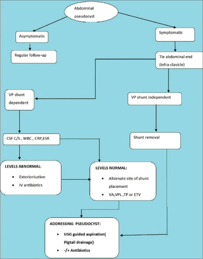
Cerebrospinal fluid (CSF) pseudocyst abdomen is a rare but well-described complication following ventriculoperitoneal (VP) shunt. This entity does exist since 1954. This is seen more commonly among pediatric population and cause of CSF pseudocyst is still debated, many theories been postulated in the literature and so are its management. We present our experience with small case series and idea is to provide an alternate management strategy for shunt-independent cases. We did retrospective study of three cases, diagnosed on the basis of clinical profile and imaging. Subclinical infection was ruled out and patients with abdominal complaints predominantly and no ventriculomegaly on Noncontrast computed tomography head were subjected to "shunt-tie" at infraclavicular region. Out of three cases, two had abdominal complaints with no features of raised ICT and no ventriculomegaly. On tying the shunt catheter infraclavicular level for 48-72 h, they did not developed raised ICT/ventriculomegaly. Cyst was drained by percutaneous ultrasound-guided PIGTAIL. Shunt assembly was removed. One patient (shunt dependent) underwent exploratory laparotomy and repositioning of the catheter but experienced shunt malfunction, ultimately VP shunt was converted to ventriculopleural shunt. On follow-ups, there is no residual cyst or recurrence of symptoms. To conclude, evaluation of shunt dependency/non-dependency is of utmost importance. For shunt-independent cases, percutaneous ultrasound-guided PIGTAIL drainage is safe, minimally invasive, and effective procedure and we may avoid many potential complications.
Keywords: Management dilemma; Pseudocyst abdomen; Shunt dependent; Ventriculoperitoneal shunt.
© 2022 Published by Scientific Scholar on behalf of Journal of Neurosciences in Rural Practice.
Conflict of interest statement
There are no conflicts of interest.
Figures


References
-
- Sena FG, De Sousa RM, Meguins LC. Abdominal cerebrospinal fluid pseudocyst: A complication of ventriculoperitoneal shunt in a Brazilian amazon woman. Case report. G Chir. 2010;31:371–3. - PubMed
-
- Bryant MS, Bremer AM, Tepas JJ, 3rd, Mollitt DL, Nquyen TQ, Talbert JL. Abdominal complications of ventriculoperitoneal shunts. Case reports and review of the literature. Am Surg. 1988;54:50–5. - PubMed
Publication types
LinkOut - more resources
Full Text Sources
Miscellaneous