

Clinical, dermoscopic and histological assessment of melanocytic lesions: a comparative study of the accuracy of the diagnostic methods
- PMID: 36743868
- PMCID: PMC9894303
Clinical, dermoscopic and histological assessment of melanocytic lesions: a comparative study of the accuracy of the diagnostic methods
Abstract
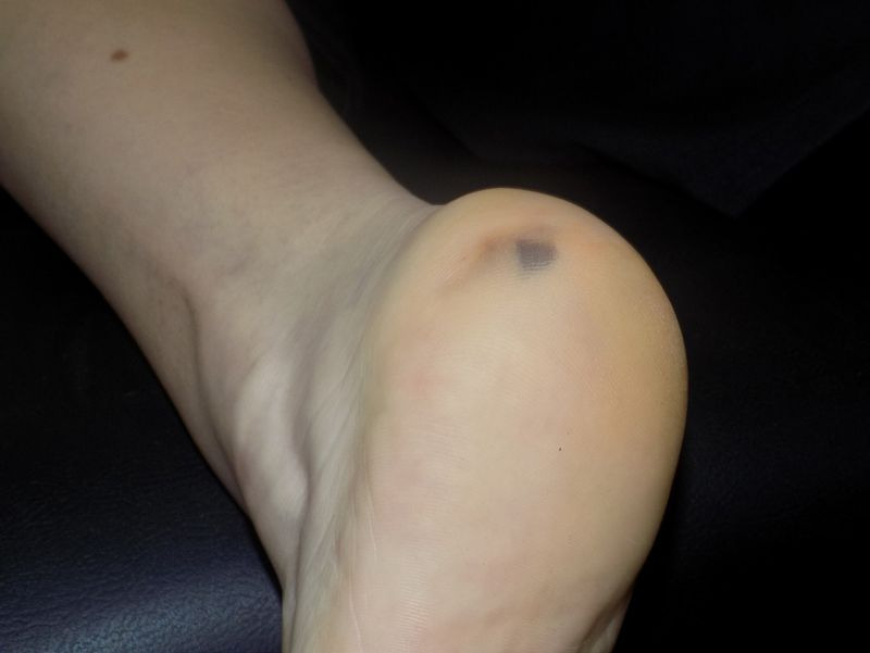
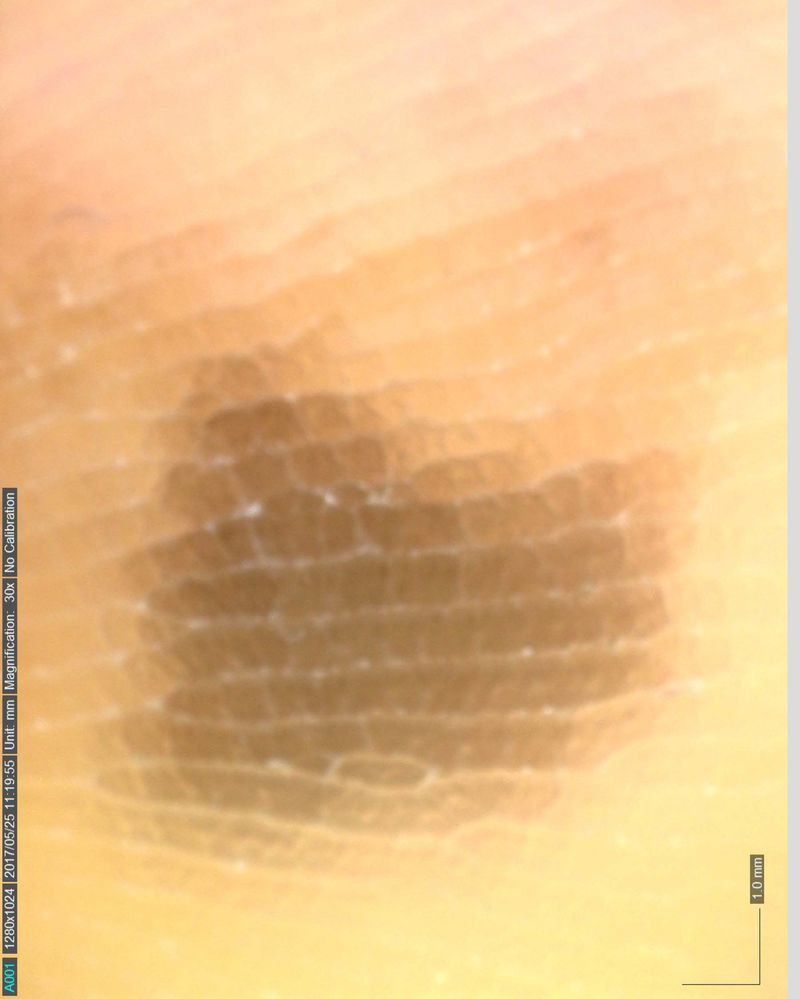
Background: Worldwide, the incidence of melanoma is increasing, while late diagnosis is related to poor prognosis. A significant risk marker for melanoma is the presence of atypical nevi; therefore, it is of outstanding importance to make accurate clinical classification of common benign nevi, atypical nevi, and melanomas. The non-invasive method of dermoscopy allowed for the visualization of structures invisible to the naked eye and undoubtedly advanced the assessment of melanocytic lesions to a new dimension. This study aimed to evaluate the sensitivity and specificity of naked-eye examination and dermoscopy in diagnosing melanocytic lesions compared to the histopathological results, constituting the gold standard of diagnosis.
Material and methods: One hundred eighteen melanocytic lesions were clinically evaluated via the naked eye and dermoscopic examination, using Pattern Analysis Methodology, and afterward, they were excised. The histopathological results were correlated with the findings.
Results: According to the final histopathological analysis, 63 common benign nevi, 41 dysplastic nevi, and 14 cutaneous melanomas were excised in total. Clinical examination via the naked eye showed 78.2 % sensitivity and 71.4 % specificity in identifying the clinical atypia, while dermoscopy demonstrated 89.1 % sensitivity and 93.7 % specificity.
Conclusions: The results of the present study indicate a higher sensitivity and specificity of dermoscopy in evaluating and diagnosing melanocytic lesions compared to the naked-eye examination. HIPPOKRATIA 2021, 25 (4):156-161.
Keywords: Melanocytic lesions; atypia; dermoscopy; dysplasia.
Copyright 2021, Hippokratio General Hospital of Thessaloniki.
Figures
Similar articles
-
Diagnostic Performance of Dermoscopy for Distinguishing Early Melanomas and Intermediate Melanocytic Lesions From Low-Grade Dysplastic Nevi.J Cutan Med Surg. 2025 Mar 12:12034754251325508. doi: 10.1177/12034754251325508. Online ahead of print. J Cutan Med Surg. 2025. PMID: 40072505
-
Sensitivity, specificity, and diagnostic accuracy of three dermoscopic algorithmic methods in the diagnosis of doubtful melanocytic lesions: the importance of light brown structureless areas in differentiating atypical melanocytic nevi from thin melanomas.J Am Acad Dermatol. 2007 May;56(5):759-67. doi: 10.1016/j.jaad.2007.01.014. Epub 2007 Feb 20. J Am Acad Dermatol. 2007. PMID: 17316894
-
Correlation between dermoscopic and histopathological diagnoses of atypical nevi in a dermatology outpatient clinic of the Medical School of São José do Rio Preto, SP, Brazil.An Bras Dermatol. 2013 Mar-Apr;88(2):199-203. doi: 10.1590/S0365-05962013000200002. An Bras Dermatol. 2013. PMID: 23739709 Free PMC article.
-
Role of In Vivo Reflectance Confocal Microscopy in the Analysis of Melanocytic Lesions.Acta Dermatovenerol Croat. 2018 Apr;26(1):64-67. Acta Dermatovenerol Croat. 2018. PMID: 29782304 Review.
-
Melanomas difficult to diagnose via dermoscopy.G Ital Dermatol Venereol. 2010 Feb;145(1):111-26. G Ital Dermatol Venereol. 2010. PMID: 20197750 Review.
Cited by
-
Clinical, Dermoscopic, and Histological Characteristics of Melanoma Patients According to the Age Groups: A Retrospective Observational Study.Life (Basel). 2023 Jun 12;13(6):1369. doi: 10.3390/life13061369. Life (Basel). 2023. PMID: 37374151 Free PMC article.
-
Co-occurrence of posterior chest wall pilonidal sinus with melanocytic nevus: a challenging presentation: a case report.J Cardiothorac Surg. 2024 Jun 12;19(1):330. doi: 10.1186/s13019-024-02802-y. J Cardiothorac Surg. 2024. PMID: 38867278 Free PMC article.
References
-
- Sung H, Ferlay J, Siegel RL, Laversanne M, Soerjomataram I, Jemal A, et al. Global Cancer Statistics 2020: GLOBOCAN Estimates of Incidence and Mortality Worldwide for 36 Cancers in 185 Countries. CA Cancer J Clin. 2021;71:209–249. - PubMed
-
- Rastrelli M, Tropea S, Rossi CR, Alaibac M. Melanoma: epidemiology, risk factors, pathogenesis, diagnosis and classification. In Vivo. 2014;28:1005–1011. - PubMed
-
- Marghoob NG, Liopyris K, Jaimes N. Dermoscopy: A Review of the Structures That Facilitate Melanoma Detection. J Am Osteopath Assoc. 2019;119:380–390. - PubMed
-
- Rao BK, Ahn CS. Dermatoscopy for melanoma and pigmented lesions. Dermatol Clin. 2012;30:413–434. - PubMed
LinkOut - more resources
Full Text Sources