

Conventional and three-dimensional photography as a tool to map distribution patterns of in-transit melanoma metastases on the lower extremity
- PMID: 36744147
- PMCID: PMC9892836
- DOI: 10.3389/fmed.2023.1089013
Conventional and three-dimensional photography as a tool to map distribution patterns of in-transit melanoma metastases on the lower extremity
Abstract
Background: In melanoma, in-transit metastases characteristically occur at the lower extremity along lymphatic vessels.
Objectives: The objective of this study was to evaluate conventional or three-dimensional photography as a tool to analyze in-transit metastasis pattern of melanoma of the lower extremity. In addition, we assessed risk factors for the development of in-transit metastases in cutaneous melanoma.
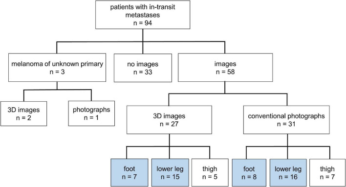
Methods: In this retrospective, monocentric study first we compared the clinical data of all evaluable patients with in-transit metastases of melanoma on the lower extremity (n = 94) with melanoma patients without recurrence of disease (n = 288). In addition, based on conventional (n = 24) and three-dimensional photography (n = 22), we defined the specific distribution patterns of the in-transit metastases on the lower extremity.
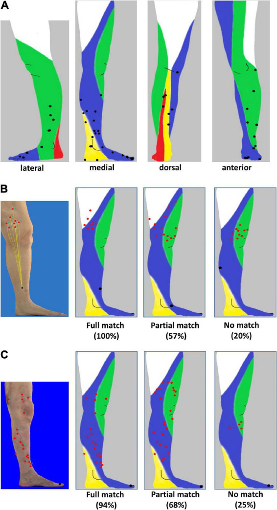
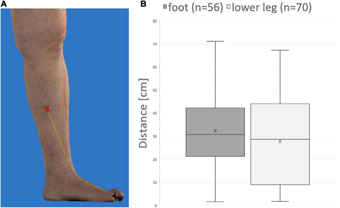
Results: Using a multivariate analysis we identified nodular melanoma, tumor thickness, and ulceration as independent risk factors to develop in-transit metastases ITM (n = 94). In patients with melanoma on the lower leg (n = 31), in-transit metastases preferentially developed along anatomically predefined lymphatic pathways. In contrast when analyzing in-transit metastases of melanoma on the foot (n = 15) no clear pattern could be visualized. In addition, no difference in distance between in-transit metastases and primary melanoma on the foot compared to the lower leg was observed using three-dimensional photography (n = 22).
Conclusion: A risk-adapted follow-up of melanoma patients to detect in-transit metastases can be applied by knowledge of the specific lymphatic drainage of the lower extremity. Our current analysis suggests a more complex lymphatic drainage of the foot.
Keywords: 3D photography; in-transit metastasis; lower extremity; lymphatic pathways; melanoma.
Copyright © 2023 Müller, Berking, Voskens, Heppt, Heinzerling, Koch, Kramer, Merkel, Schuler-Thurner, Schellerer, Steeb, Wessely and Erdmann.
Conflict of interest statement
The authors declare that the research was conducted in the absence of any commercial or financial relationships that could be construed as a potential conflict of interest.
Figures
References
-
- Luke J, Rutkowski P, Queirolo P, Del Vecchio M, Mackiewicz J, Chiarion-Sileni V, et al. Pembrolizumab versus placebo as adjuvant therapy in completely resected stage IIB or IIC melanoma (KEYNOTE-716): a randomised, double-blind, phase 3 trial. Lancet. (2022) 399:1718–29. - PubMed
-
- Kahler K, Egberts F, Gutzmer R. Palliative treatment of skin metastases in dermato-oncology. J Dtsch Dermatol Ges. (2013) 11:1041–5;quiz6. - PubMed
LinkOut - more resources
Full Text Sources