. 2023 Feb 28;51(4):1750-1765.
doi: 10.1093/nar/gkad037.

Mechanism of strand displacement DNA synthesis by the coordinated activities of human mitochondrial DNA polymerase and SSB

Affiliations

Mechanism of strand displacement DNA synthesis by the coordinated activities of human mitochondrial DNA polymerase and SSB

Ismael Plaza-G A et al. Nucleic Acids Res. .

Abstract

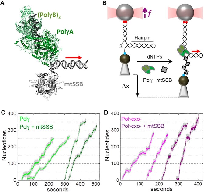
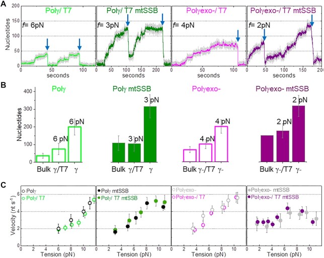
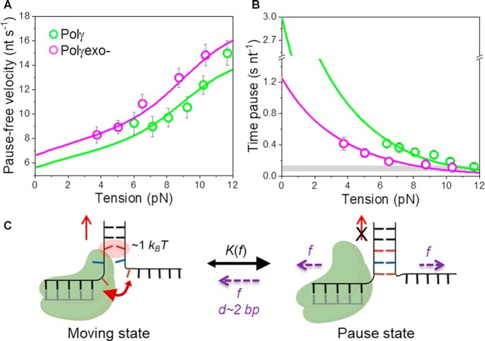
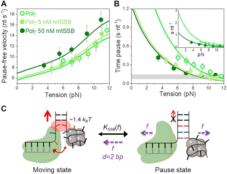
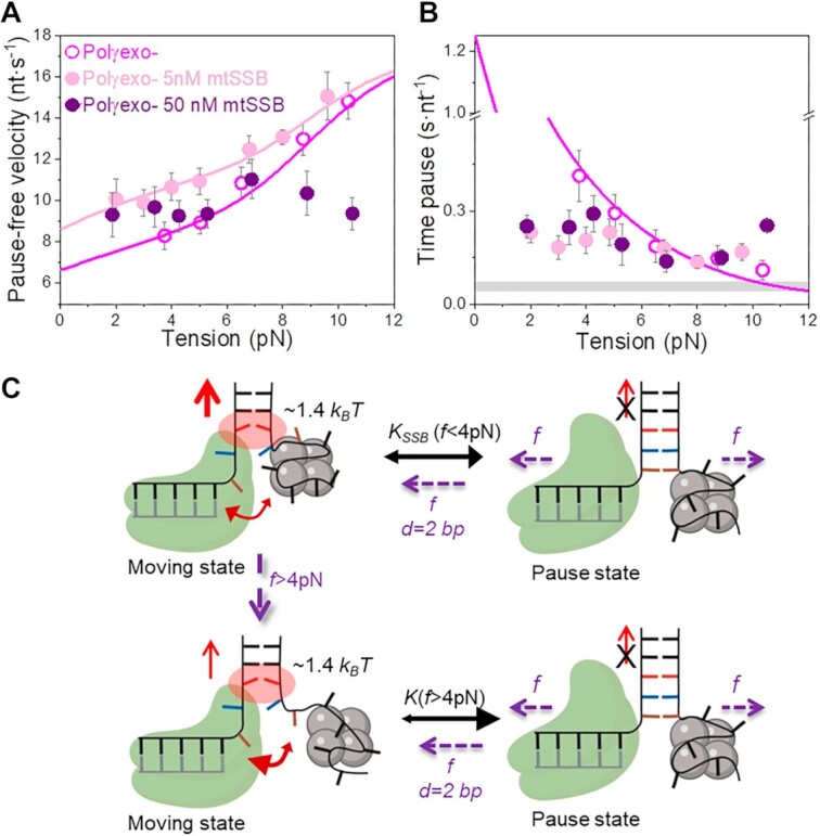
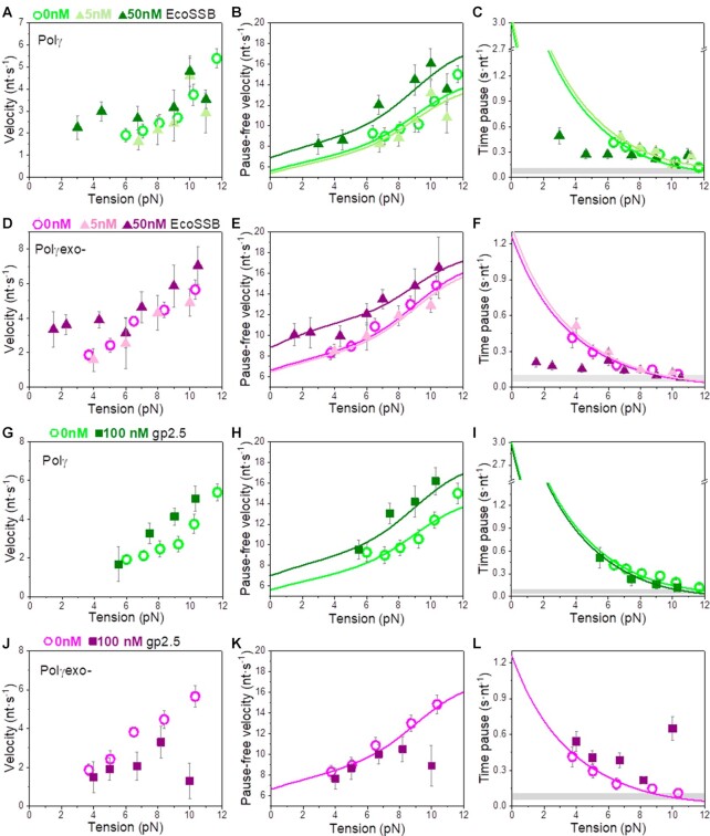
Many replicative DNA polymerases couple DNA replication and unwinding activities to perform strand displacement DNA synthesis, a critical ability for DNA metabolism. Strand displacement is tightly regulated by partner proteins, such as single-stranded DNA (ssDNA) binding proteins (SSBs) by a poorly understood mechanism. Here, we use single-molecule optical tweezers and biochemical assays to elucidate the molecular mechanism of strand displacement DNA synthesis by the human mitochondrial DNA polymerase, Polγ, and its modulation by cognate and noncognate SSBs. We show that Polγ exhibits a robust DNA unwinding mechanism, which entails lowering the energy barrier for unwinding of the first base pair of the DNA fork junction, by ∼55%. However, the polymerase cannot prevent the reannealing of the parental strands efficiently, which limits by ∼30-fold its strand displacement activity. We demonstrate that SSBs stimulate the Polγ strand displacement activity through several mechanisms. SSB binding energy to ssDNA additionally increases the destabilization energy at the DNA junction, by ∼25%. Furthermore, SSB interactions with the displaced ssDNA reduce the DNA fork reannealing pressure on Polγ, in turn promoting the productive polymerization state by ∼3-fold. These stimulatory effects are enhanced by species-specific functional interactions and have significant implications in the replication of the human mitochondrial DNA.


Figures

Figure 1.
Figure 1.
Experimental set up. (A) Schematic of Polγ (PDB: 3IKM) and mtSSB (PDB: 3ULL) at the DNA fork. Polγ holoenzyme is composed by the catalytic subunit, PolγA (dark green) and a dimer of the accessory subunit, PolγB, (light green). mtSSB (grey) binds the displaced ssDNA as a tetramer. (B) In the optical tweezers, a single DNA hairpin (559 bp) is tethered between two functionalized beads and held at constant tension (f). One strand of the hairpin is connected to the bead in the optical trap (red cone) through a ∼2.6-kb dsDNA handle via digoxigenin-antibody connections (red dots). The other strand is attached to a bead on a micropipette by biotin–streptavidin linkages (blue dot). The dsDNA handle includes a 3’ end for polymerase loading, and the 5’ end of the hairpin includes a poly-(dT)30 site for SSB binding. At constant tension, strand displacement DNA synthesis by Polγ (or Polγexo-) increases the end-to-end extension of the hairpin (Δx). Experiments were performed without and with several concentrations of cognate and noncognate SSBs (grey) in solution. In A) and B) red arrow indicates the 5’ to 3’ direction of DNA polymerase translocation along the hairpin. (C) Representative experimental traces of Polγ (2 nM) without (light green) and with (olive) mtSSB (50 nM) in solution (f= 6 pN). (D) Representative experimental traces of Polγexo- (2 nM) without (magenta) and with (purple) mtSSB (50 nM) in solution (f= 6 pN). Traces were displaced along the time axis for clarity of display.
Figure 2.
Figure 2.
Polymerase exchange is not rate limiting. (A) Representative strand displacement traces of Polγ and Polγexo- with competitor T7DNApol in solution in the absence and presence of mtSSB (50 nM). Exchange of Polγ and Polγexo- by T7DNAp was monitored as fast exo events (blue arrow) not observed in the absence of T7DNAp in solution (Figures 1C and D). Displayed traces were taken at the lowest tension (f) at which activity could be detected in each condition. (B) For all plots. First column (Bulk) shows the maximum number of replicated nucleotides obtained in single turn-over bulk experiments (Supplementary Figure S2). The second and third columns show the average number of replicated nucleotides measured in optical tweezers assays in the presence (γ/T7 and γ-/T7) and absence (γ, γ-) of T7DNApol in solution at the lowest detection tension in each case. Bulk Polγexo- mtSSB data does not include error bar because the enzyme generated full length product (150 nt) in each experiment. The average number of nucleotides replicated by Polγ and Polγexo- as a function of tension in the absence and presence of T7DNApol are shown in Figure S3. (C) Tension dependent average strand displacement rates (velocity, nt s−1) of Polγ and Polγexo- in the absence and presence of mtSSB without and with competitor T7DNApol in solution. The similarities between the average rates of Polγ (and Polγexo-) before exchange with T7DNAp with those measured in the absence of T7DNAp suggest that the polymerase exchange reaction is not rate limiting. Polymerase exchange events at f > 8 pN are shown in Figure S3. For all figures error bars show standard errors.
Figure 3.
Figure 3.
Effect of tension on Polγ and Polγexo- strand displacement kinetics. For all plots: Polγ green symbols (N = 80), Polγexo- magenta symbols (N = 71). Error bars show standard errors. (A) For both holoenzymes pause-free velocities (nt s−1) increased with tension continuously towards values measured during primer extension (Figure S1C). Green and magenta lines are the fits of the strand displacement model (SI) to Polγ and Polγexo- data, respectively. (B) Tension dependencies of the average residence times at the pause state per nucleotide (Tp(f), s nt−1). Green and magenta lines are the fits to Polγ and Polγexo- data, respectively, with a two state model (Eq. 1). Grey box show average Tp(f) values measured under primer extension conditions in the absence of mtSSB, Figure S1D and (16). (C) Diagram illustrating the two-state model in which the holoenzyme alternates between moving and pause or non-productive states during the strand displacement reaction. In the moving state, two template nucleotides (brown and blue) are bound to the pol site and the holoenzyme advances through the dsDNA destabilizing partially the first base pair of the junction (in red) with interaction energy of ΔGint ∼1 kBT per dNTP incorporation step. In the absence of tension, the regression pressure of the dsDNA fork outcompetes the holoenzyme for the template (two headed arrow), which shifts the equilibrium towards the pause or non-productive state strongly (K(0) >1, Table 1) and restricts the probability of finding Polγ and Polγexo- in the moving state to ∼4 and 12%, respectively (SI). Application of tension (f) to the hairpin decreases the rewinding kinetics and/or favors the unwinding of first ∼2 bp of the fork (d), which shifts the equilibrium towards the moving state.
Figure 4.
Figure 4.
Effect of mtSSB on the tension dependent strand displacement kinetics of Polγ. (A) 50 nM (N = 40) but not 5 nM (N = 20) mtSSB stimulated the pause-free velocity of Polγ at all tensions. Green lines correspond to the fits of the strand displacement model to data in the absence and presence of mtSSB. (B) 5 and 50 nM concentrations of mtSSB decreased average residence time at pause state per nucleotide (Tp(f), s nt−1) of Polγ at all tensions. Grey box shows average Tp(f) values obtained under primer extension conditions in the absence of mtSSB (16). Green lines are the fits of two-state model (Eq. 1) to data in the absence and presence of mtSSB. mtSSB binding to the displaced strand decreases ∼2–3 times the average residence time of Polγ at a pause or non-productive state. For both figures error bars show standard errors. Inset shows the intersection of the fits with the Y-axis. (C) Diagram illustrating the two-state model in the presence of mtSSB. Polγ alternates between moving and pause or non-productive states. In the moving state, two template nucleotides (brown and blue) are bound at the pol active site and the holoenzyme-mtSSB complex destabilizes partially the first base pair of the DNA hairpin with interaction energy a ∼40% higher than in the absence of mtSSB (ΔGint ∼1.4 kBT per dNTP incorporated). In addition, mtSSB decreases the fork regression kinetics (represented by a two-headed arrow), which in turn, increases the probability of finding the holoenzyme at the moving state from ∼4 to ∼12% (SI). Even in the presence of mtSSB, the equilibrium is shifted towards the pause or inactive-state (KSSB(0) > 1, Table 1). Destabilization of 2 base pairs (d) of the DNA junction by application of mechanical tension (f) is required to shift the equilibrium towards the moving state.
Figure 5.
Figure 5.
Effect of mtSSB on the tension dependent strand displacement kinetics of Polγexo-. Effects of 5 nM (N = 44) and 50 nM (N = 78) mtSSB on the tension dependent (A) pause-free velocity, and (B) average residence times at pause state per nucleotide (Tp(f), s nt−1) of Polγexo-. Both mtSSB concentrations stimulated the strand displacement activity of Polγexo- below 4–6pN. However, the stimulatory effects diminished above 4–6 pN. Even more, as tension increased above ∼8 pN, 50 nM mtSSB had detrimental effects on the pause-free rates (A) and residence times at pause state per nucleotide (B) of the mutant holoenzyme variant. In (A) magenta and pink lines correspond to the fits of the strand displacement model to the tension dependent pause-free rates in the absence and presence of 5 nM mtSSB, respectively. In (B), the magenta line is the fit of the two-state model (Eq.1) to the tension dependent average residence time at pause state per nucleotide in the absence of mtSSB. Grey box shows the average Tp(f) values measured under primer extension conditions in the absence of SSB, Figure S1. For (A) and (B) error bars show standard errors. (C) Diagram illustrating the effect of tension on the moving-pause state equilibrium of Polγexo- in the presence of mtSSB. At tension f< 4 pN, Polγexo- would alternate between moving and pause state with an equilibrium constant (KSSB(f< 4pN)) leading to a residence time in the pause state ∼3–4-times shorter than that in the absence of mtSSB. Application of tension above 4 pN promotes the release of ssDNA nucleotides from the mtSSB (brown and blue), which in turn could decrease its ability to counteract the fork regression kinetics. Under these conditions, the mutant holoenzyme would alternate between moving and pause states with an equilibrium constant similar to that in the absence of mtSSB (K(f > 4 pN)). In both situations, mtSSB binding energy and kinetics would help the holoenzyme to destabilize the first base pair of the DNA fork (ΔGint∼ 1.4 kBT per dNTP incorporated). Mechanical destabilization of the 2 first base pairs (d) of the DNA junction by tension (f) will further shift the equilibrium towards the moving state in the two situations.
Figure 6.
Figure 6.
Effects non-cognate SSB on Polγ and Polγexo- tension dependent strand displacement kinetics. (A–C) 5 nM EcoSSB had no apparent effects on the strand displacement kinetics of Polγ (N = 28). In contrast, 50 nM EcoSSB (N = 32) stimulated pause-free velocity at all tensions (B), and the average rates (A), and residence times at pause state, Tp(f), (C) below 5 pN. (D–F) 5 nM EcoSSB (N = 27) had no significant effects on the strand displacement kinetics of Polγexo-, whereas 50 nM EcoSSB (N = 42) stimulated the pause-free velocity at all tensions (E), and the average rates (D) and residence time at the pause state (F) at tension below 5 pN. (G–I) gp2.5 (100 nM, N = 15) stimulated the pause-free velocities of Polγ to a similar extend than 50 nM mtSSB and EcoSSB (H), but did not decrease the residence time at pause state of the wild-type holoenzyme at any tension (I). (J–L) gp2.5 (100 nM, N = 17) did not stimulate the strand displacement kinetics of Polγexo- and was inhibitory at tension above ∼8 pN. Lines correspond to the fits of the strand displacement model to the pause-free data (B, E, H, K) and Eq. 1 to the residence time at pause state data (C, F, I, L). Grey boxes show the average Tp(f) values obtained during primer extension conditions in the absence of SSBs (Figure S1 and (16)). For all plots error bars show standard errors.

References

    1. Kaguni L.S. DNA polymerase gamma, the mitochondrial replicase. Annu. Rev. Biochem. 2004; 73:293–320. - PubMed
    1. Krasich R., Copeland W.C.. DNA polymerases in the mitochondria: a critical review of the evidence. Front. Biosci. (Landmark Ed.). 2017; 22:692–709. - PMC - PubMed
    1. Lee H.R., Johnson K.A.. Fidelity of the human mitochondrial DNA polymerase. J. Biol. Chem. 2006; 281:36236–36240. - PubMed
    1. Stumpf J.D., Saneto R.P., Copeland W.C.. Clinical and molecular features of POLG-related mitochondrial disease. Cold Spring Harb. Perspect. Biol. 2013; 5:a011395. - PMC - PubMed
    1. Johnson A.A., Johnson K.A.. Fidelity of nucleotide incorporation by human mitochondrial DNA polymerase. J. Biol. Chem. 2001; 276:38090–38096. - PubMed

Publication types

MeSH terms