. 2023 May;101(5):383-396.
doi: 10.1111/imcb.12627. Epub 2023 Feb 27.

Mouse guanylate-binding protein 1 does not mediate antiviral activity against influenza virus in vitro or in vivo

Affiliations

Mouse guanylate-binding protein 1 does not mediate antiviral activity against influenza virus in vitro or in vivo

Melkamu B Tessema et al. Immunol Cell Biol. 2023 May.

Abstract

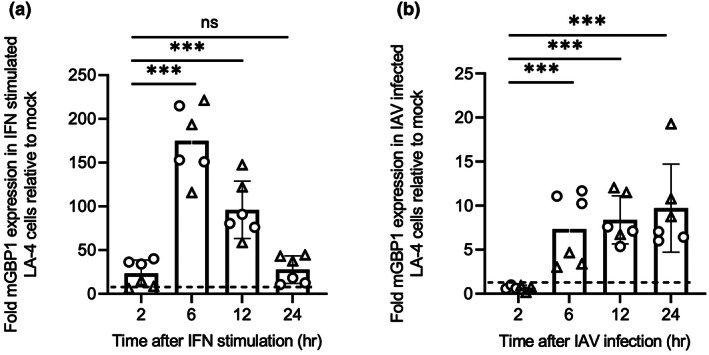
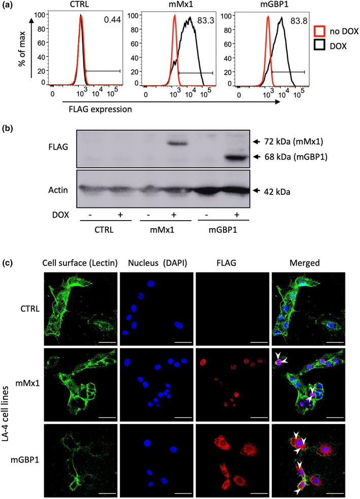
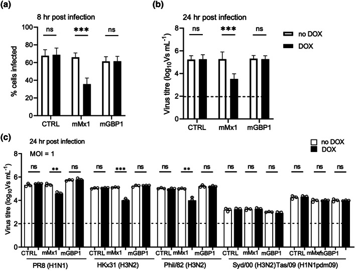
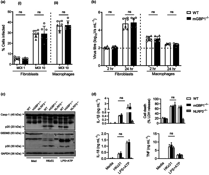
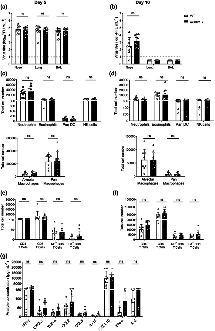
Many interferon (IFN)-stimulated genes are upregulated within host cells following infection with influenza and other viruses. While the antiviral activity of some IFN-stimulated genes, such as the IFN-inducible GTPase myxoma resistance (Mx)1 protein 1, has been well defined, less is known regarding the antiviral activities of related IFN-inducible GTPases of the guanylate-binding protein (GBP) family, particularly mouse GBPs, where mouse models can be used to assess their antiviral properties in vivo. Herein, we demonstrate that mouse GBP1 (mGBP1) was upregulated in a mouse airway epithelial cell line (LA-4 cells) following pretreatment with mouse IFNα or infection by influenza A virus (IAV). Whereas doxycycline-inducible expression of mouse Mx1 (mMx1) in LA-4 cells resulted in reduced susceptibility to IAV infection and reduced viral growth, inducible mGBP1 did not. Moreover, primary cells isolated from mGBP1-deficient mice (mGBP1-/- ) showed no difference in susceptibility to IAV and mGBP1-/- macrophages showed no defect in IAV-induced NLRP3 (NLR family pyrin domain containing 3) inflammasome activation. After intranasal IAV infection, mGBP1-/- mice also showed no differences in virus replication or induction of inflammatory responses in the airways during infection. Thus, using complementary approaches such as mGBP1 overexpression, cells from mGBP1-/- mice and intranasal infection of mGBP1-/- we demonstrate that mGBP1 does not play a major role in modulating IAV infection in vitro or in vivo.

Keywords: GTPase; influenza A virus; innate immunity; interferon-stimulated gene.


Conflict of interest statement

The authors declare no conflict of interest.

Figures

Figure 1
Figure 1
Induction of mGBP1 in LA‐4 cells in response to type I IFN or IAV infection. LA‐4 cells were seeded into 12‐well tissue culture plates (2 × 105 cells/well), cultured overnight and then (a) treated with 1000 U mL−1 IFNα or with media alone (mock) or (b) mock infected or infected with the IAV strain HKx31 (MOI = 5 PFU/cell). At indicated times, RNA was extracted from cells, cDNA synthesized and relative gene expression determined by quantitative real‐time PCR. To assess mGBP1 expression, samples were normalized to mouse GAPDH and then expressed as fold change relative to mock. Data show the mean ± standard deviation expression of triplicate samples pooled from two independent experiments, represented as circles and triangles, respectively. Statistical significance was determined by one‐way ANOVA; ***P < 0.001. The broken horizontal line represents the mGBP1 expression level in the mock. cDNA, complementary DNA; GAPDH, glyceraldehyde 3‐phosphate dehydrogenase; IAV, influenza A virus; IFN, interferon; mGBP1, murine guanylate‐binding protein 1; MOI, multiplicity of infection; PFU, plaque‐forming unit.
Figure 2
Figure 2
DOX‐inducible expression of mGBP1 and mMx1 in lentivirus‐transduced LA‐4 cells. LA‐4 cells with DOX‐inducible expression of intracellular ovalbumin (CTRL), mMx1 or mGBP1 were seeded at 1 × 105 cells/well into 12‐well tissue culture plates and incubated overnight at 37°C in 5% CO2. Media was then supplemented with or without 1 μg mL−1 DOX for 24 h. (a) After incubation, cells were detached, fixed, permeabilized and stained for intracellular FLAG expression as described in the “Methods” section. Histograms show FLAG expression after incubation in the absence (red) or presence (black) of DOX. Data are representative of three independent experiments. (b) Cell lysates were subjected to sodium dodecyl sulfate–polyacrylamide gel electrophoresis, transferred to a polyvinylidene fluoride membrane and probed with anti‐FLAG mAb (upper panel). Membranes were also probed with anti‐β‐actin mAb to confirm similar amounts of cellular protein were loaded for each sample (lower panel). Blot shown has been cropped for presentation. (c) Cells were washed, stained for cell‐surface lectin MAL II (lectin) and then fixed, permeabilized and stained for intracellular FLAG and DAPI. Representative images from confocal microscopy show cell‐surface MAL II (green; lectin stain), cell nucleus (blue; DAPI stain), expression of FLAG (red; FLAG tag stain) as well as a merged panel (merged image). The white arrowheads in the merged images indicate mGBP1 expression localizing to the cell cytoplasm and mMx1 expression in the nucleus, by colocalization with DAPI. The scale bar represents 50 μm. CTRL, control; DAPI, 4′,6‐diamidoino‐2‐phenylindole; DOX, doxycycline; FLAG, DYKDDDDK; mAb, monoclonal antibody; mGBP1, murine guanylate‐binding protein 1; mMx1, mouse Mx1.
Figure 3
Figure 3
DOX‐inducible mMx1, but not mGBP1, inhibits IAV infection and replication in LA‐4 cells. LA‐4 cells with DOX‐inducible expression of mGBP1 or mMx1, as well as CTRL cells, were seeded at 1 × 105 cells/well into 12‐well tissue culture plates and incubated overnight at 37°C in 5% CO2. Media was then supplemented with or without 1 μg mL−1 DOX for 24 h. After DOX induction, cells were incubated with (a, b) the IAV strain HKx31 (H3N2) at an MOI of 10 or 1 PFU/cell as indicated or (c) PR8 (H1N1), HKx31 (H3N2), Phil/82x (H3N2), Syd/00 (H3N2) or Tas/09 (H1N1pdm09) at an MOI of 1 for 60 min at 37°C, washed and cultured at 37°C. (a) At 8 h p.i., cells were fixed, permeabilized, stained for intracellular viral NP and analyzed by flow cytometry. (b, c) At 24 h p.i., supernatants were removed and clarified, then activated with 2 μg mL−1 TPCK‐treated trypsin and titrated on MDCK cells using the VS assay. Data from a and b show the mean ± standard deviation from three independent experiments. Data from c shows the mean ± standard deviation of triplicate samples from one of two independent experiments repeated with similar results. The dashed line in b and c indicates the detection limit of the VS assay. Statistical significance was determined by the Student's t‐test; **P < 0.01; ***P < 0.001. CTRL, control; DOX, doxycycline; IAV, influenza A virus; MDCK, Madin–Darby Canine Kidney; mGBP1, murine guanylate‐binding protein 1; mMx1, mouse Mx1; MOI, multiplicity of infection; NP, nucleoprotein; ns, not significant; PFU, plaque‐forming unit; p.i., postinfection; TPCK, tosylsulfonyl phenylalanyl chloromethyl ketone; VS, ViroSpot.
Figure 4
Figure 4
Endogenous mGBP1 does not inhibit IAV infection and replication in primary lung fibroblasts and peritoneal macrophages or IAV‐induced inflammasome activation in BMDMs in vitro. Lung primary fibroblasts (2 × 105 cells/well) and peritoneal macrophages (105 cells/well) from WT and mGBP1−/− mice were seeded into 12‐well plates or 8‐well chamber slides, respectively, and incubated overnight at 37°C. The next day cells were infected with the IAV strain HKx31 at the indicated MOI. (a) At 8 h p.i. (1) fibroblasts were detached, stained for intracellular viral NP and analyzed by flow cytometry; and (2) peritoneal macrophages were fixed, stained for intracellular viral NP and counted under a fluorescent microscope to determine the percent of infected cells. (b) At 24 h p.i., supernatants were removed, clarified and titers of infectious virus were determined by the VS assay on MDCK cells. Data for a and b show the mean ± standard deviation of triplicate samples pooled from two independent experiments, represented as circles and triangles, respectively. The dashed line represents the detection limit of the VS assay. Statistical significance was determined by the Student's t‐test. (c) Immunoblot analysis of pro‐casapse‐1 (Casp‐1) and the active caspase‐1 p20 subunit, pro‐pyroptosis effector protein GSDMD, the active GSDMD p30 subunit of WT, and NLPR3−/− and mGBP1−/− BMDMs left untreated or infected with IAV for 16 h. Blots shown have been cropped for presentation. (d) Levels of IL‐1β, IL‐18, LDH and TNF in cell culture supernatants 16 h after infection with IAV. Data show the mean ± standard deviation of three independent experiments. BMDM, bone marrow–derived macrophage; GAPDH, glyceraldehyde 3‐phosphate dehydrogenase; GSDMD, gasdermin D; IAV, influenza A virus; IL, interleukin; LDH, lactate dehydrogenase; LPS, lipopolysaccharide; MDCK, Madin–Darby Canine Kidney; mGBP1, murine guanylate‐binding protein 1; MOI, multiplicity of infection; NLRP3, NLR family pyrin domain containing 3; NP, nucleoprotein; ns, not significant; p.i., postinfection; TNF, tumor necrosis factor; VS, ViroSpot; WT, wild type.
Figure 5
Figure 5
Titers of infectious virus and inflammatory responses detected from WT and mGBP1−/− mice infected with HKx31. Mice were infected via the intranasal route with 50 μL of PBS containing 103.5 PFU of the IAV strain HKx31. At days 5 (a, c, e, g) or 10 (b, d, f) p.i., mice were killed and BAL was performed, before removal of lungs and nasal tissues. (a, b) Virus titers in homogenates prepared from nasal tissues or lungs, or in BAL, were determined by the plaque assay using MDCK cells. The broken line indicates the limit of detection of the plaque assay. (c–f) BAL cells were stained with antibodies against specific markers and flow cytometry was used to identify and quantify populations of (c, e) innate immune cells and (d, f) T cells. (g) Cell‐free BAL fluid was analyzed by flow cytometry using a multiplex CBA assay to detect 13 different inflammatory cytokines and chemokines. All data (a–f) show mean ± standard deviation values from mice pooled from two independent experiments, represented as circles and triangles, respectively (n = 8 or 11/group in total). A two‐tailed unpaired Student's t‐test was performed to compare viral titers, numbers of immune cell populations and cytokines and chemokines released between WT and mGBP1−/− mice. No significant differences were noted between WT and mGBP1−/− mice. BAL, bronchoalveolar lavage; CBA, cytometric bead array; CCL, chemokine (C–C motif) ligand; CXCL, C–X–C motif chemokine ligand; DC, dendritic cell; IAV, influenza A virus; IFN, interferon; IL, interleukin; MDCK, Madin–Darby Canine Kidney; mGBP1, murine guanylate‐binding protein 1; NP, nucleoprotein; ns, not significant; PA, polymerase acidic; PBS, phosphate‐buffered saline; PFU, plaque‐forming unit; p.i., postinfection; TNF, tumor necrosis factor; WT, wild type.

Similar articles

Cited by

References

    1. Isaacs A, Lindenmann J. Virus interference. I. the interferon. Proc R Soc Lond B Biol Sci 1957; 147: 258–267. - PubMed
    1. Man SM, Place DE, Kuriakose T, Kanneganti TD. Interferon‐inducible guanylate‐binding proteins at the interface of cell‐autonomous immunity and inflammasome activation. J Leukoc Biol 2017; 101: 143–150. - PMC - PubMed
    1. Martens S, Howard J. The interferon‐inducible GTPases. Annu Rev Cell Dev Biol 2006; 22: 559–589. - PubMed
    1. MacMicking JD. Interferon‐inducible effector mechanisms in cell‐autonomous immunity. Nat Rev Immunol 2012; 12: 367–382. - PMC - PubMed
    1. Verhelst J, Hulpiau P, Saelens X. Mx proteins: antiviral gatekeepers that restrain the uninvited. Microbiol Mol Biol Rev 2013; 77: 551–566. - PMC - PubMed

Publication types

Associated data