Review
. 2023 Jul;96(1147):20220544.
doi: 10.1259/bjr.20220544. Epub 2023 Feb 10.

Photon-counting detector CT: early clinical experience review

Affiliations
Review

Photon-counting detector CT: early clinical experience review

Thomas Sartoretti et al. Br J Radiol. 2023 Jul.

Abstract

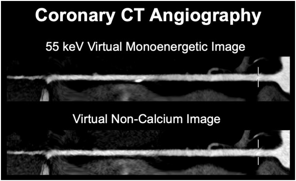
Since its development in the 1970s, X-ray CT has emerged as a landmark diagnostic imaging modality of modern medicine. Technological advances have been crucial to the success of CT imaging, as they have increasingly enabled improvements in image quality and diagnostic value at increasing radiation dose efficiency. With recent advances in engineering and physics, a novel technology has emerged with the potential to surpass several shortcomings and limitations of current CT systems. Photon-counting detector (PCD)-CT might substantially improve and expand the applicability of CT imaging by offering intrinsic spectral capabilities, increased spatial resolution, reduced electronic noise and improved image contrast. In this review we sought to summarize the first clinical experience of PCD-CT. We focused on most recent prototype and first clinically approved PCD-CT systems thereby reviewing initial publications and presenting corresponding clinical cases.


Figures

Figure 1.
Figure 1.
63-year-old male patient (body weight 77 kg) with multifocal hepatocellular carcinoma. Contrast-enhanced abdominal portal venous phase scans were acquired on a clinical PCD-CT system (NAEOTOM Alpha, Siemens Healthineers) in the spectral imaging mode at 120 kV with a tube current of 86 mAs. The CTDIvol was 6.7 mGy. Virtual monoenergetic images at 60 keV were reconstructed with a 2 mm slice thickness without Quantum Iterative Reconstruction and with all strength levels of QIR (QIR 1–4). CTDIvol, volume of CT dose index; PCD, Photon-counting detector; QIR, Quantum Iterative Reconstruction
Figure 2.
Figure 2.
65-year-old male patient (body weight 94 kg) with calcified coronary plaques in the left main and left anterior descending coronary artery with an Agatston Score of 245. Coronary CTA images were acquired in the UHR mode (z-coverage of 24 mm) on a clinical dual source PCD-CT system (NAEOTOM Alpha, Siemens Healthineers) at 120 kV with a tube current of 61 mAs. The gantry rotation time was 0.25 s, with a temporal resolution of 66 ms. The CTDIvol was 46.3 mGy. Images at different slice thicknesses and kernels were reconstructed. Note the improved sharpness of anatomical structures, vessels, and calcified coronary plaques on UHR images reconstructed with the Bv64 kernel and 0.2 mm section thickness. CTA, CT angiography; CTDIvol, volume of CT dose index; PCD, Photon-counting detector; UHR, ultra-high resolution.
Figure 3.
Figure 3.
32-year-old male patient (body weight 64 kg) presenting with a depression fracture of the talus. The fracture gap extends to the lateral part of the talus. Images were acquired in the UHR mode (z-coverage of 24 mm) on a clinical PCD-CT system (NAEOTOM Alpha, Siemens Healthineers) at 120 kV and with a tube current of 40 mAs. Radiation dose was 3.23 mGy CTDIvol. Images were reconstructed with a Br56 kernel, 2 mm section thickness and a 512 × 512 matrix size, with a Qr68 kernel, 1 mm section thickness and a 512 × 512 matrix size and with a sharp Qr72 kernel, 0.2 mm section thickness (UHR image) and 1024 × 1024 matrix size. The left image represents the default setting for bone imaging, while the right image leverages the potential of PCD-CT UHR imaging. Note the excellent visualization of bone features and trabeculae and the exquisite delineation of the small fracture gap. CTDIvol, volume of CT dose index; PCD, Photon-counting detector; UHR, ultra-high resolution.
Figure 4.
Figure 4.
47-year-old male patient (body weight 85 kg) with COVID-19-associated pneumonia presenting with ground-glass opacities and mild reticular abnormalities. Non-enhanced chest CT was acquired on a clinical PCD-CT system (NAEOTOM Alpha, Siemens Healthineers) in the UHR mode at 120 kV; the CTDIvol was 0.55 mGy. CTDIvol, volume of CT dose index; PCD, Photon-counting detector; UHR, ultra-high resolution.
Figure 5.
Figure 5.
82-year-old male patient (body weight 81 kg) undergoing CTA for follow-up after endovascular treatment of an abdominal aortic aneurysm. Images were acquired on a third generation dual-source EID-CT system (SOMATOM Force, Siemens Healthineers) with automated tube voltage selection (80 kV) and CTA with a clinical dual-source PCD-CT system (NAEOTOM Alpha, Siemens Healthineers) (120 kV) at matched radiation dose (CTDIvol 6.1 mGy) and using the same contrast media protocol. Note the reduced noise and improved contrast on PCD-CT images. CTA, CT angiography; CTDIvol, volume of CT dose index; PCD, Photon-counting detector; EID, energy-integrating detector; PCD, Photon-counting detector.
Figure 6.
Figure 6.
71-year-old female patient (body weight 75 kg) with a cyst in liver segment VII. Contrast-enhanced abdominal portal venous phase images were acquired on a third generation dual-source EID-CT system (SOMATOM Force, Siemens Healthineers) with automatic tube voltage selection (120 kV) and with a clinical PCD-CT system (NAEOTOM Alpha, Siemens Healthineers) in the spectral imaging mode at 120 kV at matched radiation dose (CTDIvol 7.24 mGy) and using the same contrast media protocol. Note the improved iodine contrast and lesion conspicuity on PCD-CT images. CTDIvol, volume of CT dose index; EID, energy-integrating detector; PCD, Photon-counting detector.
Figure 7.
Figure 7.
67-year-old male patient (body weight 71 kg) with atypical chest pain. Coronary CTA was performed on a clinical dual source PCD-CT system (NAEOTOM Alpha, Siemens Healthineers) at 120 kV (CTDIvol 10.1 mGy). Virtual monoenergetic images at 55 keV and VNCa images using a novel vascular calcium removal algorithm (PureLumen) were generated. A calcified plaque can be seen in the distal right coronary artery, which is subtracted on a dual-energy basis in the VNCa (PureLumen) images. CTA, CT angiography; CTDIvol, volume of CT dose index; PCD, Photon-counting detector; VNCa, virtual non-calcium.
Figure 8.
Figure 8.
54-year-old male patient (body weight 76 kg) with hepatocellular carcinoma and vascular invasion. Late arterial scans were performed with a clinical PCD-CT (NAEOTOM Alpha, Siemens Healthineers) in the spectral mode with reconstruction of virtual monoenergetic images at 70 keV, virtual non-contrast images, and iodine maps from a single acquisition (CTDIvol3.6 mGy). Although tumor enhancement is seen also on the monoenergetic images, the iodine maps allow for a better appreciation of the carcinoma along with the possibility of quantification of iodine uptake. CTDIvol, volume of CT dose index; PCD, Photon-counting detector.
Figure 9.
Figure 9.
59-year-old male patient (body weight 81 kg) with chronic occlusion of the right pulmonary artery and partial occlusion of left pulmonary artery branches resulting in severely reduced perfusion of the right and, to a lesser extent, of the left lung. Axial and coronal thick maximum intensity projection images and coronal PBV map computed from a routine contrast-enhanced chest CT scan acquired on a clinical PCD-CT (NAEOTOM Alpha, Siemens Healthineers) at 120 kV (CTDIvol 2.37 mGy) illustrate both the anatomical and functional situation in the lungs. CTDIvol, volume of CT dose index; PBV, perfused blood volume; PCD, Photon-counting detector.

References

    1. Smith-Bindman R, Kwan ML, Marlow EC, Theis MK, Bolch W, Cheng SY, et al. . Trends in use of medical imaging in US health care systems and in Ontario, Canada, 2000-2016. JAMA 2019; 322: 843–56. doi: 10.1001/jama.2019.11456 - DOI - PMC - PubMed
    1. Alkadhi H, Euler A. The future of computed tomography: personalized, functional, and precise. Invest Radiol 2020; 55: 545–55. doi: 10.1097/RLI.0000000000000668 - DOI - PubMed
    1. Wildberger JE, Prokop M. Hounsfield’s legacy. Invest Radiol 2020; 55: 556–58. doi: 10.1097/RLI.0000000000000680 - DOI - PubMed
    1. Lell MM, Wildberger JE, Alkadhi H, Damilakis J, Kachelriess M. Evolution in computed tomography: the battle for speed and dose. Invest Radiol 2015; 50: 629–44. doi: 10.1097/RLI.0000000000000172 - DOI - PubMed
    1. Flohr T, Petersilka M, Henning A, Ulzheimer S, Ferda J, Schmidt B. Photon-Counting CT review. Phys Med 2020; 79: 126–36: S1120-1797(20)30273-8. doi: 10.1016/j.ejmp.2020.10.030 - DOI - PubMed