. 2023 Feb 16;14(6):1485-1493.
doi: 10.1021/acs.jpclett.2c03110. Epub 2023 Feb 6.

Reaction Dynamics in the Chrimson Channelrhodopsin: Observation of Product-State Evolution and Slow Diffusive Protein Motions

Affiliations

Reaction Dynamics in the Chrimson Channelrhodopsin: Observation of Product-State Evolution and Slow Diffusive Protein Motions

Ivo H M van Stokkum et al. J Phys Chem Lett. .

Abstract

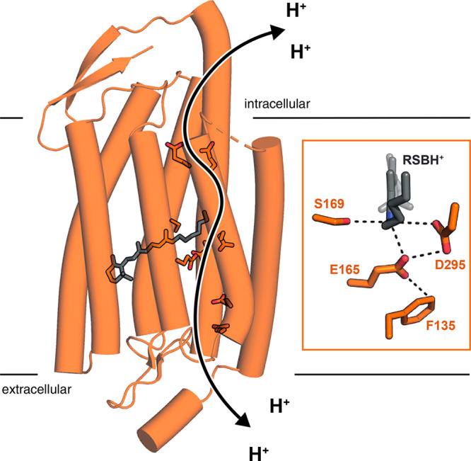
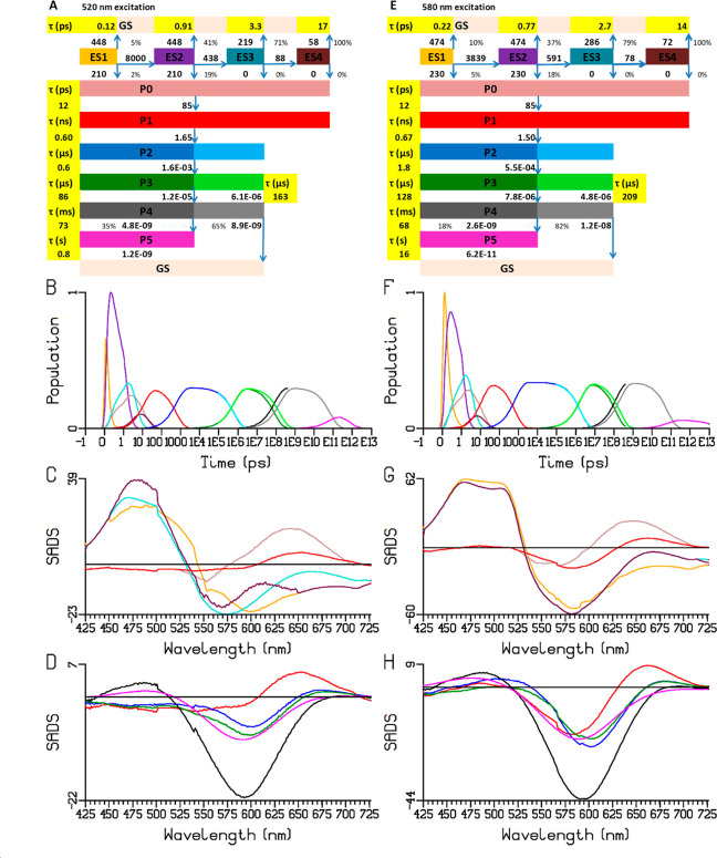
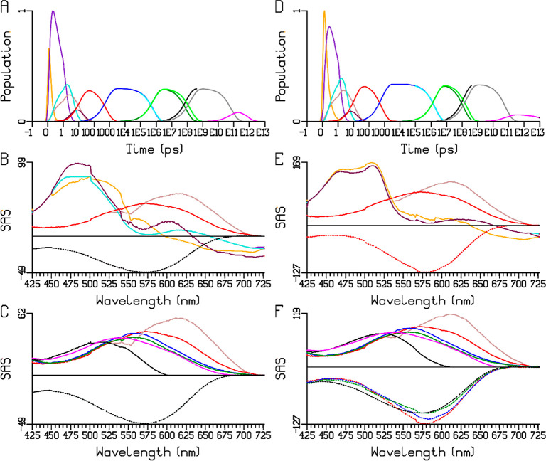
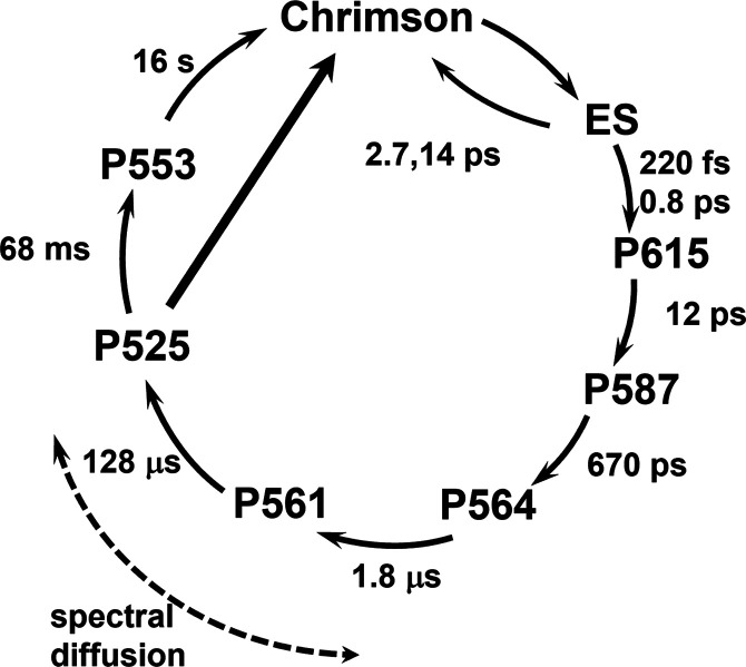
Chrimson is a red-light absorbing channelrhodopsin useful for deep-tissue optogenetics applications. Here, we present the Chrimson reaction dynamics from femtoseconds to seconds, analyzed with target analysis methods to disentangle spectrally and temporally overlapping excited- and product-state dynamics. We found multiple phases ranging from ≈100 fs to ≈20 ps in the excited-state decay, where spectral features overlapping with stimulated emission components were assigned to early dynamics of K-like species on a 10 ps time scale. Selective excitation at the maximum or the blue edge of the absorption spectrum resulted in spectrally distinct but kinetically similar excited-state and product-state species, which gradually became indistinguishable on the μs to 100 μs time scales. Hence, by removing specific protein conformations within an inhomogeneously broadened ensemble, we resolved slow protein backbone and amino acid side-chain motions in the dark that underlie inhomogeneous broadening, demonstrating that the latter represents a dynamic interconversion between protein substates.


Conflict of interest statement

The authors declare no competing financial interest.

Figures

Figure 1
Figure 1
Three-dimensional structure of Chrimson (pdb 5zih) showing the retinal chromophore, pore lining glutamates E1′ to E5′, and the counterion complex. The putative proton pathway is indicated by the black arrow. (inset) Enlarged illustration of the active site with the protonated retinal Schiff base (RSBH+) and its direct hydrogen-bond interaction partners.
Figure 2
Figure 2
Sequential analysis of the Chrimson reaction dynamics at pH 5.0 upon 520 (A–C) and 580 (E–G) nm excitation. (A, E) Populations of the components, with the kinetic schemes indicated at the top, the lifetimes are in the yellow highlighted cells. Note that the time axis is linear until 1 ps (after the maximum of the instrument response function, IRF) and logarithmic thereafter. (B, C, F, G) EADS (in mOD), in C and G starting from S4. The black dotted curve represents the sign-inverted ground-state absorption spectrum, scaled by 0.2 in C and G. Overlays of scaled EADS of (D) S1–S4, (H) S5–S8, key: 520 (solid) and 580 nm (dotted) excitation.
Figure 3
Figure 3
Target analysis of the Chrimson reaction dynamics at pH 5.0 upon 520 (A–D) and 580 (E–H) nm excitation according to the kinetic scheme (3) (A, E) in which the SADS of ES2, ES3, and ES4 are assumed to be virtually identical, and a product P0 (brown SADS) is introduced that precedes P1. (B, F) Populations of the species. Note that the time axis is linear until 1 ps (after the maximum of the IRF) and logarithmic thereafter. (C, G) SADS (in mOD) of ES1–ES4, P0, and P1. (D,H) SADS of P1–P5. Key: ES1–ES4: orange, purple, turquoise, maroon; P0–P5: brown, red, blue, dark green, black, magenta. Cyan, green, gray, and magenta in (B, F) refer to flash photolysis populations.
Figure 4
Figure 4
Target analysis including the GSB according to the kinetic scheme (3) in Figure 3A,E of the Chrimson reaction dynamics at pH 5.0 upon 520 (A–C) and 580 (D–F) nm excitation. (A, D) Populations of the species. Note that the time axis is linear until 1 ps (after the maximum of the IRF) and logarithmic thereafter. (B, E) GSB (dotted) and SAS (solid, in mOD) of ES1–ES4, P0, and P1. (C, F) GSB (dotted) and SAS (solid) of P0–P5. Key: ES1–ES4: orange, purple, turquoise, maroon; P0–P5: brown, red, blue, dark green, black, magenta. Cyan, green, gray, and magenta in (A, D) refer to flash photolysis populations.
Figure 5
Figure 5
Chrimson photocycle at pH 5, describing the sequential and nonsequential interconversions between spectroscopic intermediates and their lifetimes (with 580 nm excitation), and the occurrence of spectral diffusion phenomena. See text for details.

Similar articles

Cited by

References

    1. Ernst O. P.; Lodowski D. T.; Elstner M.; Hegemann P.; Brown L. S.; Kandori H. Microbial and animal rhodopsins: structures, functions, and molecular mechanisms. Chem. Rev. 2014, 114 (1), 126–63. 10.1021/cr4003769. - DOI - PMC - PubMed
    1. Govorunova E. G.; Sineshchekov O. A.; Li H.; Spudich J. L.; Kornberg R. D. Microbial Rhodopsins: Diversity, Mechanisms, and Optogenetic Applications. Annu. Rev. Biochem. 2017, 86, 845–872. 10.1146/annurev-biochem-101910-144233. - DOI - PMC - PubMed
    1. Zhang F.; Vierock J.; Yizhar O.; Fenno L. E.; Tsunoda S.; Kianianmomeni A.; Prigge M.; Berndt A.; Cushman J.; Polle J.; Magnuson J.; Hegemann P.; Deisseroth K. The Microbial Opsin Family of Optogenetic Tools. Cell 2011, 147 (7), 1446–1457. 10.1016/j.cell.2011.12.004. - DOI - PMC - PubMed
    1. Hochbaum D. R.; Zhao Y.; Farhi S. L.; Klapoetke N.; Werley C. A.; Kapoor V.; Zou P.; Kralj J. M.; Maclaurin D.; Smedemark-Margulies N.; Saulnier J. L.; Boulting G. L.; Straub C.; Cho Y. K.; Melkonian M.; Wong G. K. S.; Harrison D. J.; Murthy V. N.; Sabatini B. L.; Boyden E. S.; Campbell R. E.; Cohen A. E. All-optical electrophysiology in mammalian neurons using engineered microbial rhodopsins. Nat. Methods 2014, 11 (8), 825–833. 10.1038/nmeth.3000. - DOI - PMC - PubMed
    1. Klapoetke N. C.; Murata Y.; Kim S. S.; Pulver S. R.; Birdsey-Benson A.; Cho Y. K.; Morimoto T. K.; Chuong A. S.; Carpenter E. J.; Tian Z.; Wang J.; Xie Y.; Yan Z.; Zhang Y.; Chow B. Y.; Surek B.; Melkonian M.; Jayaraman V.; Constantine-Paton M.; Wong G. K.-S.; Boyden E. S. Independent optical excitation of distinct neural populations. Nat. Methods 2014, 11 (3), 338–46. 10.1038/nmeth.2836. - DOI - PMC - PubMed

Substances