. 2023 May 23;7(10):2053-2065.
doi: 10.1182/bloodadvances.2022008405.

TIM-3 signaling hijacks the canonical Wnt/β-catenin pathway to maintain cancer stemness in acute myeloid leukemia

Affiliations

TIM-3 signaling hijacks the canonical Wnt/β-catenin pathway to maintain cancer stemness in acute myeloid leukemia

Teppei Sakoda et al. Blood Adv. .

Abstract

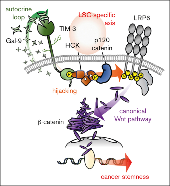
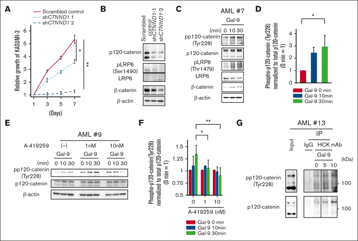
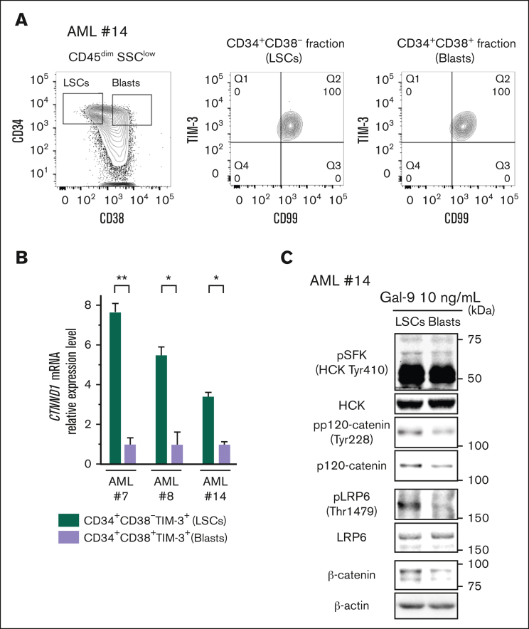
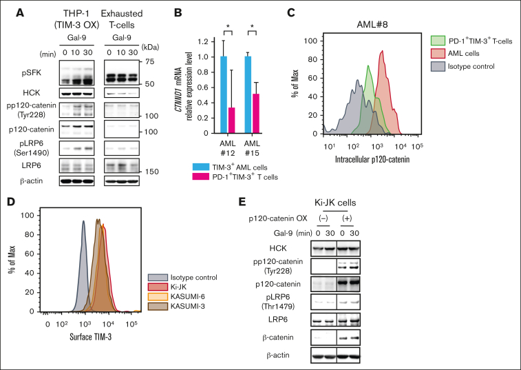
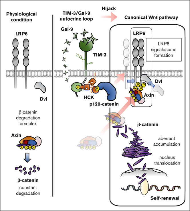
The activation of β-catenin plays critical roles in normal stem cell function, and, when aberrantly activated, the maintenance and enhancement of cancer stemness in many solid cancers. Aberrant β-catenin activation is also observed in acute myeloid leukemia (AML), and crucially contributes to self-renewal and propagation of leukemic stem cells (LSCs) regardless of mutations in contrast with such solid tumors. In this study, we showed that the AML-specific autocrine loop comprised of T-cell immunoglobulin mucin-3 (TIM-3) and its ligand, galectin-9 (Gal-9), drives the canonical Wnt pathway to stimulate self-renewal and propagation of LSCs, independent of Wnt ligands. Gal-9 ligation activates the cytoplasmic Src homology 2 domain of TIM-3 to recruit hematopoietic cell kinase (HCK), a Src family kinase highly expressed in LSCs but not in HSCs, and HCK phosphorylates p120-catenin to promote formation of the LDL receptor-related protein 6 (LRP6) signalosome, hijacking the canonical Wnt pathway. This TIM-3/HCK/p120-catenin axis is principally active in immature LSCs compared with TIM-3-expressed differentiated AML blasts and exhausted T cells. These data suggest that human AML LSCs constitutively activates β-catenin via autocrine TIM-3/HCK/p120-catenin signaling, and that molecules related to this signaling axis should be critical targets for selective eradication of LSCs without impairing normal HSCs.


Conflict of interest statement

Conflict-of-interest disclosure: The authors declare no competing financial interests.

Figures

None
Graphical abstract
Figure 1.
Figure 1.
Identification of the canonical Wnt pathway as major downstream signaling of TIM-3/Gal-9 autocrine loop. (A) Fluorescence-activated cell sorting (FACS) analysis of surface TIM-3 expression (left) and intracellular Gal-9 expression (right) in KASUMI-3 cells (B) Percentage of BM chimerism in NSG mice with xenografts with TIM-3 KD KASUMI-3 cells (n = 6) and scrambled control cells (n = 6) (left panel), and human Gal-9 concentration in serum of mice with xenografts (right panel). ∗∗P < .01 vs scrambled control. These data were obtained at 13 weeks after xenotransplantation. (C-D) Enrichment plots for gene sets significantly enriched in scrambled control cells compared with KASUMI-3 cells transfected with shHAVCR2-1 and shHAVCR2-2. (C) HSC- and LSC-related genes. (D) Canonical Wnt pathway–related genes (BIOCARTA). (E) Quantification of nucleus translocation of β-catenin evaluated by ArrayScan system. Left panels show the representative images for localization of β-catenin in scrambled control and TIM-3 KD KASUMI-3 cells. Scale bar represents 10 μm. Right panel shows the quantification of β-catenin (green) translocation to the nucleus calculated from the fluorescence intensity and area overlapped with nucleus (blue), which were analyzed in TIM-3 KD KASUMI-3 cells and scrambled control cells. Data are presented as mean ± SEM, ∗∗P < .01 vs scrambled control.
Figure 2.
Figure 2.
Gal-9 ligation to TIM-3 induces LRP6 signalosome formation and subsequent β-catenin accumulation in AML. (A) WB analysis of β-catenin accumulation induced by stimulation with Gal-9 in the presence of inhibitors in KASUMI-3 cells. KASUMI-3 cells were stimulated with Gal-9 in the presence of dimethyl sulfoxide, 10 μM of U0126 (MEK1/2 inhibitor), 10 μM of LY294002 (PI3K inhibitor), or 200 ng/mL of DKK-1, and their total cell lysates subjected to WB analysis. (B) Summarized data of Gal-9 stimulation–induced changes in the accumulation of β-catenin from 3 independent experiments. (C) Extent of β-catenin translocation to the nucleus evaluated using the ArrayScan system compared with that observed in nonstimulated controls. Data in panels B and C are presented as mean ± SEM, ∗P < .05. (D) Immunoblotting analysis of total lysates (left) and IP lysates (right) of Gal-9–stimulated primary TIM-3+ AML samples. Cells were lysed and subjected to IP with an anti-LRP6 antibody or normal mouse IgG. A representative result of TIM-3+ primary AML cells out of 3 independent cases (AML#4, #9, and #13) is shown. (E) WB analysis of Gal-9 stimulation–induced phosphorylation of LRP6 at Thr1479 in mock-transfected and TIM-3-OX THP-1 cells. (F) WB analysis of cell lysates from TIM-3+ AML cells (left) and TIM-3 AML cells (right). TIM-3+ AML cells were stimulated with Gal-9 in the absence or presence of DKK-1 (200 ng/mL). Representative results out of 4 independent TIM-3+ AML cells (AML#1, #2, #4, and #16) and 2 TIM-3 AML cells (AML#10 and #11) are shown. n.s., no significant difference.
Figure 3.
Figure 3.
HCK is a critical signal transducing molecule involved in TIM-3–induced canonical Wnt pathway activation. (A) WB analysis of Gal-9–stimulated KASUMI-6 cells in the presence of SFK inhibitors: AMGT, PP1, PP2, and A-419259 (potent HCK inhibitor). Inhibitable members of SFKs at each concentration of inhibitors are listed. Because KASUMI-6 cells showed obvious response to SFKs inhibitors, we presented the representative data of KASUMI-6 cells. (B) WB analysis of cell lysates from Gal-9–stimulated TIM-3+ primary samples in the presence of 0, 1, and 10 nM of A-419259. Cells were preincubated with A-419259 for 2 hours before stimulation with Gal-9. Representative results out of 4 independent TIM-3+ AML cases (AML#6, #9, #14, and #17) are shown. (C) Images of KASUMI-3 cells captured from representative fields obtained by ArrayScan analysis. These cells were stimulated with or without Gal-9 in the presence or absence of A-419259 for 20 hours. Scale bar represents 10 μm. (D) Extent of β-catenin translocation to the nucleus evaluated using the ArrayScan system compared with nonstimulated control. Data are presented as mean ± SEM, ∗P < .05 and ∗∗P < .01. (E) Immunoblotting analysis with HCK, pSFK, and TIM-3 antibodies of total cell lysates (left) and immunoprecipitated lysates (right). Cells were stimulated with Gal-9 for 0, 5, and 10 minutes and subsequently lysed and subjected to IP with an HCK antibody or normal rabbit IgG. Representative results (AML#13) out of 3 independent experiments are shown here.
Figure 4.
Figure 4.
p120-catenin plays a crucial role for bridging TIM-3/Gal-9 signaling to the canonical Wnt pathway in AML cells. (A) Cellular growth of KASUMI-3 cells transfected with scrambled control and shRNA-mediated CTNND1 KD vectors. Two kinds of shRNA (shCTNND1-1: target sites in the 3' untranslated region, and shCTNND1-2: target sites in the coding sequence) were used. Data are presented as mean ± SEM, ∗P < .05 and ∗∗P < .01 vs scrambled control. (B) WB analysis of the canonical Wnt pathway–related protein using cell lysates from KASUMI-3 cells transfected with scrambled control and 2 kinds of shRNA-targeting CTNND1. (C) WB analysis of the phosphorylation status of p120-catenin at Tyr228 induced by stimulation with Gal-9 (AML#7). (D) Quantification of the levels of phosphorylated p120-catenin at Tyr228 by Gal-9 stimulation in KASUMI-3 cells. Results of 4 independent experiments (AML#1, #7, #9, and #16) are summarized. Data are presented as mean ± SEM, ∗P < .05 vs nonstimulated control. (E) WB analysis of the inhibitory effect of A-419259 (0, 1, or 10 nM) on Gal-9 stimulation–induced phosphorylation of p120-catenin using a primary AML sample (AML#9). (F) Quantification of the levels of phosphorylated p120-catenin induced by Gal-9 stimulation in the presence of each concentration of A-419259 (0, 1, and 10 nM). Data are presented as mean ± SEM, ∗P < .05 and ∗∗P < .01. Results from 4 independent AML experiments (AML#6, #9, #14, and #17) are summarized. (G) Immunoblotting analysis with total p120-catenin and phosphorylated p120-catenin (Tyr228) antibodies of total cell lysates (left), and immunoprecipitated lysates (right). Cells were stimulated with Gal-9 for 0, 5, and 10 minutes, and subsequently lysed and subjected to IP with an anti-HCK antibody or normal rabbit IgG. Representative results out of 3 independent experiments are shown here.
Figure 5.
Figure 5.
TIM-3 signaling induces β-catenin accumulation in accordance with p120-catenin expression level in AML. (A) FACS analysis of the expression of TIM-3 on CD34+CD38 (middle; LSCs-enriched fraction) and CD34+CD38+ (right; main blasts–enriched fraction) AML cells from the CD45dimSSClow fraction. Representative FACS plots of 4 independent AML samples (AML#7, #8, #13, and #14) are shown. (B) RT-qPCR analysis of the expression of CTNND1 mRNA in LSCs compared with blasts from AML samples of 3 independent AML cases. Results were normalized to the mRNA expression of GAPDH. Data are presented as mean ± SEM, ∗∗P < .01 and ∗P < .05. (C) WB analysis of cell lysate from Gal-9–stimulated CD34+CD38 AML cells (LSCs) and CD34+CD38+ cells (blasts) (AML#14). Of note, LSCs with higher p120-catenin expression exhibited profound β-catenin accumulation.
Figure 6.
Figure 6.
The expression of p120-catenin results in a striking difference in the signal transduction of TIM-3 between AML and exhausted T cells. (A) WB analysis of Gal-9–stimulated TIM-3-OX THP-1 and PD-1+TIM-3+ exhausted T cells. Of note, Gal-9 ligation to TIM-3 induced LRP6 phosphorylation in TIM-3-OX THP-1 cells but not in the exhausted T cells devoid of p120-catenin expression. (B) RT-qPCR analysis of CTNND1 mRNA in FACS-purified AML cells and the exhausted (PD-1+TIM-3+) T cells from 2 patients with AML (AML#12 and #15). Results were normalized to the mRNA expression of GAPDH. Data are presented as mean ± SEM, ∗P < .05. (C) Intracellular FACS analysis of p120-catenin expression in TIM-3+ AML cells and PD-1+TIM-3+ exhausted T cells in the identical patient with AML. A representative result out of 3 AML samples (AML#8, #12, and #15) is shown. (D) FACS analysis of surface TIM-3 expression in Ki-JK cells. TIM-3 expression of KASUMI-3 and KASUMI-6 are shown as positive controls. (E) The results of WB analysis using mock-transfected (left panels) and p120-catenin-OX Ki-JK cells (right panels) stimulated with Gal-9 are shown.
Figure 7.
Figure 7.
Schematic summary of a novel molecular mechanisms of the hijacking of the canonical Wnt pathway by TIM-3 signaling in AML LSCs. The schema shows how TIM-3 signaling induces constitutively canonical Wnt pathway activation and aberrant accumulation of β-catenin in AML-LSCs. As shown in the left panel, β-catenin is constantly destroyed by the β-catenin degradation complex in the absence of canonical Wnt pathway activation. As shown in the right panel, in AML, a TIM-3/Gal-9 autocrine loop constitutively recruits and activates HCK, leading to the induction of p120-catenin phosphorylation. The activated p120-catenin initiates LRP6 signalosome formation to inhibit the function of the β-catenin degradation complex, leading to the aberrant accumulation of β-catenin in AML cells. Through the use of LSCs-specific molecules such as TIM-3, HCK, and p120-catenin, the AML LSCs hijack the canonical Wnt pathway.

Similar articles

Cited by

References

    1. Kikushige Y, Miyamoto T, Yuda J, et al. A TIM-3/Gal-9 autocrine stimulatory loop drives self-renewal of human myeloid leukemia stem cells and leukemic progression. Cell Stem Cell. 2015;17(3):341–352. - PubMed
    1. Kikushige Y, Shima T, Takayanagi Si, et al. TIM-3 is a promising target to selectively kill acute myeloid leukemia stem cells. Cell Stem Cell. 2010;7(6):708–717. - PubMed
    1. Jan M, Chao MP, Cha AC, et al. Prospective separation of normal and leukemic stem cells based on differential expression of TIM3, a human acute myeloid leukemia stem cell marker. Proc Natl Acad Sci U S A. 2011;108(12):5009–5014. - PMC - PubMed
    1. Monney L, Sabatos CA, Gaglia JL, et al. Th1-specific cell surface protein Tim-3 regulates macrophage activation and severity of an autoimmune disease. Nature. 2002;415(6871):536–541. - PubMed
    1. Wolf Y, Anderson AC, Kuchroo VK. TIM3 comes of age as an inhibitory receptor. Nat Rev Immunol. 2020;20(3):173–185. - PMC - PubMed

Publication types

MeSH terms