. 2023 Sep;33(3):669-676.
doi: 10.1007/s00062-022-01260-0. Epub 2023 Feb 6.

Benefit of Advanced 3D DSA and MRI/CT Fusion in Neurovascular Pathology

Affiliations

Benefit of Advanced 3D DSA and MRI/CT Fusion in Neurovascular Pathology

Tomas Dobrocky et al. Clin Neuroradiol. 2023 Sep.

Abstract

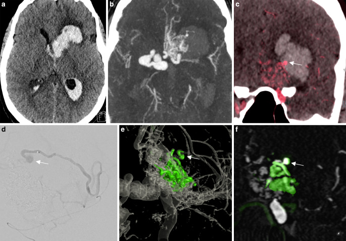
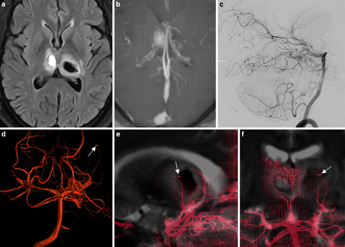
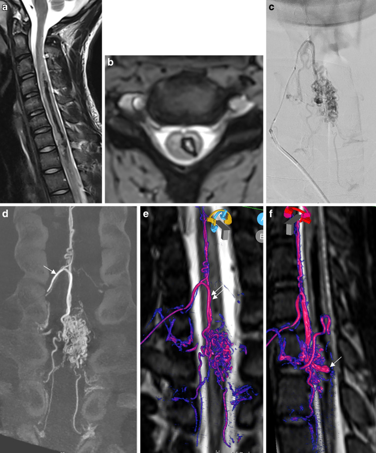
Digital subtraction angiography provides excellent spatial and temporal resolution; however, it lacks the capability to depict the nonvascular anatomy of the brain and spinal cord.A review of the institutional database identified five patients in whom a new integrated fusion workflow of cross-sectional imaging and 3D rotational angiography (3DRA) provided important diagnostic information and assisted in treatment planning. These included two acutely ruptured brain arteriovenous malformations (AVM), a small superficial brainstem AVM after radiosurgery, a thalamic microaneurysm, and a spine AVM, and fusion was crucial for diagnosis and influenced further treatment.Fusion of 3DRA and cross-sectional imaging may help to gain a deeper understanding of neurovascular diseases. This is advantageous for planning and providing treatment and, most importantly, may harbor the potential to minimize complication rates. Integrating image fusion in the work-up of cerebrovascular diseases is likely to have a major impact on the neurovascular field in the future.

Keywords: AVM; Aneurysm; Image fusion; Intracerebral hemorrhage; Rotational angiography.


Conflict of interest statement

T. Dobrocky, M. Matzinger, E.I. Piechowiak, J. Kaesmacher, S. Pilgram-Pastor, J. Goldberg, D. Bervini, T. Klail, V.M. Pereira, W. Z’Graggen, A. Raabe, P. Mordasini and J. Gralla declare that they have no competing interests.

Figures

Fig. 1
Fig. 1
Ruptured arteriovenous malformation (AVM) with an intranidal aneurysm. a Nonenhanced computed tomography (CT) in the axial plane demonstrating a deep left basal ganglia hemorrhage with intraventricular rupture. b Computed tomography angiography (CTA) demonstrating a nidus in the left orbitofrontal cortex with several enlarged venous pouches on the contralateral side overlying the carotid terminus. c Three-dimensional rotational angiography (3DRA) + CT fusion in the coronal plane demonstrating a small intranidal aneurysm (arrow) centered in the basal ganglia hemorrhage as the bleeding source. d Selective catheterization of the deep perforating branch supplying the aneurysm (arrow). e,f Baseline 3DRA + postembolization 3DRA fusion demonstrating the onyx cast (green). Note that the inferior and lateral part of the AVM with the aneurysm (arrow) have been embolized. Supplementary video demonstrates the fusion of baseline 3DRA + postembolization 3DRA in axial slides
Fig. 2
Fig. 2
Ruptured thalamo-perforating branch micro-aneurysm. a Baseline magnetic resonance imaging (MRI) demonstrating a bithalamic hemorrhage. b No sign of central venous thrombosis. c Digital subtraction angiography (DSA) run of the posterior circulation in the lateral projection with normal findings. d Three-dimensional rotational angiography (3DRA) showing a small aneurysm (white arrow) in the course of a left thalamo-perforating branch. e,f 3DRA + MRI fusion clearly demonstrates the aneurysm located within the left thalamic hemorrhage as the bleeding source
Fig. 3
Fig. 3
Ruptured spinal arteriovenous malformation (AVM) with a large intranidal aneurysm. a T2W sagittal image demonstrating extensive left-sided hematomyelia. b Hemoflash demonstrating the extension of the hematoma within the left side of the spinal cord. c (DSA anterio posterior projection). d (coronal reformated 3DRA) Superselective injection of the main radiculomedullary artery of the cervical enlargement (artery of Lazorthes, arrow) demonstrating a large nidus and multiple draining radicular veins. ef Fusion of the three-dimensional rotational angiography (3DRA) and T2W spine magnetic resonance imaging (MRI) demonstrating the feeding radiculomedullary artery which supplies the anterior spinal artery (double arrow) which than feeds the AVM (red/pink for the artery and purple or blue for the draing vein). The intranidal aneurysm is localized within the central part of the myelon and the tip of the aneurysm is pointing towards the hematoma. Supplementary video demonstrates the fusion with an embedded 3D model of the AVM
Fig. 4
Fig. 4
Small arteriovenous malformation (AVM) on the superficial surface of the mesencephalon after radiosurgery. a The baseline computed tomography (CT) scan demonstrating a left thalamic hemorrhage. b Digital subtraction angiography (DSA) showing the nidus and en-passant feeders from the left P3 segment and deep venous drainage. c Three years after radiosurgery, 7‑Tesla magnetic resonance imaging (MRI) does not show evidence of any abnormal vessels on the arterial time-of-flight sequence. d DSA demonstrates a residual arteriovenous shunt with early arterial filling of the posterior segment of the basal vein of Rosenthal (arrow). e Fusion of the follow-up 7‑Tesla and three-dimensional rotational angiography (3DRA) volumes clearly demonstrates the small nidus localized on the superficial surface of the posterolateral mesencephalon with a single draining vein corresponding to the posterior segment of the basal vein of Rosenthal (purple). Supplementary video demonstrates the fusion with an embedded 3D model of the AVM
Fig. 5
Fig. 5
Ruptured periventricular arteriovenous malformation (AVM) with an intranidal aneurysm. a The non-enhanced computed tomography (CT) demonstrates a hemorrhage in the left caudate nucleus (arrow) with intraventricular rupture. b The digital subtraction angiography (DSA) shows a left basal ganglia nidus supplied by the anterior choroidal artery, draining via a single vein into the deep venous system. c,d Fusion of the non-enhanced baseline CT scan and three-dimensional rotational angiography (3DRA) shows a small intranidal aneurysm centered in the left caudate nucleus as the bleeding source. e Fusion of the baseline CT and superselective 3DRA with the microcatheter placed in the feeding artery supplying the aneurysm and the anterior part of the nidus

References

    1. van Rooij WJ, Sprengers ME, de Gast AN, Peluso JPP, Sluzewski M. 3D rotational angiography: The new gold standard in the detection of additional intracranial aneurysms. AJNR Am J Neuroradiol. 2008;29:976–979. doi: 10.3174/ajnr.A0964. - DOI - PMC - PubMed
    1. Jalali A, Srinivasan VM, Chinnadurai P, Kan P, Arthur A, Duckworth EAM. Two-color 3D-3D fusion of selective rotational cerebral angiograms: a novel approach to imaging in cerebrovascular neurosurgery. J Neurointerv Surg. 2016;8:1056–1060. doi: 10.1136/neurintsurg-2015-011963. - DOI - PubMed
    1. Raz E, Shapiro M, Mir O, Nossek E, Nelson PK. Arterial and venous 3d fusion av-3d-dsa: A novel approach to cerebrovascular neuroimaging. AJNR Am J Neuroradiol. 2021;42:1282–1284. doi: 10.3174/ajnr.A7103. - DOI - PMC - PubMed
    1. Suzuki H, Shimizu S, Maki H, Maeda M, Sakaida H, Trousset Y, et al. Role of image fusion combining three-dimensional digital subtraction angiography with magnetic resonance imaging in evaluation of unruptured cerebral aneurysms. Neurol Res. 2007;29:58–63. doi: 10.1179/174313206X153806. - DOI - PubMed
    1. Suzuki H, Maki H, Taki W. Evaluation of cerebral arteriovenous malformations using image fusion combining three-dimensional digital subtraction angiography with magnetic resonance imaging. Turk Neurosurg. 2012;22:341–345. - PubMed

MeSH terms