. 2023 Feb 6;18(2):e0281404.
doi: 10.1371/journal.pone.0281404. eCollection 2023.

Denuded Descemet's membrane supports human embryonic stem cell-derived retinal pigment epithelial cell culture

Affiliations

Denuded Descemet's membrane supports human embryonic stem cell-derived retinal pigment epithelial cell culture

Elena Daniele et al. PLoS One. .

Abstract

Recent clinical studies suggest that retinal pigment epithelial (RPE) cell replacement therapy may preserve vision in retinal degenerative diseases. Scaffold-based methods are being tested in ongoing clinical trials for delivering pluripotent-derived RPE cells to the back of the eye. The aim of this study was to investigate human embryonic stem cell-derived retinal pigment epithelial (hESC-RPE) cells survival and behaviour on a decellularized Descemet's Membrane (DM), which may be of clinical relevance in retinal transplantation. DMs were isolated from human donor corneas and treated with thermolysin. The DM surface topology and the efficiency of the denudation method were evaluated by atomic force microscope, scanning electron microscopy and histology. hESC-RPE cells were seeded onto the endothelial-side surface of decellularized DM in order to determine the potential of the membrane to support hESC-RPE cell culture, alongside maintaining their viability. Integrity of the hESC-RPE monolayer was assessed by measuring transepithelial resistance. RPE-specific gene expression and growth factors secretion were assessed to confirm maturation and functionality of the cells over the new substrate. Thermolysin treatment did not affect the integrity of the tissue, thus ensuring a reliable method to standardize the preparation of decellularized DM. 24 hours post-seeding, hESC-RPE cell attachment and initial proliferation rate over the denuded DM were higher than hESC-RPE cells cultured on tissue culture inserts. On the new matrix, hESC-RPE cells succeeded in forming an intact monolayer with mature tight junctions. The resulting cell culture showed characteristic RPE cell morphology and proper protein localization. Gene expression analysis and VEGF secretion demonstrate DM provides supportive scaffolding and inductive properties to enhance hESC-RPE cells maturation. Decellularized DM was shown to be capable of sustaining hESC-RPE cells culture, thus confirming to be potentially a suitable candidate for retinal cell therapy.


Conflict of interest statement

The authors have declared that no competing interests exist.

Figures

Fig 1
Fig 1. Assessment of Descemet’s Membrane denudation technique.
(A) Representative stereoscopic microscope images of the DM before (left panel) and after (right panel) the decellularization process. (B) Immunofluorescence showing the expression of tight junction protein ZO1 (left panels; green) and sodium-potassium pump NaK (right panels; red) on DM before (upper panels) and after (lower panels) the decellularization process. (C) Immunostaining of ZO1 (green), type IV collagen (red) and Laminin α5 (yellow) on DM cryosections before (upper panels) and after (lower panels) the decellularization process. The bright-field images show the entire intact (upper panels) and denuded (lower panels) tissues. Dotted rectangles indicate the regions that are magnified in the adjacent panels. All nuclei were stained with DAPI (blue). Scale bars = 100 μm. DM, Descemet’s membrane; NaK, Na+/K+ ATPase.
Fig 2
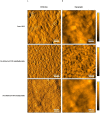
Fig 2. AFM analysis of Descemet’s membrane’s surface before and after decellularization.
AFM images representing (A) deflection and (B) topography of intact and denuded tissue. Image scale: 3 x 3 μm. The coloured bars depict the height. Scale bars = 500nm. DM, Descemet’s membrane.
Fig 3
Fig 3. SEM analysis of decellularized Descemet’s membrane’s surface morphology.
SEM images of dDM showing a dense membrane with several folds interspersed along the surface: (A) X 30, (B) X 1000.
Fig 4
Fig 4. Decellularized Descemet’s membrane provides optimal cues for hESC-RPE cell initial attachment and proliferation.
(A) Immunofluorescence for Calcein AM (green) allows the visualisation of viable hESC-RPE cells 24 hours post-seeding on dDM and in control condition. Scale bars = 100 μm. (B) Percentage of initial cell attachment on dDM compared to control. Each box represents mean ± s.d. for fourfold experiments. *p = 0.028. (C) The graph shows the percentage of proliferative BrdU-positive cells on dDM and on TC insert at different time points during the first week of culture. Each time point represents mean ± s.d. for triplicate experiments. *p = 0.032.
Fig 5
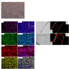
Fig 5. hESC-RPE cell culture over the denuded Descemet’s membrane.
(A) Phase-contrast image of hESC-RPE cells cultured for 4 weeks on dDM. Scale bar = 100 μm. (B) Immunostaining for typical RPE markers ZO1 (magenta), MITF (green), BEST1 (red) and OTX2 (yellow) in hESC-RPE cells cultured on dDM after 1 month of culture. Dotted rectangles indicate the regions that are magnified in the adjacent panels. Scale bars = 100 μm. (C) Immunofluorescence of hESC-RPE cells cryosections cultured on dDM after 1 month of culture, demonstrating the development of a confluent monolayer of hESC-RPE cells over the denuded ocular membrane. hESC-RPE cells express premelanosome protein PMEL (green). Denuded DM shows positive expression for CIV (red) and LAMα5 (yellow). Dotted rectangles indicate the regions that are magnified in the adjacent panels. All nuclei shown in blue were counterstained using DAPI. Scale bars = 100 μm. Best1, bestrophin1; CIV, type IV collagen; LAMα5, laminin α5.
Fig 6
Fig 6. hESC-RPE cells monolayer evaluation over the natural scaffold.
(A) Na+/K+ ATPase immunostaining shows proper polarization of the cells when cultured on dDM. Scale bar = 100 μm. (B) ELISA assay was used to quantify VEGF secretion in spent medium from hESC-RPE cells cultured on dDM. VEGF levels were then compared with the amount of VEGF secreted by control on pre-coated TC insert. Values are given as the mean ± s.d. for triplicate experiments. *p = 0.016. (C) TER measurements of hESC-RPE cell sheets on dDM at 1 month after seeding, indicating an increase in resistance following proper tight junction development. Each line represents records from four separate cultures of hESC-RPE cells grown on dDM and on TC inserts, respectively. dDM, denuded Descemet’s membrane; NaK, Na+/K+ ATPase; TER, transepithelial resistance.
Fig 7
Fig 7. Phagocytic function of pluripotent-derived RPE cells on dDM.
(A) Latex beads uptake (green) in hESC-RPE cells over dDM. Actin filaments were stained with phalloidin (red). Scale bar = 50 μm. (B) Percentage of hESC-RPE cells internalizing the fluorescent beads when cultured on the dDM and under control condition, respectively. Latex beads counts were obtained from five individual field if views, with each field containing ≥ 80 cells. Data represent mean ± s.d. *p = 0.062. dDM, denuded Descemet’s membrane.
Fig 8
Fig 8. Quantitative analysis of gene expression of RLBP1, MERTK and RPE65 by real-time PCR.
The bar graph in A shows the gene expression comparison between hESC-RPE cells cultured on dDM and hESC-RPE cells cultured on pre-coated TC inserts used as control. In B, the same comparison is carried out between adult RPE cells cultured on dDM and adult RPE cells cultured on Synthemax II-coated multiwells. Measurements in each graph were normalized to the expression level of related control condition. Values are given as the mean ± s.d. for triplicate experiments. *p = 0.027 and **p = 0.020. hESC, human embryonic stem cells; RPE, retinal pigment epithelium; dDM, denuded Descemet’s membrane.

References

    1. Causes of blindness and vision impairment in 2020 and trends over 30 years, and prevalence of avoidable blindness in relation to VISION 2020: the Right to Sight: an analysis for the Global Burden of Disease Study. Lancet Glob Health. 2020;9: e144–e160. doi: 10.1016/S2214-109X(20)30489-7 - DOI - PMC - PubMed
    1. Galvin O, Chi G, Brady L, Hippert C, Del Valle Rubido M, Daly A, et al.. The Impact of Inherited Retinal Diseases in the Republic of Ireland (ROI) and the United Kingdom (UK) from a Cost-of-Illness Perspective. Clin Ophthalmol. 2020;14: 707–719. doi: 10.2147/OPTH.S241928 - DOI - PMC - PubMed
    1. Chen Y, Han X, Gordon I, Safi S, Lingham G, Evans J, et al.. A systematic review of clinical practice guidelines for myopic macular degeneration. J Glob Health. 12: 04026. doi: 10.7189/jogh.12.04026 - DOI - PMC - PubMed
    1. Schwartz SD, Pan CK, Klimanskaya I, Lanza R. Chapter 68—Retinal Degeneration. In: Lanza R, Langer R, Vacanti J, editors. Principles of Tissue Engineering (Fourth Edition). Boston: Academic Press; 2014. pp. 1427–1440.
    1. Coffey PJ, Girman S, Wang SM, Hetherington L, Keegan DJ, Adamson P, et al.. Long-term preservation of cortically dependent visual function in RCS rats by transplantation. Nat Neurosci. 2002;5: 53–56. doi: 10.1038/nn782 - DOI - PubMed

Publication types