. 2023 Feb 6;19(2):e1011132.
doi: 10.1371/journal.ppat.1011132. eCollection 2023 Feb.

Innate sensing of picornavirus infection involves cGAS-STING-mediated antiviral responses triggered by mitochondrial DNA release

Affiliations

Innate sensing of picornavirus infection involves cGAS-STING-mediated antiviral responses triggered by mitochondrial DNA release

Huisheng Liu et al. PLoS Pathog. .

Abstract

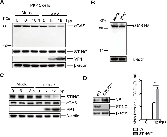
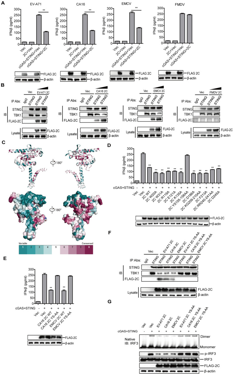
Cyclic GMP-AMP synthase (cGAS) plays a key role in the innate immune responses to both DNA and RNA virus infection. Here, we found that enterovirus 71 (EV-A71), Seneca Valley virus (SVV), and foot-and-mouth disease virus (FMDV) infection triggered mitochondria damage and mitochondrial DNA (mtDNA) release in vitro and vivo. These responses were mediated by picornavirus 2B proteins which induced mtDNA release during viral replication. SVV infection caused the opening of mitochondrial permeability transition pore (mPTP) and led to voltage-dependent anion channel 1 (VDAC1)- and BCL2 antagonist/killer 1 (Bak) and Bak/BCL2-associated X (Bax)-dependent mtDNA leakage into the cytoplasm, while EV-A71 and FMDV infection induced mPTP opening and resulted in VDAC1-dependent mtDNA release. The released mtDNA bound to cGAS and activated cGAS-mediated antiviral immune response. cGAS was essential for inhibiting EV-A71, SVV, and FMDV replication by regulation of IFN-β production. cGAS deficiency contributed to higher mortality of EV-A71- or FMDV-infected mice. In addition, we found that SVV 2C protein was responsible for decreasing cGAS expression through the autophagy pathway. The 9th and 153rd amino acid sites in 2C were critical for induction of cGAS degradation. Furthermore, we also show that EV-A71, CA16, and EMCV 2C antagonize the cGAS-stimulator of interferon genes (STING) pathway through interaction with STING, and highly conserved amino acids Y155 and S156 were critical for this inhibitory effect. In conclusion, these data reveal novel mechanisms of picornaviruses to block the antiviral effect mediated by the cGAS-STING signaling pathway, which will provide insights for developing antiviral strategies against picornaviruses.


Conflict of interest statement

The authors have declared that no competing interests exist.

Figures

Fig 1
Fig 1. Mitochondrial damage induced by picornavirus infection.
HeLa cells were mock-infected and infected with EV-A71 (MOI 1) for 0, 12, and 24 h. PK-15 cells were mock-infected and infected with SVV (MOI 1) or FMDV (MOI 0.5) for 0, 6, and 12 h. The ΔΨm (A), mitochondrial Ca2+ concentration (B), mitochondrial ROS (C), and mPTP opening (D) were detected using JC-1, Rhod-2 AM, mitoSOX, and calcein-AM, respectively. Data for CCCP (A) and lono (D) positive controls are shown. Error bars show standard deviation. **, P<0.01.
Fig 2
Fig 2. Picornavirus induced mtDNA release.
HeLa cells were mock-infected or infected with EV-A71 (MOI 1) for 0, 12, and 24 h, while PK-15 cells were mock-infected or infected with SVV (MOI 1) or FMDV (MOI 0.5) for 0, 6, and 12 h. The lysates were immunoprecipitated with anti-cGAS antibody. The mtDNA and gDNA in cGAS pulldown samples were detected and analyzed by qPCR. The EGFP DNA was used as an internal control (A). HeLa cells were mock-infected or infected with EV-A71 (MOI 1) for 24 h. PK-15 cells were mock-infected or infected with SVV (MOI 1) or FMDV (MOI 0.5) for 12 h. The mtDNA release was evaluated by IFA. Cells were double-immunostained for detection of Tom20 (red) and dsDNA (green); cellular nuclei were counterstained with 4’,6-diamidino-2-phenylindole (DAPI) (blue) (B, C, and D). Scale bar, 10 μm. Oropharyngeal tonsils collected from pigs infected with SVV (E) or FMDV (F) were analyzed for mtDNA release. Error bars show standard deviation. **, P<0.01.
Fig 3
Fig 3. Picornavirus induced mtDNA release into the cytoplasm via mPTP.
HT-29 WT and PPID-/- cells were mock-infected or infected with EV-A71 (MOI 1) for 24 h, while PK-15 WT or PPID-/- cells were mock-infected or infected with SVV (MOI 1) or FMDV (MOI 0.5) for 12 h. The mtDNA release was evaluated by qPCR (A). The expression of the IFN-β protein was detected by ELISA kit, and the expression of PPID was confirmed by Western blotting (B). (C) HT-29 cells were mock-infected or infected with EV-A71 (MOI 1), while PK-15 cells were mock-infected or infected with SVV (MOI 1) or FMDV (MOI 0.5). After 1 h of incubation of the virus or DMEM, cells were treated with DMSO or VBIT4 inhibitor (10 uM) for 24 or 12 h. The mtDNA release was then detected by qPCR. The expression of Bax protein in the WT and Bax-/- cells was confirmed by western blotting (D) and these cells were used to measure mtDNA release after infection with EV-A71 (HT-29 cells at MOI 1) or SVV (PK-15 cells at MOI 1) or FMDV (PK-15 cells at MOI 0.5) for 12 h (E). Error bars show standard deviation. **, P<0.01.
Fig 4
Fig 4. Picornavirus 2B proteins promoted mtDNA release.
PK-15 cells were transfected with 2 μg of empty vector, the indicated SVV protein expressing plasmids (A) or FMDV protein expressing plasmids (B), and HeLa cells were transfected with 2 μg of empty vector or EV-A71 protein expressing plasmids (C). At 24 hpt, the mtDNA release was analyzed by qPCR. The expression of viral proteins was determined by Western blotting. (D) HeLa cells were transfected with 2 μg of empty vector, EMCV 2B or CA16 2B expressing plasmids for 24 h. The mtDNA release was detected by qPCR. (E) HT-29 WT and PPID-/- cells were transfected with 2 μg of empty vector, EMCV 2B, CA16 2B, or EV-A71 2B expressing plasmids for 24 h, and PK-15 WT or PPID-/- cells were transfected with 2 μg of empty vector, SVV 2B or FMDV 2B expressing plasmids for 24 h. The mtDNA release was detected by qPCR. Error bars show standard deviation. **, P<0.01.
Fig 5
Fig 5. Effect of cGAS deficiency on picornaviruses replication in vitro.
IFN-β and ISG54 mRNA expression in cGAS-/- HeLa cells infected with SVV (A), and cGAS-/- HT-29 cells infected with EV-A71 (B); IFN-β protein expression is also shown (C). Western blotting was used to detect the expression of SVV VP1 protein (D) and EV-A71 3C protein (E) and viral titers were determined by TCID50 assay. Bone marrow-derived macrophages from the WT or cGAS-/- mice were infected with FMDV and IFN-β protein in the supernatant was determined by ELISA (F). The expression of FMDV VP1 protein was detected by Western blotting, and the viral titers were determined by TCID50 assay (G). WT and ρ0 PK-15 cells were infected with SVV or FMDV and WT and ρ0 HT-29 cells were infected with EV-A71. IFN-β protein expression level was determined by ELISA (H), and the viral titers were determined by TCID50 assay (I). Error bars show standard deviation. **, P<0.01.
Fig 6
Fig 6. cGAS deficiency promoted EV-A71- and FMDV-induced mice death.
(A) The 3-weeks-old WT and cGAS-/- mice were showed, and the expression of cGAS in the WT and cGAS-/- mice was detected by Western blotting. (B and C) The three-day-old WT mice were subcutaneously inoculated with FMDV (108 TCID50) or EV-A71 (108 TCID50) for 0, 1, or 3 d (n = 3/group), the mice carcasses without the head, tail, limbs, and viscera were collected for detection of mtDNA release (B) and IFN-β mRNA expression (C). (D and E) The three-day-old WT (n = 8) and cGAS-/- (n = 8) mice were subcutaneously inoculated with FMDV (108 TCID50) or EV-A71 (108 TCID50). The IFN-β mRNA level in the carcasses without the head, tail, limbs, and viscera from FMDV-infected mice was measured and compared at 3 dpi by qPCR (D). The IFN-β mRNA level in the carcasses without the head, tail, limbs, and viscera from EV-A71-infected mice was measured and compared at 1 dpi by qPCR (E). FMDV (F) and EV-A71 (G) titers in the mice carcasses without the head, tail, limbs, and viscera were determined at 2 dpi by TCID50 assay. The mortality of WT (n = 10) and cGAS-/- (n = 10) mice infected by FMDV (F) or EV-A71 (G) was determined, respectively. Error bars show standard deviation. **, P<0.01.
Fig 7
Fig 7. The impact of picornaviruses infection on cGAS and STING expression.
(A) PK-15 cells were mock-infected or infected with SVV (MOI 1) for 0, 8, or 16 h. The expression of cGAS and STING proteins was detected by Western blotting. (B) HEK-293T cells were transfected with 1.5 μg of cGAS-HA expressing plasmids. At 24 hpt, the cells were mock-infected or infected with SVV (MOI 1) for 12 h. Expression of cGAS-HA protein was detected by Western blotting. (C) PK-15 cells were mock-infected or infected with FMDV (MOI 0.5) for 0, 6, or 12 h. Expression of cGAS and STING proteins was detected by Western blotting. (D) WT and STING-/- PK-15 cells were infected with FMDV (MOI 0.5) for 12 h. The expression of FMDV VP1 protein was detected by Western blotting, and viral titers were determined by TCID50 assay. Error bars show standard deviation. **, P<0.01.
Fig 8
Fig 8. SVV 2C protein was responsible for cGAS reduction.
PK-15 cells were transfected with 2 μg of plasmids expressing various SVV (A) and FMDV (B) proteins. The expression of endogenous cGAS or STING proteins was detected by Western blotting. (C) HEK-293T cells were transfected with 1 μg of empty vector or FLAG-L- and Myc-2A-expressing plasmids, and 1 μg of empty vector or HA-cGAS plus HA-STING expressing plasmids. At 24 hpt, the IFN-β protein expression level was determined by ELISA. (D) PK-15 cells were transfected with 0, 1, or 2 μg of plasmids expressing FLAG-2C protein. At 24 hpt, expression of endogenous cGAS protein was determined by Western blotting. (E and F) HEK-293T cells were transfected with 0.1 μg of empty vector or FLAG-2C-expressing plasmids, and 0.1 μg of empty vector or HA-cGAS plus HA-STING expressing plasmids, together with 0.1 μg of IFN-β-Luc, and 0.01 μg of pRL-TK plasmid. At 24 hpt, the promoter activation of IFN-β was determined by the dual-specific luciferase assay kit, and the expression of cGAS, STING, and 2C was confirmed by Western blotting (E). The mRNA expression levels of IFN-β and ISG54 were determined by qPCR assay (F).(G) PK-15 cells were transfected with 2 μg of empty vector or FLAG-2C-expressing plasmids. At 24 hpt, the cells were infected with SVV (MOI 1) for 12 h. Viral VP1 protein and titers were examined by Western blotting and TCID50 assay, respectively. Error bars show standard deviation. **, P<0.01.
Fig 9
Fig 9. Identification of the pathways and 2C functional sites invovled in induction of cGAS reduction during SVV infection.
(A) PK-15 cells were mock-infected or infected with SVV (MOI 1). At 1 hpi, the cells were maintained in the fresh medium in the presence or absence of MG132 (20 μM), 3-MA (1 or 2.5 mM), or CQ (100 μM). At 12 hpi, the expression of cGAS protein was determined by Western blotting. (B) PK-15 cells were transfected with 1.5 μg of FLAG vector or FLAG-2C-expressing plasmid. At 6 hpt, the cells were maintained in the fresh medium in the presence or absence of MG132 (20 μM), 3-MA (1 mM), or CQ (100 μM) for 18 h. Expression of cGAS protein was determined by Western blotting. (C) Expression of ATG7 protein in ATG7 WT and ATG7-/- cells was detected by Western blotting. (D) ATG7 WT and ATG7-/- cells were mock-infected or infected with SVV (MOI 1), or transfected with empty vector or FLAG-2C-expressing plasmids. At 12 hpi or 24 hpt, expression of cGAS, ATG7, and VP1 proteins was determined by Western blotting. (E) Schematic representation of a series of FLAG-tagged truncated 2C mutants. (F) PK-15 cells were transfected with empty vector, FLAG-2C- or the indicated FLAG-2C-mutant-expressing plasmids. At 24 hpt, expression of cGAS and LC3 proteins was determined by Western blotting. (G and H) PK-15 cells were mock-infected or infected with SVV WT (MOI 1) or SVV 2C-W9A (MOI 1) for 12 h. The expression of cGAS was detected by Western blotting (G). The IFN-β protein expression in the supernatant was determined by ELISA (H). Error bars show standard deviation. **, P<0.01.
Fig 10
Fig 10. EV-A71, CA16, and EMCV 2C proteins antagonized cGAS-STING signaling pathway activation by impairing the interaction between STING and TBK1.
(A) HEK-293T cells were transfected with 1 μg of empty vector or the indicated FLAG-2C-expressing plasmids, along with 1 μg of empty vector or HA-cGAS plus HA-STING expressing plasmids. At 24 hpt, the IFN-β protein amount in the supernatant was determined by ELISA, and the expression of 2C was confirmed by Western blotting. (B) STING-HEK-293T cells were transfected with increasing amount (0, 3, or 6 μg) of the indicated FLAG-2C-expressing plasmids. At 24 hpt, cells were transfected with poly(dA:dT) (2 μg/ml) for 12 h. The cells lysates were immunoprecipitated with anti-STING antibody. The antibody-antigen complexes were detected using anti-STING, TBK1, and FLAG antibodies, respectively. (C) Schematic representation of the structure and conserved functional sites in EV-A71 2C protein. The redder the color was, the more conservative the sites were. (D, E) HEK-293T cells were transfected with 1 μg of empty vector or EV-A71 (D), CA16 (E), and EMCV (E) FLAG-2C- or FLAG-2C mutants-expressing plasmids, along with 1 μg of empty vector or HA-cGAS plus HA-STING expressing plasmids. At 24 hpt, the IFN-β protein amount in the supernatant was determined by ELISA, and the expression of 2C was confirmed by Western blotting. (F) STING-HEK-293T cells were transfected with 6 μg of empty vector, FLAG-2C- or FLAG-2C mutants-expressing plasmids. At 24 hpt, cells were transfected with poly(dA:dT) (2 μg/ml) for 12 h. The cells lysates were immunoprecipitated with anti-STING antibody. The antibody-antigen complexes were detected using anti-STING, TBK1, and FLAG antibodies, respectively. (G) HEK-293T cells were transfected with 1 μg of empty vector, FLAG-2C- or FLAG-2C mutants-expressing plasmids, and 1 μg of HA-cGAS plus HA-STING expressing plasmids. At 24 hpt, expression of IRF3, p-IRF3, and FLAG-2C protein was determined by Western blotting. The IRF3 dimerization was detected using native PAGE. Error bars show standard deviation. **, P<0.01.
Fig 11
Fig 11. Schematic representation of the model of cGAS-mediated antiviral function in EV-A71, SVV, and FMDV replication.
In this model, cytosolic mtDNA released after EV-A71, SVV, and FMDV infection binds to cGAS to activate cGAS-mediated signal transduction, resulting in the antiviral responses. Virus immune evasion strategies include SVV 2C induced the reduction of cGAS by activation of autophagy and FMDV 2B and 3Cpro proteins which inhibit STING expression to block the antiviral response. Furthermore, EV-A71, CA16, and EMCV 2C can antagonize activation of the cGAS-STING signaling pathway by impairing the interaction of STING with TBK1, while FMDV Lpro and EV-A71 2A proteins inhibit cGAS-STING-induced IFN-β protein expression.

References

    1. Jiang P, Liu Y, Ma HC, Paul AV, Wimmer E. Picornavirus morphogenesis. Microbiol Mol Biol Rev. 2014; 78(3): 418–437. doi: 10.1128/MMBR.00012-14 - DOI - PMC - PubMed
    1. Luo Z, Su R, Wang W, Liang Y, Zeng X, Shereen MA, et al.. EV71 infection induces neurodegeneration via activating TLR7 signaling and IL-6 production. PLoS Pathog. 2019; 15(11): e1008142. doi: 10.1371/journal.ppat.1008142 - DOI - PMC - PubMed
    1. Netea MG, Joosten LA, Latz E, Mills KH, Natoli G, Stunnenberg HG, et al.. Trained immunity: A program of innate immune memory in health and disease. Science. 2016; 352(6284): aaf1098. doi: 10.1126/science.aaf1098 - DOI - PMC - PubMed
    1. Ablasser A, Hur S. Regulation of cGAS- and RLR-mediated immunity to nucleic acids. Nat Immunol. 2020; 21(1): 17–29. doi: 10.1038/s41590-019-0556-1 - DOI - PubMed
    1. You H, Lin Y, Lin F, Yang M, Li J, Zhang R, et al.. beta-catenin is Required for cGAS/STING Signaling Pathway but Antagonized by HSV-1 US3 Protein. J Virol. 2020; 94(5):e01847–19. doi: 10.1128/JVI.01847-19 - DOI - PMC - PubMed

Publication types

MeSH terms

Supplementary concepts