. 2023 Feb 14;120(7):e2217831120.
doi: 10.1073/pnas.2217831120. Epub 2023 Feb 6.

Limb girdle muscular disease caused by HMGCR mutation and statin myopathy treatable with mevalonolactone

Affiliations

Limb girdle muscular disease caused by HMGCR mutation and statin myopathy treatable with mevalonolactone

Yuval Yogev et al. Proc Natl Acad Sci U S A. .

Abstract

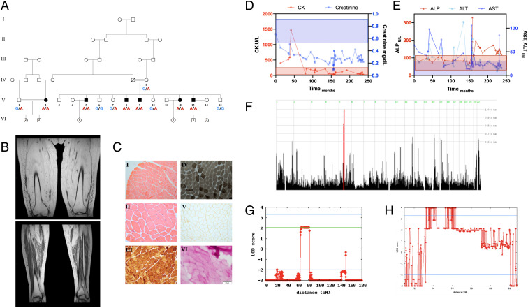
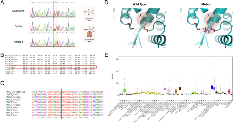
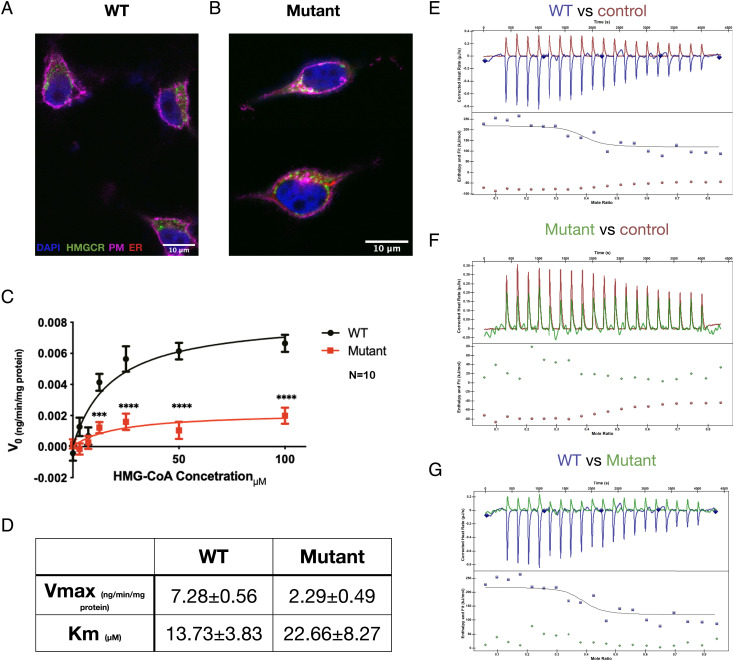
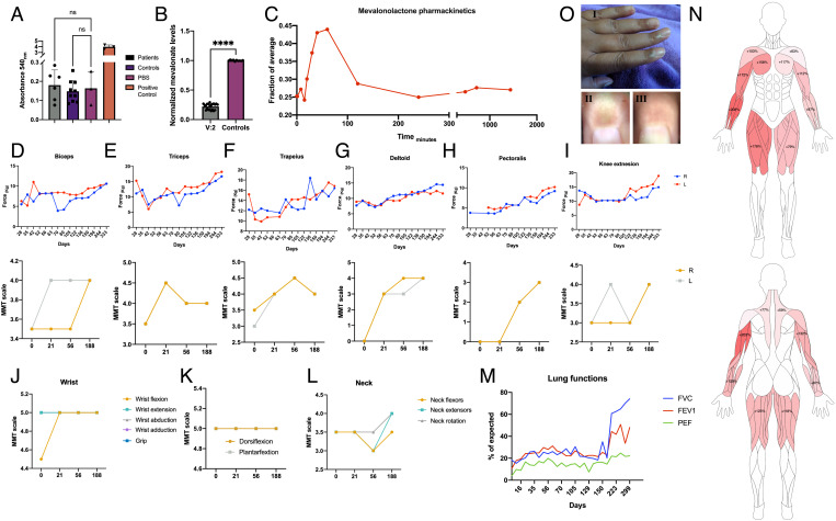
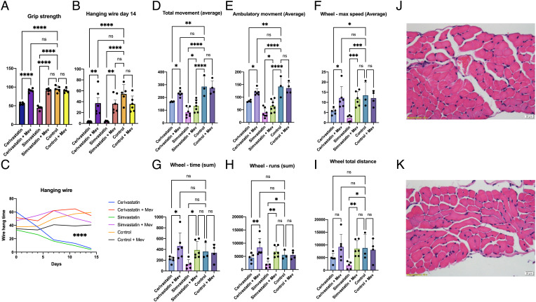
Myopathy is the main adverse effect of the widely prescribed statin drug class. Statins exert their beneficial effect by inhibiting HMG CoA-reductase, the rate-controlling enzyme of the mevalonate pathway. The mechanism of statin myopathy is yet to be resolved, and its treatment is insufficient. Through homozygosity mapping and whole exome sequencing, followed by functional analysis using confocal microscopy and biochemical and biophysical methods, we demonstrate that a distinct form of human limb girdle muscular disease is caused by a pathogenic homozygous loss-of-function missense mutation in HMG CoA reductase (HMGCR), encoding HMG CoA-reductase. We biochemically synthesized and purified mevalonolactone, never administered to human patients before, and establish the safety of its oral administration in mice. We then show that its oral administration is effective in treating a human patient with no significant adverse effects. Furthermore, we demonstrate that oral mevalonolactone resolved statin-induced myopathy in mice. We conclude that HMGCR mutation causes a late-onset severe progressive muscular disease, which shows similar features to statin-induced myopathy. Our findings indicate that mevalonolactone is effective both in the treatment of hereditary HMGCR myopathy and in a murine model of statin myopathy. Further large clinical trials are in place to enable the clinical use of mevalonolactone both in the rare orphan disease and in the more common statin myopathy.

Keywords: HMGCR; limb girdle muscular dystrophy; mutation; statins.


Conflict of interest statement

The authors have patent filings to disclose, A patent has been filed by Y.Y. and O.S.B. Otherwise, the authors have declared that no conflict of interests exists.

Figures

Fig. 1.
Fig. 1.
Pedigree, phenotype, and linkage analysis. (A) Pedigree of a Bedouin kindred from the Negev region of Israel, showing six individuals affected by adult-onset LGMD, (B) T1 MRI imaging of the thighs and legs of patient V:2 at age 49, showing complete atrophy of skeletal muscles of the thigh with fatty replacement, and partial sparing of the distal muscles of the legs. Additional imaging can be seen in SI Appendix, Fig. S5. (C) Deltoid muscle histology of patient V:2 at age 34, when muscles showed marked weakness; blood tests and EMG indicated myopathy. I–II: H&E staining showing normal morphology with no necrosis, fibrosis or inflammation, III: COX stain showing normal COX activity, IV: ATPase pH 9.4 stain showing mild deficiency of type II muscle fibers, V: Dystrophin III stain showing normal distribution, VI: PAS stain showing no accumulation of glycogen. Other stains did not demonstrate marked features, as described. (D and E) Serum enzymes of patient V:2 over 21 y, starting with disease onset at age 31. Shading represents the reference range of the color-matched test. (D) As the CK levels dropped into the normal range and eventually below, creatinine levels dropped as well, indicating very low muscle mass. (E) ALT and AST levels decreased over the course of disease, following muscle damage, while ALP levels rose late. Acute spikes in ALT, AST, and ALP levels coincided with hospital and ICU admissions, possibly indicating disease flare-ups. (F) Homozygosity mapping showing a homozygous locus in chromosome 5 shared by all affected individuals but not other tested family members. (G) Reduced complexity multipoint linkage analysis chart of chromosome 5, showing the shared locus (H) Two-point linkage analysis chart of the 3.2 Mbp locus and adjacent regions, showing a LOD score higher than four.
Fig. 2.
Fig. 2.
The HMGCR mutation. (A) Sanger sequencing of an HMGCR amplicon of an unaffected individual (V:6), an obligatory carrier (IV:1) and an affected individual (V:13). The mutation causes a glycine to aspartate nonconservative substitution at position 822. (B) Precent identity matrix of HMGCR produced using Clustal-Omega, showing high homology between the human HMGCR and orthologs. (C) Multiple alignment of human HMGCR and orthologs produced using Clustal-Omega, showing that the substituted amino acid is highly conserved throughout evolution. (D) Structural model of HMG CoA-reductase protein in the WT and mutated form, based on 1DQ8. The substitution presumably forms a new H-bond with a distal residue, compromises the helix dipole, and causes electrostatic repulsion. (E) Expression of HMGCR across various tissues, obtained using the GTEx database.
Fig. 3.
Fig. 3.
HMGCR mutation impairs protein function. (A and B) Subcellular localization of WT (A) and CRISPR-KI mutant (B) HMGCR in SH-SY5Y cells. Both WT and mutant HMGCR protein (green) are located in clusters in the cytoplasm with some relation to the endoplasmic reticulum (red). (C and D) HMGCR enzymatic activity. The velocity of enzymatic reduction of different concentrations of HMG CoA was measured by spectrophotometric NADPH oxidation assay of the WT (n = 9) and mutant (n = 12) forms of HMGCR. Analyzed using Michaelis–Menten analysis and multiple t-tests. (EG) Mutant HMGCR shows decreased affinity to pravastatin, a protein inhibitor that binds to the same catalytic pocket as HMG CoA (n = 8). As seen, mutant kinetics are almost identical to the no protein control. PM, plasma membrane; ER, endoplasmic reticulum.
Fig. 4.
Fig. 4.
Oral mevalonolactone treatment in human HMGCR-LGMD. (A) Anti-HMGCR autoantibodies titer in patients (n = 6) and healthy controls (n = 10). (B) Plasma mevalonolactone levels of patients V:2 (n = 20 on multiple occasions) and healthy controls (n = 10), normalized to average level of controls. (C) Mevalonolactone levels in peripheral blood of V:2 after an oral dose of 16 mg/kg mevalonolactone, normalized to average level of controls. (DI) Evaluation of muscle strength of patient V:2 throughout the treatment period by dynamometry (Top) and manual muscle test (MMT; Bottom) by an experienced neurologist. (JL) Evaluation of distal muscles throughout the treatment by an experienced neurologist. (M) Lung functions throughout the treatment period assessed by spirometry. (N) Muscle strength improvement "heat-map" by dynamometry, precent improvement from weakest state. (O) Pigmentation in the proximal nail fold which appeared occasionally following treatment. I: Gross appearance; II: Dermatoscopic image of the index finger showing pigmentation; and III: Dermatoscopic image of the same index finger several days later, pigmentation has resolved. Statistical analysis using t-test.
Fig. 5.
Fig. 5.
Oral mevalonolactone treatment in murine statin-induced myopathy. Mice were treated daily with intraperitoneal injections of either Cerivastatin, Simvastatin, or 0.9% saline solution for 14 d, with or without 200 mg/kg oral mevalonolactone. n = 4 in each group. Mice were housed in PhenoMaster cages for the last 3 d of treatment. (A) Grip strength test on day 14. (B) Hanging wire test on day 14. (C) Hanging wire test throughout the study. (DI) Measurements of strength and endurance from PhenoMaster cages. (JK) H&E stained diaphragm muscles of a subject from the Cerivastatin group (J) and the Cerivastatin + mevalonolactone group (K). No signs of fibrosis, necrosis, or inflammation were evident. Diaphragm, gastrocnemius, and quadriceps muscles were examined from two subjects in every group. Statistical analysis using one-way ANOVA with multiple comparisons.

Comment in

References

    1. Mercuri E., Bönnemann C. G., Muntoni F., Muscular dystrophies. Lancet 394, 2025–2038 (2019). - PubMed
    1. Murphy A. P., Straub V., The classification, natural history and treatment of the limb girdle muscular dystrophies. J. Neuromuscul. Dis. 2, S7–S19 (2015). - PMC - PubMed
    1. Pozsgai E., et al. , Unmet needs and evolving treatment for limb girdle muscular dystrophies. Neurodegener. Dis. Manag. 11, 411–429 (2021). - PubMed
    1. Taylor F., Statins for the primary prevention of cardiovascular disease. Cochrane Database Syst. Rev. 2017, CD004816 (2013). - PMC - PubMed
    1. Baigent C., et al. , Efficacy and safety of cholesterol-lowering treatment: Prospective meta-analysis of data from 90,056 participants in 14 randomised trials of statins. Lancet 366, 1267–78 (2005). - PubMed

Publication types

MeSH terms