. 2023 Feb 14;120(7):e2218516120.
doi: 10.1073/pnas.2218516120. Epub 2023 Feb 6.

Allosteric mechanism of transcription inhibition by NusG-dependent pausing of RNA polymerase

Affiliations

Allosteric mechanism of transcription inhibition by NusG-dependent pausing of RNA polymerase

Rishi K Vishwakarma et al. Proc Natl Acad Sci U S A. .

Abstract

NusG is a transcription elongation factor that stimulates transcription pausing in Gram+ bacteria including B. subtilis by sequence-specific interaction with a conserved pause-inducing -11TTNTTT-6 motif found in the non-template DNA (ntDNA) strand within the transcription bubble. To reveal the structural basis of NusG-dependent pausing, we determined a cryo-EM structure of a paused transcription complex (PTC) containing RNA polymerase (RNAP), NusG, and the TTNTTT motif in the ntDNA strand. The interaction of NusG with the ntDNA strand rearranges the transcription bubble by positioning three consecutive T residues in a cleft between NusG and the β-lobe domain of RNAP. We revealed that the RNAP swivel module rotation (swiveling), which widens (swiveled state) and narrows (non-swiveled state) a cleft between NusG and the β-lobe, is an intrinsic motion of RNAP and is directly linked to trigger loop (TL) folding, an essential conformational change of all cellular RNAPs for the RNA synthesis reaction. We also determined cryo-EM structures of RNAP escaping from the paused transcription state. These structures revealed the NusG-dependent pausing mechanism by which NusG-ntDNA interaction inhibits the transition from swiveled to non-swiveled states, thereby preventing TL folding and RNA synthesis allosterically. This motion is also reduced by the formation of an RNA hairpin within the RNA exit channel. Thus, the pause half-life can be modulated by the strength of the NusG-ntDNA interaction and/or the stability of the RNA hairpin. NusG residues that interact with the TTNTTT motif are widely conserved in bacteria, suggesting that NusG-dependent pausing is widespread.

Keywords: Cryo-EM; NusG; RNA polymerase; transcription; transcription pausing.


Conflict of interest statement

The authors declare no competing interest.

Figures

Fig. 1.
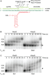
Fig. 1.
B. subtilis NusG stimulates TTNTTT motif-specific pausing of B. subtilis and Mtb RNAPs. (A) DNA sequence used for promoter-initiated in vitro pausing assays. The −35 and extended −10 promoter elements are underlined. A 29-nucleotides C-less region from a transcription start site (+1) is in cyan and the predicted coaA pause hairpin is in green. The TTNTTT motif and the pause position (arrowhead) are in magenta. (B) Representative gels showing RNAs generated from promoter-initiated single-round in vitro transcription-pausing assays. Reactions were performed with the coaA DNA template shown in A, B. subtilis NusG and either B. subtilis (Left) or Mtb (Right) RNAP. Transcription reactions were performed with 150 μM of each NTP ± 1 μM NusG. Reactions were stopped at the times shown above each lane. Chase reactions (Ch) were extended for an additional 10 min at 37 °C. Positions of the coaA pause (P and arrow), and runoff (RO) transcripts are indicated. Additional pause, terminated, or arrested RNA species (P/T) were observed between P and RO. Calculated pause half-lives are shown at the bottom of the gel. (C) Representative gels showing RNAs from the promoter-initiated single-round in vitro transcription reactions using a DNA template in which the TTNTTT motif was replaced with AACAAA. Values at the bottom of the gels are averages ± SD (n = 3). (DMtb NusG stimulates pausing of Mtb RNAP at the same position as B. subtilis NusG. Reactions were performed in the absence (−NusG) and in the presence of either B. subtilis or Mtb NusG as indicated. Transcription reactions were performed with 150 μM of each NTP except Adenosine 5'-triphosphate (ATP) (10 µM). The DNA template used for this analysis contained the RNA hairpin sequence used for reconstruction of the PTC and TEC (Fig. 2A) rather than the natural coaA hairpin sequence (green in A).
Fig. 2.
Fig. 2.
Reconstitution of active PTCs with a nucleic acid scaffold, Mtb RNAP and B. subtilis NusG. (A) Nucleic acid scaffold used for reconstitution of the active PTC and TEC. ntDNA for preparing the PTC contained the TTNTTT motif (underlined). The last three T residues of the TTNTTT motif (positions −8 to −6) were replaced with A residues (TTCAAA) for preparing the TEC. upDNA, upstream DNA; downDNA, downstream DNA. (B) Representative gels showing the results of single-round in vitro pause escape assays using the PTC nucleic acid scaffold. Reactions were stopped at the times shown above each lane. Chase reactions (Ch) were extended for an additional 10 min at 37 °C. (C) Same as panel B except the nucleic acid scaffold for the TEC was used. Values at the bottom of the gels are averages ± SD (n = 3).
Fig. 3.
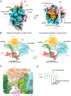
Fig. 3.
Cryo-EM structures of the TEC and NusG-dependent PTC. (A) Orthogonal views of the cryo-EM density map of the PTC. Subunits and domains of RNAP, DNA, RNA and NusG are colored and labeled. downDNA, downstream DNA; upDNA, upstream DNA; β′-i1, lineage-specific insertion. (B) Cryo-EM densities (transparent) of DNA, RNA, β-lobe domain and NusG are overlayed with the PTC structure (Left) and TEC structure (Right) revealing the rearrangement of the transcription bubble preceding the upstream DNA in the PTC. The 5′ and 3′ ends of the RNA, as well as positions −10 and −6 of the ntDNA are indicated. The cryo-EM density maps and the structure are colored according to A. (C) A magnified view of the ntDNA interactions with NusG and RNAP (same orientation as in the B). Amino acid residues of NusG and the β′ subunit interacting with ntDNA are indicated as stick models. DNA strands (tDNA, dark green; ntDNA, light green) are shown as sphere models and thymine residues in the TTNTTT motif are colored (red, oxygen; blue, nitrogen; green, carbon). (D) Schematic representations of the base pairing distortion of the upstream DNA duplex in the PTC.
Fig. 4.
Fig. 4.
Mtb RNAP swiveling. (A) Structure of the Mtb RNAP-B. subtilis NusG TEC indicating the modules and domains. The rotation axis of the swivel module is shown as yellow circles (Left and Middle). (B) Cryo-EM densities (transparent) of DNA, RNA, NusG, β-lobe and β-protrusion domains are overlayed with the swiveled (Left) and non-swiveled states (Right) of the TEC. Rotation of the NusG/swivel module from the swiveled to non-swiveled states is indicated by a black arrow (Right).
Fig. 5.
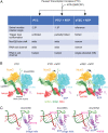
Fig. 5.
RNAP escaping from paused transcription state. (A) Experimental scheme of the transcription complex preparation from the NusG-dependent PTC mixed with the incoming NTP. Three transcription complexes were formed, the angles of the swivel module relative to the eTEC + NTP are shown, and the conformations of the TL are indicated. (B) Comparison of the three transcription complexes. Cryo-EM densities (transparent) of DNA, RNA, NusG, β-lobe and TL are overlayed with the final model. Rotation of NusG with the swivel module toward the downstream DNA are indicated by black arrows. Pushing the ntDNA out of the NusG/β-lobe cavity in the eTEC + NTP is indicated by a black arrow. (C) Comparison of the TL conformations in the three transcription complexes. Mg2+ ions at the active centers are shown as red spheres (MgA and MgB), while the TL residues M1012 and H1016 are shown as stick models.
Fig. 6.
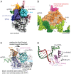
Fig. 6.
RNAP swiveling and TL conformational changes of Mtb RNAP during the nucleotide addition cycle. (A) The cryo-EM density map of the TEC containing Mtb RNAP and Mtb NusG with NTP. Modules and domains of RNAP, DNA, RNA and NusG are colored and labeled. The N-terminal extension of the Mtb NusG is indicated by a red arrow. (B) Model of Mtb NusG interacting with the ntDNA for the NusG-dependent PTC. The interaction between Mtb NusG and ntDNA in the PTC is modeled by superposing Mtb NusG on B. subtilis NusG of the PTC structure. Mtb NusG is depicted as a surface representation with a ribbon model. Amino acid residues that interact with ntDNA bases are indicated as stick models and labeled. The N-terminal extension of the Mtb NusG is indicated in red. (C and D) Comparison of the structures of the Mtb RNAP-Mtb NusG TEC in the swiveled (without NTP, black) and non-swiveled (with NTP, color) states, showing the rotation of the NusG/swivel module (C) and folding of the TL (D) upon NTP binding at the active site (indicated by black arrows). Mg2+ ions at active centers are shown as red spheres (MgA and MgB) and the TL residues M1012 and H1016 are shown as stick models that contact the nucleobase and triphosphate group of the NTP, respectively.
Fig. 7.
Fig. 7.
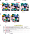
RNA exit channels of Mtb and E. coli RNAPs. (A) Comparison of the RNA exit channels of the Mtb TEC without an RNA hairpin (Left), the Mtb PTC with an RNA hairpin (Middle), and the E. coli TEC without an RNA hairpin (Right). The structures are depicted as surface representations (RNAP and NusG) and stick (DNA and RNA) models. The C-terminus of the E. coli β subunit (E1342, yellow) is positioned below the Zn binding domain (ZBD) of the β′ subunit (Right). The C-terminal region of the Mtb β subunit (E1151 to N1169, yellow) forms a bridge between the Zn binding and dock domains of the β′ subunit, making the RNA exit channel narrower (Left), but it is disordered in the PTC to accommodate the RNA hairpin (Middle). (B) Cryo-EM density maps of the TEC (Left) and PTC (Right). Densities of RNA are omitted. (C) Sequence alignment of the C-terminus of the β subunit (βC) of representative bacterial RNAPs (M. tub, M. tuberculosis; B. sub, B. subtilis; S. aur, Staphylococcus aureus; S. elo, Synechococcus elongatus; C. dif, Clostridioides difficile; T. the, Thermus thermophilus; B. bac, Bdellovibrio bacteriovorus; N. men, Neisseria meningitidis, C. cre; Caulobacter crescentus. C. jej, Campylobacter jejuni). The C-terminal tail of the Mtb β subunit and C-terminus of the E. coli β subunit are highlighted.
Fig. 8.
Fig. 8.
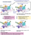
RNAP conformations associated with transcription elongation, NusG-dependent pausing, and escape from paused transcription. A series of transcription complexes with NusG determined in this study are depicted as surface, ribbon and stick model representations, and the modules and domains are indicated. Top panels represent the TEC, while the Bottom panels represent the PTC (Left) and a complex escaping from the pause (eTEC) (Right).

References

    1. Saba J., et al. , The elemental mechanism of transcriptional pausing. Elife 8, e40981 (2019). - PMC - PubMed
    1. Yakhnin A. V., Kashlev M., Babitzke P., NusG-dependent RNA polymerase pausing is a frequent function of this universally conserved transcription elongation factor. Crit Rev Biochem. Mol. Biol. 55, 716–728 (2020). - PMC - PubMed
    1. Pan T., Artsimovitch I., Fang X. W., Landick R., Sosnick T. R., Folding of a large ribozyme during transcription and the effect of the elongation factor NusA. Proc. Natl. Acad. Sci. U. S. A. 96, 9545–9550 (1999). - PMC - PubMed
    1. Pan T., Sosnick T., RNA folding during transcription. Annu. Rev. Biophys. Biomol. Struct. 35, 161–175 (2006). - PubMed
    1. Steinert H., et al. , Pausing guides RNA folding to populate transiently stable RNA structures for riboswitch-based transcription regulation. Elife 6, e21297 (2017). - PMC - PubMed

Publication types

LinkOut - more resources