. 2023 Sep 1;78(3):878-895.
doi: 10.1097/HEP.0000000000000303. Epub 2023 Feb 9.

The outcome of boosting mitochondrial activity in alcohol-associated liver disease is organ-dependent

Affiliations

The outcome of boosting mitochondrial activity in alcohol-associated liver disease is organ-dependent

Naroa Goikoetxea-Usandizaga et al. Hepatology. .

Abstract

Background and aims: Alcohol-associated liver disease (ALD) accounts for 70% of liver-related deaths in Europe, with no effective approved therapies. Although mitochondrial dysfunction is one of the earliest manifestations of alcohol-induced injury, restoring mitochondrial activity remains a problematic strategy due to oxidative stress. Here, we identify methylation-controlled J protein (MCJ) as a mediator for ALD progression and hypothesize that targeting MCJ may help in recovering mitochondrial fitness without collateral oxidative damage.

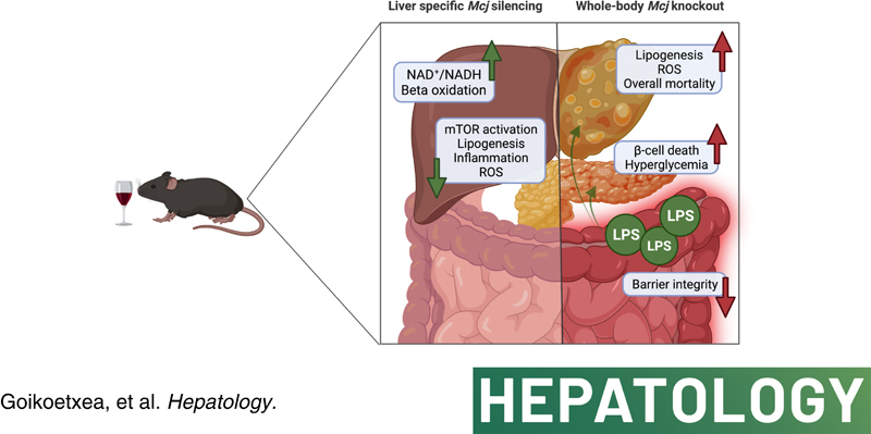
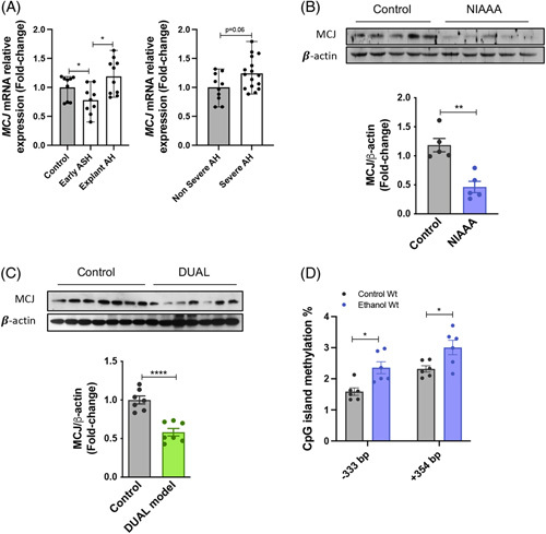
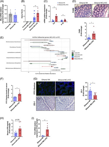
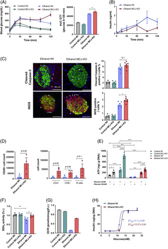
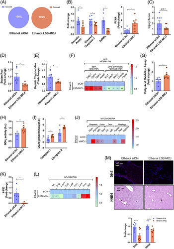
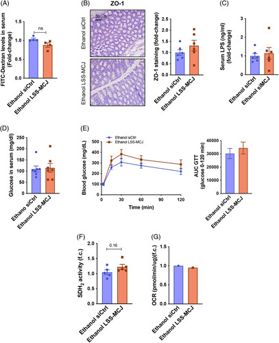
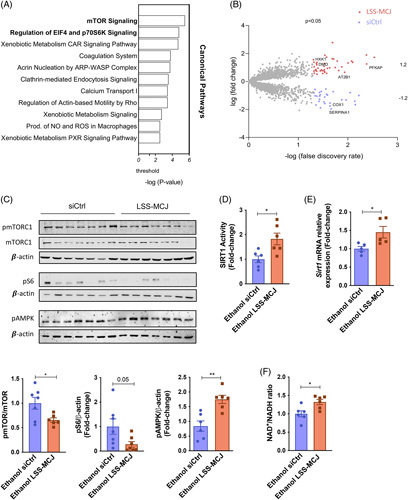
Approach and results: C57BL/6 mice [wild-type (Wt)] Mcj knockout and Mcj liver-specific silencing (MCJ-LSS) underwent the NIAAA dietary protocol (Lieber-DeCarli diet containing 5% (vol/vol) ethanol for 10 days, plus a single binge ethanol feeding at day 11). To evaluate the impact of a restored mitochondrial activity in ALD, the liver, gut, and pancreas were characterized, focusing on lipid metabolism, glucose homeostasis, intestinal permeability, and microbiota composition. MCJ, a protein acting as an endogenous negative regulator of mitochondrial respiration, is downregulated in the early stages of ALD and increases with the severity of the disease. Whole-body deficiency of MCJ is detrimental during ALD because it exacerbates the systemic effects of alcohol abuse through altered intestinal permeability, increased endotoxemia, and dysregulation of pancreatic function, which overall worsens liver injury. On the other hand, liver-specific Mcj silencing prevents main ALD hallmarks, that is, mitochondrial dysfunction, steatosis, inflammation, and oxidative stress, as it restores the NAD + /NADH ratio and SIRT1 function, hence preventing de novo lipogenesis and improving lipid oxidation.

Conclusions: Improving mitochondrial respiration by liver-specific Mcj silencing might become a novel therapeutic approach for treating ALD.


Conflict of interest statement

This work was supported by grants from Ministerio de Ciencia e Innovación, Programa Retos-Colaboración RTC2019-007125-1 (for Jorge Simon and Maria Luz Martinez-Chantar); Ministerio de Economía, Industria y Competitividad, Retos a la Sociedad AGL2017-86927R (for F.M.); Instituto de Salud Carlos III, Proyectos de Investigación en Salud DTS20/00138 and DTS21/00094 (for Jorge Simon and Maria Luz Martinez-Chantar, and Asis Palazon. respectively); Instituto de Salud Carlos III, Fondo de Investigaciones Sanitarias co-founded by European Regional Development Fund/European Social Fund, “Investing in your future” PI19/00819, “Una manera de hacer Europa” FIS PI20/00765, and PI21/01067 (for Jose J. G. Marin., Pau Sancho-Bru,. and Mario F. Fraga respectively); Departamento de Industria del Gobierno Vasco (for Maria Luz Martinez-Chantar); Asturias Government (PCTI) co-funding 2018-2023/FEDER IDI/2021/000077 (for Mario F. Fraga.); Ministerio de Ciencia, Innovación y Universidades MICINN: PID2020-117116RB-I00, CEX2021-001136-S PID2020-117941RB-I00, PID2020-11827RB-I00 and PID2019-107956RA-100 integrado en el Plan Estatal de Investigación Científica y Técnica y Innovación, cofinanciado con Fondos FEDER (for Maria Luz Martinez-Chantar, Francisco J Cubero., Yulia A Nevzorova and Asis Palazon); Ayudas Ramón y Cajal de la Agencia Estatal de Investigación RY2013-13666 and RYC2018-024183-I (for Leticia Abecia and Asis Palazon); European Research Council Starting Grant 804236 NEXTGEN-IO (for Asis Palazon); The German Research Foundation SFB/TRR57/P04, SFB1382-403224013/A02 and DFG NE 2128/2-1 (for Francisco J Cubero and Yulia A Nevzorova); National Institute of Health (NIH)/National Institute of Alcohol Abuse and Alcoholism (NIAAA) 1U01AA026972-01 (For Pau Sancho-Bru); Junta de Castilla y León SA074P20 (for Jose J. G. Marin); Junta de Andalucía, Grupo PAIDI BIO311 (for Franz Martin); CIBERER Acciones Cooperativas y Complementarias Intramurales ACCI20-35 (for Mario F. Fraga); Ministerio de Educación, Cultura y Deporte FPU17/04992 (for Silvia Ariño); Fundació Marato TV3 201916-31 (for Jose J. G. Marin.); Ainize Pena-Cearra is a fellow of the University of the Basque Country (UPV/EHU); BIOEF (Basque Foundation for Innovation and Health Research); Asociación Española contra el Cáncer (Maria Luz Martinez-Chantar and Teresa C. Delgado.); Fundación Científica de la Asociación Española Contra el Cáncer (AECC Scientific Foundation) Rare Tumor Calls 2017 (for Maria Luz Martinez-Chantar); La Caixa Foundation Program (for Maria Luz Martinez-Chantar); Proyecto Desarrollo Tecnologico CIBERehd (for Maria Luz Martinez-Chantar); Ciberehd_ISCIII_MINECO is funded by the Instituto de Salud Carlos III.

María Luz Martínez-Chantar advises for Mitotherapeutix LLC. Guadalupe Sabio received grants from Mitotherapeutix LLC. The remaining authors have nothing to report.

Figures

None
Graphical abstract
FIGURE 1
FIGURE 1
MCJ expression is regulated in alcohol-associated liver injury. (A) Relative MCJ mRNA levels in liver biopsies from patients with Early ASH (n=11), Nonsevere AH (n=11), Severe AH (n=18), and Explanted AH (n=11), together with healthy control individuals (n=10). Values represented as median ± range. U test was used to compare 2 groups. (B) Western blotting and densitometric quantification of MCJ in Wt liver extracts with control (n=5) and NIAAA diet (n=5) and (C) with control (n=7) and DUAL (n=7) diet. β-actin as a loading control. (D) CpG islands methylation status analyzed by bisulfite pyrosequencing. *p<0.05, **p<0.01, and ****p<0.0001 versus control.
FIGURE 2
FIGURE 2
Whole body MCJ-KO show increased liver injury after ethanol consumption. (A) Survived and Deceased Wt (Left panel) and MCJ-KO (Right panel) mice following the NIAAA model. (B) Hepatic histopathological evaluation. (C) Quantification of Necrotic Areas (based on HE staining), Cleaved Caspase-3 and TUNEL stained liver sections. (D) ALT and AST plasmatic levels. (E) Quantification of Sudan Red stained liver sections. (F) Hepatic triglyceride content. (G) Hepatic fatty acid oxidation assay. (H) Heatmap showing the liver mRNA relative expression of genes involved in alcohol metabolism: Adh1, Aldh2, and Cyp2e1. (I) Quantification and representative DHE and 4HNE stained liver sections. (J) Hepatic SDH2 activity. (K) Oxygen consumption rate of mitochondrial complex I and complex II in freshly isolated hepatic mitochondria. (L) Heatmap showing the liver mRNA relative expression of mitochondrial quality control genes: Tfam, Pgc1a, Mfn, Opa1, Fis1, Mff, Prkn, Pink1, and Sqstm. (M) Hepatic LPS content. (N) Hepatic mRNA relative expression of genes involved in LPS recognition: Tlr, Ap-1, and Hamp. *p<0.05, **p<0.01, and ***p<0.001 versus Wt.
FIGURE 3
FIGURE 3
Increased intestinal permeability and translocation in ethanol-fed MCJ-KO mice. (A) Relative Mcj mRNA expression in gut tissue from control and ethanol-fed Wt mice. (B) Gut histopathological evaluation. (C) Gut mRNA relative expression of Tnf and Il-1b. (D) Quantification and representative F4/80 stained gut sections. (E) Central Log-Ratio (CLR) transformed abundance for the significant genera (FDR <10%) found for the comparison between the gut microbiome of MCJ-KO mice versus Wt mice. For each genus, the distribution of the CLR-transformed is represented for each of the 4 study groups using a combination of a violin plot for the general distribution and a boxplot for the summary distribution, colored accordingly. (F) Serum FITC-Dextran levels. (G) Immunohistochemical representation of intestinal FITC-Dextran levels and quantification and representative Zonula occludens stained gut sections. (H) Serum LPS content. (I) Gut mRNA relative expression of Tlr4. *p<0.05, **p<0.01, and ***p<0.001 versus Wt.
FIGURE 4
FIGURE 4
Augmented pancreatic injury and hyperglycaemia in ethanol-fed MCJ-KO mice. (A) Blood glucose levels and the resulting AUC during the IPGTT (B) Insulin levels during the IPGTT. (C) Quantification and representative Cleaved caspase-3 and iNOS stained pancreatic beta cell sections. (D) Pancreatic infiltrating total CD45+ cells and further characterization of CD4+, CD8+, and B cells populations using FACS. (E) ATP content in isolated pancreatic islets. (F) Pancreatic SDH2 activity (G) A representative measurement of the oxygen consumption rate of freshly isolated pancreatic islets. (H) In vitro static insulin release assay, using freshly isolated pancreatic islets. *p<0.05, **p<0.01, and ****p<0.0001 versus Wt.
FIGURE 5
FIGURE 5
Liver specific silencing of Mcj reduces liver damage and steatosis following the NIAAA model. (A) Survived and Deceased siCtrl (Left panel) and MCJ-LSS (Right panel) mice. (B) Quantification of Necrotic Areas (based on HE staining), Cleaved Caspase-3, TUNEL, and PCNA stained liver sections. (C) Hepatic histopathological evaluation. (D) Quantification of Sudan Red stained liver sections. (E) Hepatic triglyceride content. (F) Heatmap showing the liver mRNA relative expression of genes involved in lipid metabolism: Fatp2, Cpt1a, Ppara, Acadl, Acc, FasN, Srebp, Chrebp, and Pparg. (G) Hepatic fatty acid oxidation assay. (H) Hepatic SDH2 activity. (I) Oxygen Consumption Rate of mitochondrial Complex I and Complex II in freshly isolated mitochondria. (J) Heatmap showing the liver mRNA relative expression of mitochondrial quality control genes: Tfam, Pgc1a, Mfn, Opa1, Fis1, Mff, Prkn, Pink1, and Sqstm. (K) Quantification of F4/80 stained liver sections. (L) Heatmap showing the liver mRNA relative expression of genes involved in inflammation: Tnf, Il-1b, Cxcl1, Ccl2, Ccl5, Ccr2, Ccr5, Il-10, and Ho-1. (M) Quantification and representative DHE and 4HNE stained liver sections. *p<0.05, **p<0.01, and ***p<0.001 versus Wt.
FIGURE 6
FIGURE 6
Liver-specific silencing of Mcj does not induce a systemic alcohol injury. (A) Serum FITC-Dextran levels. (B) Quantification and representative Zonula occludens stained gut sections. (C) Serum LPS content. (D) Serum glucose levels. (E) Blood glucose levels and the resulting AUC during the IPGTT. (F) Pancreatic SDH2 activity. (G) A representative measurement of the oxygen consumption rate of freshly isolated pancreatic islets.
FIGURE 7
FIGURE 7
Silencing of MCJ inhibits mTOR activation via increased NAD+ and improved Sirt1 activity. (A) Ingenuity pathway analysis of top canonical pathways. (B) Volcano plot showing all the identified hepatic proteins. Statistically significant proteins are shown in the corresponding colors, and the highlighted proteins were identified as mTOR interactors. (C) Activated and total hepatic mTOR protein levels, together with activated S6 protein levels, by western blotting and densitometric quantification. ß-actin as a loading control. (D) Hepatic SIRT1 activity. (E) Sirt1 hepatic mRNA relative expression. (F) Hepatic NAD+/NADH ratio. *p<0.05 versus Wt.

References

    1. Rehm J, Samokhvalov AV, Shield KD. Global burden of alcoholic liver diseases. J Hepatol. 2013;59:160–8. - PubMed
    1. Pimpin L, Cortez-Pinto H, Negro F, Corbould E, Lazarus JV, Webber L, Sheron N. EASL HEPAHEALTH Steering Committe. Burden of liver disease in Europe: epidemiology and analysis of risk factors to identify prevention policies. J Hepatol. 2018;69:718–35. - PubMed
    1. Mathurin P, Lucey MR. Liver transplantation in patients with alcohol-related liver disease: current status and future directions. Lancet Gastroenterol Hepatol. 2020;5:507–14. - PubMed
    1. Teschke R. Alcoholic liver disease: alcohol metabolism, cascade of molecular mechanisms, cellular targets, and clinical aspects. Biomedicines. 2018;6:106. - PMC - PubMed
    1. Zhong Z, Ramshesh VK, Rehman H, Liu Q, Theruvath TP, Krishnasamy Y, et al. . Acute ethanol causes hepatic mitochondrial depolarization in mice: role of ethanol metabolism. PLoS One. 2014;9:e91308. - PMC - PubMed

Publication types