

A retrospective analysis of Stewart-Treves syndrome in the context of chronic lymphedema
- PMID: 36746732
- PMCID: PMC10173066
- DOI: 10.1016/j.abd.2022.04.011
A retrospective analysis of Stewart-Treves syndrome in the context of chronic lymphedema
Abstract
Background: stewart-treves syndrome (STS) is an angiosarcoma associated with chronic lymphedema.
Objectives: This article analyses the characteristics of twenty-two patients and proposes active intervention in lymphedema and the early diagnosis of STS.
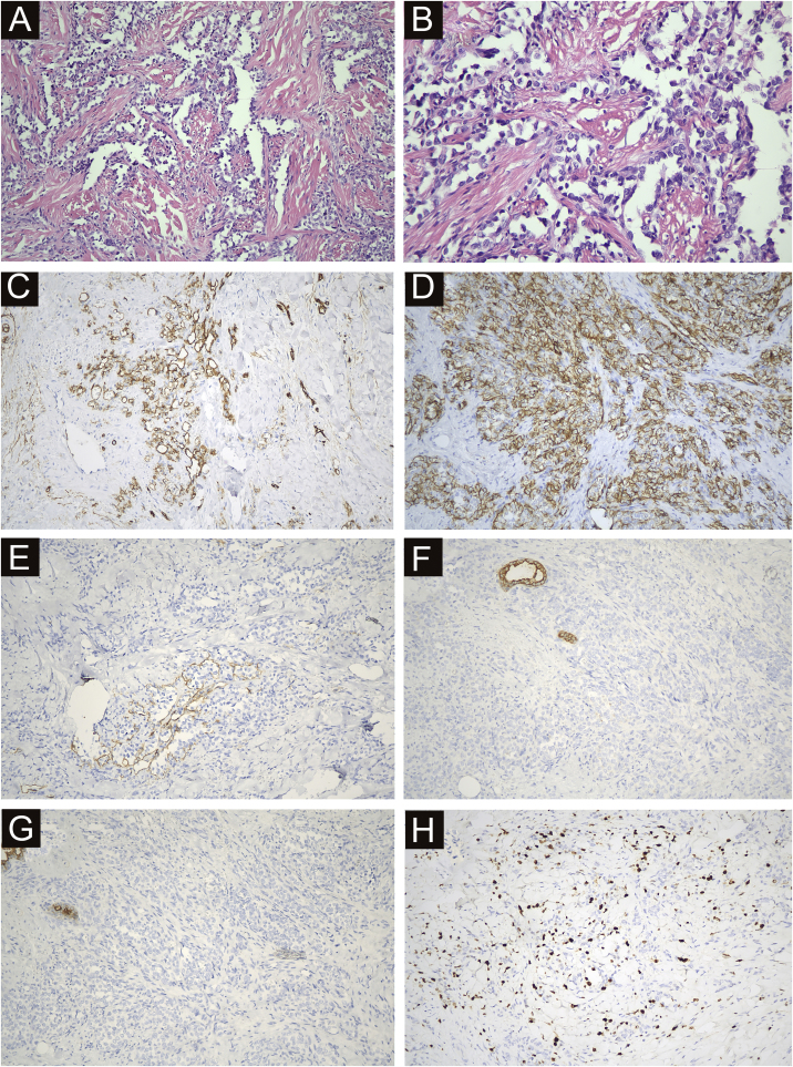
Methods: Twenty-two patients with STS were diagnosed at the centre over an 11-year period. Clinical manifestations, a series of conventional analyses, and histopathology were used to study these cases retrospectively.
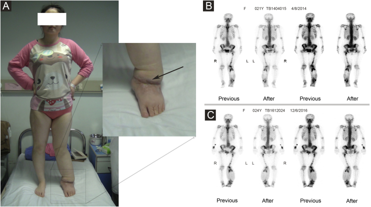
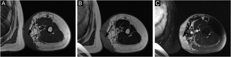
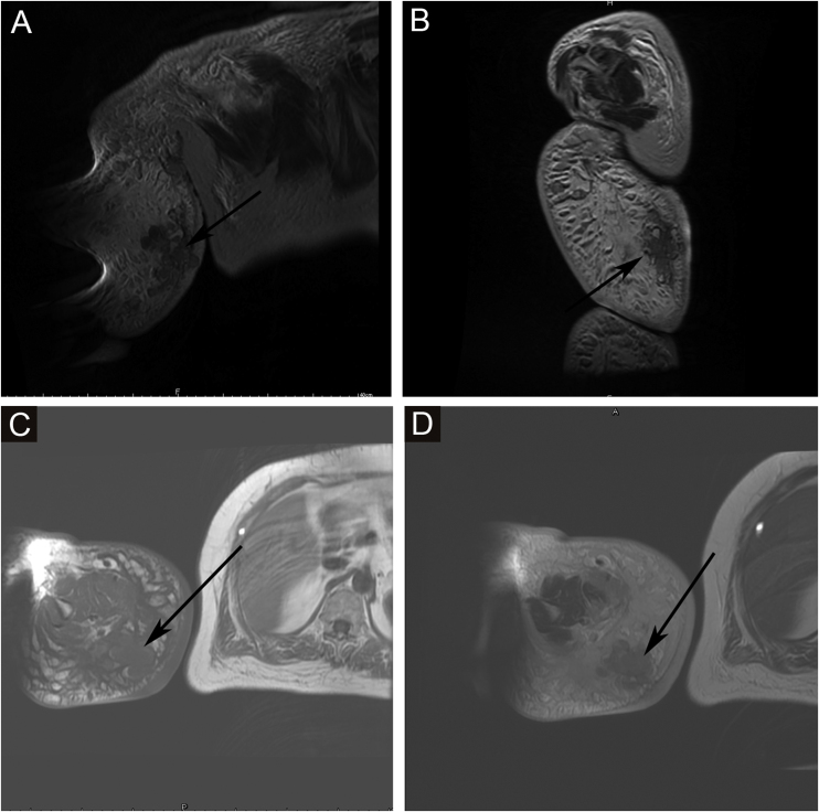
Results: The age range of 22 patients with STS was 15 to 78 years. The main clinical manifestations included multiple skin and subcutaneous nodules and scattered red or purplish-red rashes in the lymphoedematous limbs. These patients often showed clinical symptoms such as lymphedema, weakness, emaciation, pain, mass, lymphadenopathy and so on. The positive rates of ultrasonography, MRI and radionuclide imaging were 66.7% (6/9), 92.3% (12/13) and 18.2% (2/11), respectively. The main points regarding active intervention in lymphedema and early diagnosis of STS were summarized.
Study limitations: Since this was a retrospective study, the main points summarized by the author need to be further quantified in clinical work to guide the diagnosis of this kind of disease more conveniently. In addition, further clinical trials are needed to evaluate the role of lymphedema in the occurrence and development of malignant tumors.
Conclusions: STS can appear in lymphoedematous tissue many years after lymphedema onset. To avoid delays in the diagnosis and therapy of STS, physicians should actively look for signs or symptoms of malignant lymphedema during the follow-up period and promptly manage patients developing problems.
Keywords: Lymphangiosarcoma; Lymphedema; Stewart Treves syndrome.
Copyright © 2023 Sociedade Brasileira de Dermatologia. Published by Elsevier España, S.L.U. All rights reserved.
Figures
References
-
- Ruocco V., Brunetti G., Puca R.V., Ruocco E. The immunocompromised district: a unifying concept for lymphooedematous, herpes-infected and otherwise damaged sites. J Eur Acad Dermatol Venereol. 2009;23:1364–1373. - PubMed
-
- Ruocco V., Schwartz R.A., Ruocco E. Lymphedema: an immunologically vulnerable site for development of neoplasms. J Am Acad Dermatol. 2002;47:124–127. - PubMed
-
- Marando A., Bernasconi B., Sabatino D., Militti L., Capella C. Complex karyotype in a case of cutaneous lymphangiosarcoma associated with chronic lymphedema of the lower limb. Pathol Res Pract. 2014;210:1138–1141. - PubMed
-
- González-Vela M.C., González-López M.A., Val-Bernal J.F., Fernández-Llaca H. Cutaneous diffuse large B-cell lymphoma of the leg associated with chronic lymphedema. Int J Dermatol. 2008;47:174–177. - PubMed
-
- Sharma A., Schwartz R.A. Stewart-Treves syndrome: pathogenesis and management. J Am Acad Dermatol. 2012;67:1342–1348. - PubMed
MeSH terms
Supplementary concepts
LinkOut - more resources
Full Text Sources
Medical
