. 2023 Feb 6;14(1):643.
doi: 10.1038/s41467-023-36314-5.

Small-molecule-mediated OGG1 inhibition attenuates pulmonary inflammation and lung fibrosis in a murine lung fibrosis model

Affiliations

Small-molecule-mediated OGG1 inhibition attenuates pulmonary inflammation and lung fibrosis in a murine lung fibrosis model

L Tanner et al. Nat Commun. .

Abstract

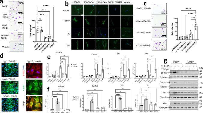
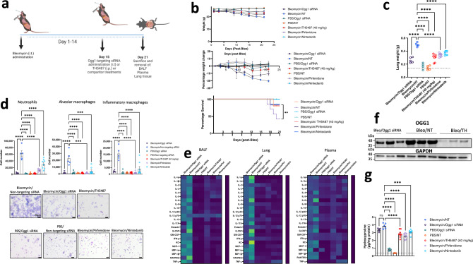
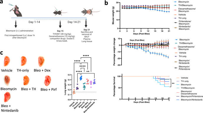
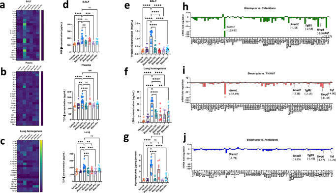
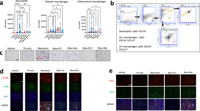
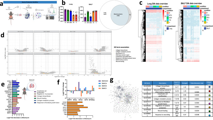
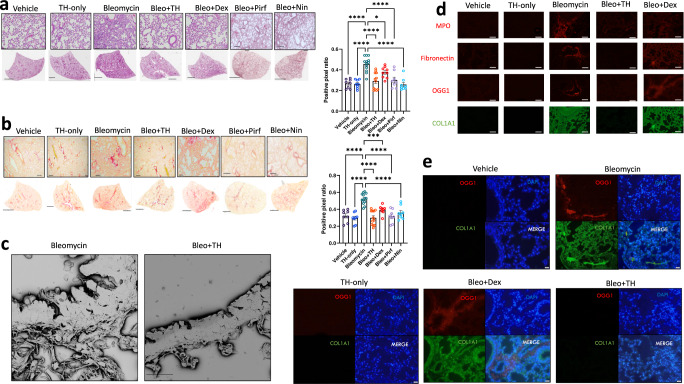
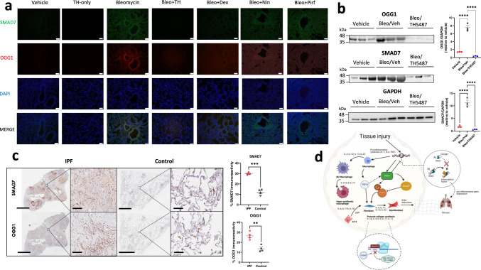
Interstitial lung diseases such as idiopathic pulmonary fibrosis (IPF) are caused by persistent micro-injuries to alveolar epithelial tissues accompanied by aberrant repair processes. IPF is currently treated with pirfenidone and nintedanib, compounds which slow the rate of disease progression but fail to target underlying pathophysiological mechanisms. The DNA repair protein 8-oxoguanine DNA glycosylase-1 (OGG1) has significant roles in the modulation of inflammation and metabolic syndromes. Currently, no pharmaceutical solutions targeting OGG1 have been utilized in the treatment of IPF. In this study we show Ogg1-targeting siRNA mitigates bleomycin-induced pulmonary fibrosis in male mice, highlighting OGG1 as a tractable target in lung fibrosis. The small molecule OGG1 inhibitor, TH5487, decreases myofibroblast transition and associated pro-fibrotic gene expressions in fibroblast cells. In addition, TH5487 decreases levels of pro-inflammatory mediators, inflammatory cell infiltration, and lung remodeling in a murine model of bleomycin-induced pulmonary fibrosis conducted in male C57BL6/J mice. OGG1 and SMAD7 interact to induce fibroblast proliferation and differentiation and display roles in fibrotic murine and IPF patient lung tissue. Taken together, these data suggest that TH5487 is a potentially clinically relevant treatment for IPF but further study in human trials is required.


Conflict of interest statement

T.H. is listed as inventor on a provisional U.S. patent application no. 62/636983, covering OGG1 inhibitors. The patent is fully owned by a nonprofit public foundation, the Helleday Foundation, and T.H. is a member of the foundation board developing OGG1 inhibitors toward the clinic. An inventor reward scheme is under discussion. The remaining authors declare no other competing interests.

Figures

Fig. 1
Fig. 1. OGG1-dependent cell migration and fibrotic gene expression.
a Migration of human primary lung fibroblasts (pHLF) post-TGF-β1 induction for 24 h, with significantly more myofibroblast cells appearing than in mock- and inhibitor(s)-treated wells. Data were analyzed using a one-way ANOVA followed by a Dunnett’s post hoc test unless otherwise specified: TGF-β1 vs TGF-β1/TH5487 (P = 0.0001); TGF-β1 vs TGF-β1/Dex (P = 0.6954); TGF-β1 vs TGF-β1/Nin (P = 0.0001); TGF-β1 vs TGF-β1/Veh (P < 0.0001). ns: not significant. Source data are provided as Source data file. Data are representative of 4 independent experiments containing 3 biological replicates (scale bar = 180 μm). Data are presented as the mean ± standard error of the mean (a, c, e, f). b Immunostaining of pHLF cells (green) following 24 h of TGF-β1 treatment, with TH5487 treatment displaying visually reduced levels of collagen (COL1A1), fibronectin (FN), vimentin (VIM), α-smooth muscle actin (α-SMA). Scale bar = 50 μm. Results shown from 3 independent experiments. c Migration of pHLF post-TGF-β1 induction for 24 h, and siRNA transfection targeting OGG1 or scrambled sequence (control). Data were analyzed using a one-way ANOVA followed by a Dunnett’s post hoc test: si Control/TGF-β1 vs si OGG1/TGF-β1 (P = 0.0008); si Control/TGF-β1 vs si OGG1/vehicle and si Control/TGF-β1 vs si Control/vehicle (P < 0.0001). Data are representative of 4 independent experiments containing 3 biological replicates (scale bar = 180 μm). Data are presented as the mean ± standard error of the mean. d Stress fiber formation was seen in response to TGF-β1 in Ogg1+/+, but not Ogg1−/− MF cells (left panel, F-actin). Expression and colocalization of α-SMA (red) with F-actin (green) are shown (right panels, scale bar = 90 μm). Results shown from 3 independent experiments. e The effects of TH5487 on transcription of α-Sma, Fn1, Vim, and Col1A1 in TGF-β1-stimulated MF cells as determined by qRT-PCR. TH5487 (10 μM), significantly decreased mRNA levels of all genes with compiled data representative of 3 independent experiments. Data were analyzed by one-way ANOVA followed by a Dunnett’s post hoc test. f TGF-β1-induced expression of αSma, Col1a1, Fn1, and Vim was decreased in Ogg1−/− but not in Ogg1+/+ MF cells. Data representative of 3 independent experiments. g Immunoblot analysis of TGF-β1-stimulated pHLF cells showing decreased levels of α-SMA, COL1A1, FN1, and VIM levels in OGG1-depleted cells by siRNA. TGF-β1, 2 ng/mL; TH5487, 10 μM; Nintedanib (Nin), 10 μM; Dexamethasone (Dex), 10 μM. Results shown from 3 independent experiments.
Fig. 2
Fig. 2. In vivo bleomycin model highlighting OGG1 as a therapeutic target.
a Bleomycin (Bleo, 2.5 U/kg) was intratracheally-administered to C57Bl/6J mice, with mice left to develop fibrosis over a period of 21 days. Subsequent Ogg1-targetting or non-targeting siRNA (NT) administration occurred at day 14, with the inclusion of TH5487 (TH; 40 mg/kg; i.p.; daily for 5 days), Nin (60 mg/kg; p.o.; 5 daily doses), and pirfenidone (300 mg/kg; p.o.; daily for 5 days) treatment groups. Data are presented as means ± SEM (b, d, g). ns: not significant. b Murine weights, survival, and percentage total weight loss for the Bleo administered groups displayed similar trends until day 14, thereafter the Ogg1 siRNA and TH5487-treated groups displayed maintenance of weight until the end of the study (Bleo/Ogg1 siRNA n = 5, Bleo/NT n = 5, Phosphate buffered saline (PBS)/Ogg1 siRNA n = 5, PBS/NT n = 5, Bleo/TH5487 n = 9, Bleo/Pirfenidone n = 8, Bleo/Nintedanib n = 8). c Murine total lung weight. Significant differences were seen between TH5487 and Ogg1 siRNA-treated groups compared to the NT siRNA group (P < 0.0001). d Flow cytometry conducted on murine bronchoalveolar lavage fluid (BALF) indicates a significant decrease in inflammatory cell recruitment following Ogg1 siRNA or TH5487 treatment (P < 0.0001; (Bleo/Ogg1 siRNA n = 5, Bleo/NT n = 5, PBS/Ogg1 siRNA n = 4, PBS/NT n = 5, Bleo/TH5487 n = 9, Bleo/Pirfenidone n = 8, Bleo/Nintedanib n = 8)). Murine BALF samples showing cell morphology for each treatment condition. Scale bar = 20 µm. e Cytokine expression (day 21) following Bleo administration and subsequent treatment (mean-normalized values shown, with yellow indicating high values and blue indicating low values). f Lung homogenate samples were analyzed by SDS-PAGE (n = 3; 1 from each independent experiment), followed by immunoblotting using an antibody specific to OGG1. Results shown from 3 independent experiments. g Hydroxyproline levels display significant decreases in both siRNA and drug (TH5487, Nin, pirfenidone)-treated groups, indicative of reduced levels of collagen in these lungs (Bleo/Ogg1 siRNA n = 5, Bleo/NT n = 5, PBS/Ogg1 siRNA n = 5, PBS/NT n = 5, Bleo/TH5487 n = 9, Bleo/Pirfenidone n = 8, Bleo/Nintedanib n = 8). Source data are provided as a Source data file. Elements of this figure were created with BioRender.com.
Fig. 3
Fig. 3. TH5487 murine dosing strategy and weights.
a Mice received intratracheally-administered bleomycin (Bleo; 2.5 U/kg) and were subsequently dosed intraperitoneally (i.p.) with TH5487 (TH), nintedanib (Nin), pirfenidone (Pirf), or dexamethasone (Dex) 1 h post-Bleo administration. Drug treatment occurred five times per week, over the course of 21 days, followed by euthanasia and removal of BALF, plasma, and lung tissues (Bleo n = 15, Bleo/TH n = 14, Bleo/Dex n = 9, Vehicle n = 9, TH only n = 9, Bleo/Pirf n = 7, Bleo/Nin n = 7). Data are presented as means ± SEM (b, c). Data were analyzed using a one-way ANOVA followed by a Dunnett’s post hoc test unless otherwise specified. b Mice receiving Bleo showed weight loss up until day 10, where after those dosed with TH5847 picked up significant amounts of weight compared to the vehicle/bleomycin group. c Representative images of murine lungs (right lobes) removed after 21 days, with lung weights shown alongside, Bleomycin vs Bleo+TH (P < 0.0001), Bleo vs Bleo+Pirf (P < 0.0001), Bleo vs Bleo+Nintedanib (P = 0.0056) (Bleo n = 8, Bleo/TH n = 8, Bleo/Dex n = 5, Vehicle n = 5, TH only n = 5, Bleo/Pirf n = 7, Bleo/Nin n = 7). Source data are provided as a Source data file. Elements of this figure were created with BioRender.com.
Fig. 4
Fig. 4. Significantly decreased cytokine levels following TH5487 administration, with accompanying reduction in markers of lung damage.
Heatmaps (ac) showing the differences in cytokine levels, as measured by multiplex assay, in murine bronchoalveolar lavage fluid (BALF), plasma, and lung homogenate (mean-normalized values shown, with yellow indicating high values and blue indicating low values). Cytokine values were compared to the vehicle/Bleo group using a one-way ANOVA (*P < 0.05; **P < 0.01; ***P < 0.005; ****P < 0.0001). ns: not significant. TGF-β1 ELISA (d) conducted on murine BALF, plasma, and lung homogenate revealed significantly decreased TGF-β1 levels in all three murine sample types with values compared to the vehicle/Bleo group using a one-way ANOVA: Bleo vs Bleo+TH (BALF: P = 0.0022; plasma: P < 0.0001, Lung homogenate: P = 0.0001). Data are presented as means ± SEM (dg). Murine albumin content (e) and Lactate dehydrogenase (LDH) (f) were measured in BALF samples as markers for lung damage and plasma leakage, with TH5487 treatment significantly decreasing both albumin content (P < 0.0001) and LDH levels in the BALF (P = 0.0022) compared to the vehicle/Bleo group. g Murine lung collagen content was measured using a hydroxyproline assay with TH5487 significantly reducing collagen levels (P < 0.0001) compared to Bleo control lungs (dg: (Bleo n = 14, Bleo/TH n = 13, Bleo/Dex n = 8, Vehicle n = 8, TH only n = 8, Bleo/Pirf n = 8, Bleo/Nin n = 8). hj qRT-PCR arrays specific for mouse fibrotic genes were used to analyze murine lung samples (n = 5 pooled samples) following treatment using TH5487 (TH), nintedanib (Nin), and pirfenidone (Pirf). Source data are provided as a Source data file.
Fig. 5
Fig. 5. Inflammatory cell influx measured in murine BALF.
Murine BALF was assessed for neutrophils, alveolar macrophages, and inflammatory macrophages (a) using flow cytometry, with the representative gating (b) strategy depicted alongside (Bleo n = 14, Bleo/TH n = 13, Bleo/Dex n = 8, Vehicle n = 8, TH only n = 8, Bleo/Pirf n = 8, Bleo/Nin n = 8). Data are presented as means ± SEM. ns: not significant. a Decreased numbers of neutrophils (P < 0.0001) and inflammatory macrophages (P = 0.004) were detected in response to TH5487 (i.p.) treatment (TH), with no significant difference reported between the neutrophils and inflammatory macrophages of mice treated with dexamethasone (Dex) or nintedanib (Nin). Almost no significant differences were seen for alveolar macrophage numbers, aside from pirfenidone (Pirf; P = 0.0237). Inflammatory cell numbers were compared to the vehicle/Bleo group using a one-way ANOVA (*P < 0.05). c Giemsa-Wright stained cytospin slides showing vehicle/Bleo BALF samples containing enlarged inflammatory macrophages, with TH5487 treatment reducing the presence of inflammatory macrophages, while the corticosteroid dexamethasone similarly reduced inflammatory macrophage influx comparable to vehicle-treated control samples. Results shown from 3 independent experiments. Scale bar = 20 μm. Immunofluorescent staining of murine BALF samples measured inflammatory macrophage (d) and neutrophil (e) content using CD206 (red)/F4/80 (green) and LY6G (red)/MPO (green), respectively. (DAPI, blue; scale bar = 50 μm. Source data are provided as a Source data file.
Fig. 6
Fig. 6. Proteome of lungs and BALF.
a Experimental workflow showing analysis of protein extracts by mass spectrometry. b Unique and overlapping protein identifications subdivided by treatment condition in lungs and BALF by biological fluid. c Response profile heatmaps displaying protein expression across treatment conditions in each biological fluid. d Volcano plot depicting differentially expressed proteins in BALF (top panel) and lung (bottom panel) with proteins associated with significant gene ontology (GO) terms highlighted in each plot. e, f Specific lung proteins downregulated following Bleo/TH5487 treatment compared to the Bleo alone condition. Proteins highlighted in these plots are specifically associated with e fibrotic-related changes or f SMAD (top panel) and nucleotide base repair (bottom panel). g Network analysis (StringDB) comparing the Bleo/Bleo+TH treatment condition displaying significant biological GO terms, protein counts in the network, strength, and FDR. For LCMS/MS analyses, murine samples were analyzed by single-shot DIA-MS for cohorts Bleo (B) n = 5, bleomycin/TH5487 (BTH) n = 5, bleomycin/dexamethasone (DEX) n = 4, TH5487 only (TH) n = 4 and vehicle only (V) n = 4, respectively. Significance criteria; absolute fold-change ≥2 & FDR, Benjamini–Hochberg-corrected p-value ≤0.05 unless indicated otherwise. Source data are provided as a Source data file. Elements of this figure were created with BioRender.com.
Fig. 7
Fig. 7. Murine lung staining, scanning electron microscopy, and immunofluorescence show reduced levels of fibrotic-related lung damage following Bleo/TH5487 treatment compared to Bleo/vehicle samples.
a TH5487 significantly decreased lung damage in Bleo-treated mice (H&E) and b collagen deposition (picrosirius red) in both macroscopic and microscopic structures compared to vehicle/Bleo lungs and was confirmed by positive pixel analysis of whole-lung scanned images (scale bar of microscopic image =100 μm; scale bar of whole lung scan = 2 mm). Results shown from 3 independent experiments. Statistical analyses were conducted using a one-way ANOVA (*P < 0.05; **P < 0.01; ***P < 0.005). ns: not significant. Murine samples included in these analyses: Bleo n = 13, Bleo/TH n = 13, Bleo/Dex n = 8, Vehicle n = 8, TH only n = 9, Bleo/Pirf n = 7, Bleo/Nin n = 7). Bleo vs Bleo+TH displayed significantly reduced levels of H&E and picrosirius red staining (P < 0.0001). Data are presented as means ± SEM (a, b). c TH5487 /Bleo scanning electron microscopy images show reduced collagen deposition in the alveolar borders compared to Bleo-treated controls (scale bar = 20 μm). Immunofluorescent staining of murine lung sections (d) revealed decreased levels of MPO (red), fibronectin (red), OGG1 (red), and COL1A1 (green) following TH5487 treatment compared to both vehicle/Bleo and dexamethasone/Bleo groups (scale bar = 50 μm). e Co-stained murine lung sections revealed corresponding increases in OGG1 (red) and COL1A1 (green) following Bleo administration (DAPI counterstain, blue), with reduced levels of both OGG1 and COL1A1 in TH5487-treated samples (scale bar = 50 μm). Source data are provided as a Source data file.
Fig. 8
Fig. 8. Lower expression of OGG1 and SMAD7 are seen in both TH5487-treated mice and human explanted control lungs.
a Murine lung immunofluorescence analysis showed decreased levels of immunoreactivity for OGG1 (red)/SMAD7 (green) in TH5487 (TH)-treated mice compared to bleomycin (Bleo) controls (DAPI, blue; scale bar = 50 μm), results shown from 3 independent experiments, b with mouse lung homogenate samples analyzed by SDS-PAGE, followed by immunoblotting using rabbit antisera specific to OGG1 and SMAD7 (protein levels quantified inset n = 3 per treatment; statistical analyses were conducted using a one-way ANOVA (****P < 0.0001 for comparisons between all groups tested)). Data are presented as means ± SEM (b, c). c This result translated into human patient samples, with healthy control lung tissue displaying significantly decreased levels of OGG1 and SMAD7 immunoreactivity (brown staining; P = 0.0024 and P = 0.0002, respectively), as compared using an unpaired t test with Welch’s correction (two-sided), n = 4 patients per group. Scale bar = 6 mm and 100 μm for inset images. d Excessive OGG1 production facilitates pro-inflammatory gene expression, promoting inflammatory cell recruitment, leading to further exacerbation of the fibrotic lung environment. In addition, OGG1 promotes the phosphorylation of SMAD 2/3 by SMAD7 interaction, promoting TGF-β-driven FMT and EMT and excessive ECM deposition. Therefore, decreased OGG1 expression and binding to DNA by TH5487 inhibits downstream pro-inflammatory gene expression and pulmonary fibrosis. In this study, OGG1 and SMAD7 levels were both reduced after the administration of TH5487, as shown by SDS-page/immunoblotting. Source data are provided as a Source data file. Elements of this figure were created with BioRender.com.

References

    1. Raghu G, et al. An official ATS/ERS/JRS/ALAT statement: idiopathic pulmonary fibrosis: evidence-based guidelines for diagnosis and management. Am. J. Respir. Crit. Care Med. 2011;183:788–824. - PMC - PubMed
    1. Chang X, et al. Monocyte-derived multipotent cell delivered programmed therapeutics to reverse idiopathic pulmonary fibrosis. Sci. Adv. 2020;6:eaba3167. - PMC - PubMed
    1. Raghu G, et al. Use of a molecular classifier to identify usual interstitial pneumonia in conventional transbronchial lung biopsy samples: a prospective validation study. Lancet Respir. Med. 2019;7:487–496. - PubMed
    1. Martinez FJ, et al. Idiopathic pulmonary fibrosis. Nat. Rev. Dis. Prim. 2017;3:17074. - PubMed
    1. Kim HJ, Perlman D, Tomic R. Natural history of idiopathic pulmonary fibrosis. Respir. Med. 2015;109:661–670. - PubMed

Publication types