

Choroidal vascularity index in hereditary optic neuropathies
- PMID: 36747110
- PMCID: PMC10482917
- DOI: 10.1038/s41433-023-02383-5
Choroidal vascularity index in hereditary optic neuropathies
Abstract
Purpose: To assess the choroidal vascularity index (CVI) in patients affected by Leber hereditary optic neuropathy (LHON) compared to patients affected by dominant optic atrophy (DOA) and healthy subjects.
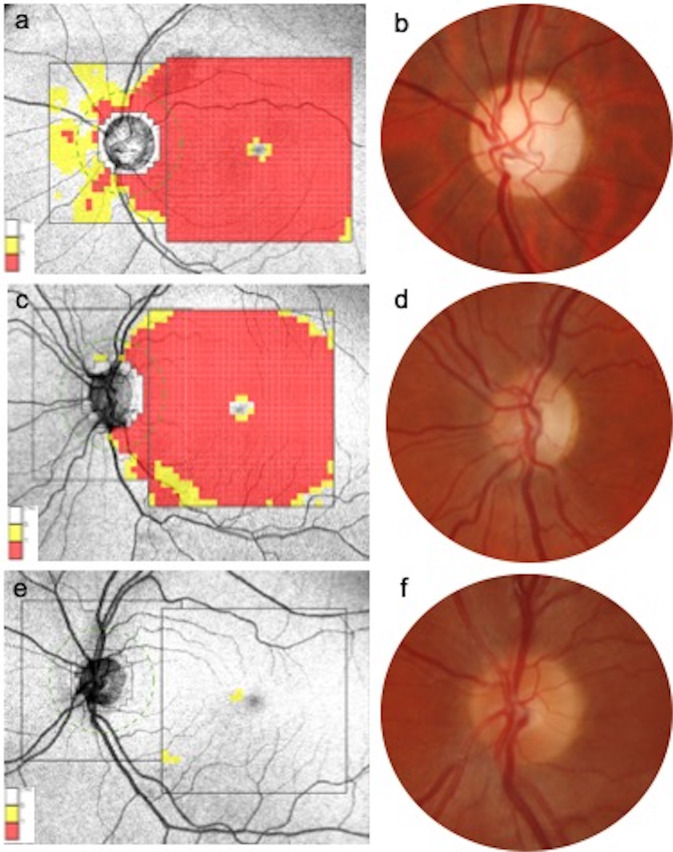
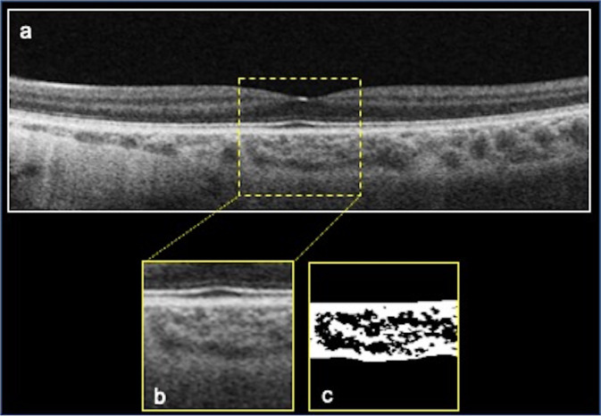
Methods: In this retrospective study, we considered three cohorts: LHON eyes (48), DOA eyes (48) and healthy subjects' eyes (48). All patients underwent a complete ophthalmologic examination, including best-corrected visual acuity (BCVA) and optical coherence tomography (OCT) acquisition. OCT parameters as subfoveal choroidal thickness (Sub-F ChT), mean choroidal thickness (ChT), total choroidal area (TCA), luminal choroidal area (LCA) were calculated. CVI was obtained as the ratio of LCA and TCA.
Results: Subfoveal ChT in LHON patients did not show statistically significant differences compared to controls, while in DOA a reduction in choroidal thickness was observed (p = 0.344 and p = 0.045, respectively). Mean ChT was reduced in both LHON and DOA subjects, although this difference reached statistical significance only in DOA (p = 0.365 and p = 0.044, respectively). TCA showed no significant differences among the 3 cohorts (p = 0.832). No changes were detected in LCA among the cohorts (p = 0.389), as well as in the stromal choroidal area (SCA, p = 0.279). The CVI showed no differences among groups (p = 0.898): LHON group was characterized by a similar CVI in comparison to controls (p = 0.911) and DOA group (p = 0.818); the DOA group was characterized by a similar CVI in comparison to controls (p = 1.0).
Conclusion: CVI is preserved in DOA and LHON patients, suggesting that even in the chronic phase of the neuropathy the choroidal structure is not irreversibly compromised.
© 2023. The Author(s), under exclusive licence to The Royal College of Ophthalmologists.
Conflict of interest statement
The authors declare no competing interests.
Figures
References
-
- Moster SJ, Moster ML, Bryan MS, Sergott RC. Retinal ganglion cell and inner plexiform layer loss correlate with visual acuity loss in LHON: A longitudinal, segmentation OCT analysis. Investig Ophthalmol Vis Sci. 2016. 10.1167/iovs.15-17328. - PubMed
MeSH terms
LinkOut - more resources
Full Text Sources
Research Materials
