

Relevant factors of posterior mandible lingual plate perforation during immediate implant placement: a virtual implant placement study using CBCT
- PMID: 36747164
- PMCID: PMC9903431
- DOI: 10.1186/s12903-022-02696-z
Relevant factors of posterior mandible lingual plate perforation during immediate implant placement: a virtual implant placement study using CBCT
Abstract
Background: To explore the influence of cross-sectional type and morphological parameters at the mandibular molar sites on lingual plate perforation (LPP) during the immediate implant placement (IIP).
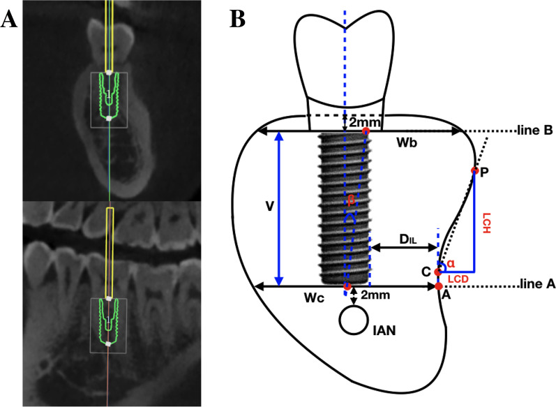
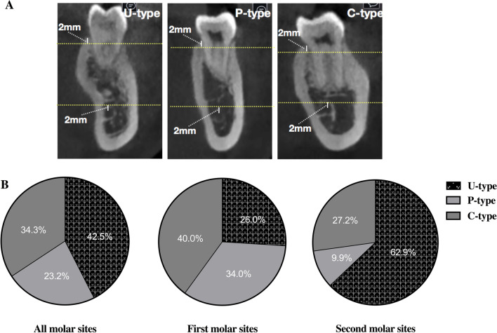
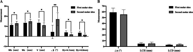
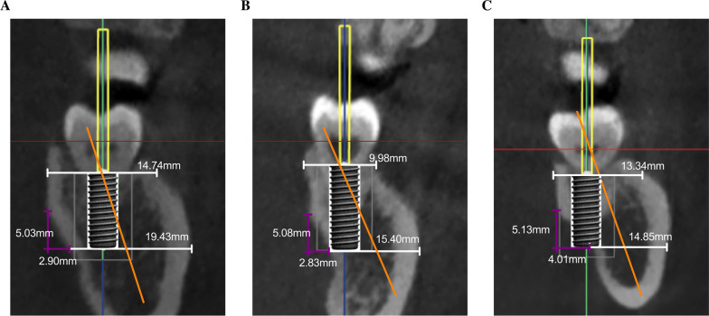
Methods: 181 implants were virtually placed in the mandibular molar sites on the cone beam computed tomography (CBCT). Each cross-section of the implantation site was divided into the Undercut (U)/Parallel (P)/Convex (C) types. Morphologically relevant parameters were measured on the cross-sections, including width of the upper end (Wb), width of the lower end (Wc), vertical height (V), angle between the natural crown axis and the alveolar bone axis (∠β), LC depth (LCD), LC height, and angle between the horizontal line and the line connecting the most prominent point and the most concave point of lingual plate (∠α). Besides, the distance from the end of the virtual implant and the lingual bone plate of the cross-section (DIL) was calculated. Relationships between all the morphologically relevant parameters and the DIL were further analyzed.
Results: A total of 77 (42.5%) cross-sections were classified as U-type, which was the most common one, accounting for 63% of the second molar regions. All LPP cases and most of the nearly LPP (87.9%) cases occurred at the U-type cross-sections, and the relationship between the DIL and the morphological parameters can be expressed by a multivariate linear equation.
Conclusions: The occurrence rate of U-type cross-sections in the second molar region was very high, and the risk of LPP should be considered during IIP. Except for the U-type, significant large LCD, small Wc, and large ∠β were the important relevant factors. CBCT and multivariate linear equations could help to assess the LPP risk and provide a reference for implant placement design pre-surgery.
Keywords: Complication; Cone-beam computed tomography; Immediate implant placement; Mandible; Molar.
© 2023. The Author(s).
Conflict of interest statement
No relevant conflicts of interest to declare.
Figures
References
-
- Bernabe E, Marcenes W, Hernandez C, Bailey J, Abreu L, Alipour V, et al. Global, regional, and national levels and Trends in burden of oral conditions from 1990 to 2017: a systematic analysis for the global burden of disease 2017 study. J Dent Res. 2020;99(4):362–373. doi: 10.1177/0022034520908533. - DOI - PMC - PubMed
-
- Di Tinco R, Bertani G, Pisciotta A, Bertoni L, Bertacchini J, Colombari B, et al. Evaluation of antimicrobial effect of air-polishing treatments and their influence on human dental pulp stem cells seeded on titanium disks. Int J Mol Sci. 2021;22(2):865. doi: 10.3390/ijms22020865. - DOI - PMC - PubMed
Publication types
MeSH terms
Substances
Grants and funding
LinkOut - more resources
Full Text Sources
