. 2023 May 29;35(6):1671-1707.
doi: 10.1093/plcell/koad026.

Rock, scissors, paper: How RNA structure informs function

Affiliations

Rock, scissors, paper: How RNA structure informs function

Sarah M Assmann et al. Plant Cell. .

Abstract

RNA can fold back on itself to adopt a wide range of structures. These range from relatively simple hairpins to intricate 3D folds and can be accompanied by regulatory interactions with both metabolites and macromolecules. The last 50 yr have witnessed elucidation of an astonishing array of RNA structures including transfer RNAs, ribozymes, riboswitches, the ribosome, the spliceosome, and most recently entire RNA structuromes. These advances in RNA structural biology have deepened insight into fundamental biological processes including gene editing, transcription, translation, and structure-based detection and response to temperature and other environmental signals. These discoveries reveal that RNA can be relatively static, like a rock; that it can have catalytic functions of cutting bonds, like scissors; and that it can adopt myriad functional shapes, like paper. We relate these extraordinary discoveries in the biology of RNA structure to the plant way of life. We trace plant-specific discovery of ribozymes and riboswitches, alternative splicing, organellar ribosomes, thermometers, whole-transcriptome structuromes and pan-structuromes, and conclude that plants have a special set of RNA structures that confer unique types of gene regulation. We finish with a consideration of future directions for the RNA structure-function field.


Conflict of interest statement

Conflict of interest statement. None declared.

Figures
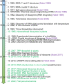
Figure 1.
Figure 1.
Timeline for seminal discoveries in RNA structural biology. Related Nobel Prizes are in purple font and important plant discoveries are in green font. LNP, lipid nanoparticle. Associated references are 1. Holley et al. (1965), 2. Kim et al. (1973), 3. Berget et al. (1977) and Chow et al. (1977a), 4. Peattie (1979) and Peattie and Gilbert (1980), 5. Kruger et al. (1982) and Guerrier-Takada et al. (1983), 6. Greider and Blackburn (1985), 7. Altuvia et al. (1989), 8. Ellington and Szostak (1990) and Robertson and Joyce (1990) and Tuerk and Gold (1990), 9. Grundy and Henkin (1993), 10. Daros and Flores (1995), 11. Shen et al. (1999), 12. Ban et al. (2000), Wimberly et al. (2000), Harms et al. (2001), and Yusupov et al. (2001), 13. Nocker et al. (2001), 14. Mironov et al. (2002) and Winkler et al. (2002), 15. Wachter et al. (2007), 16. Lee et al. (2009), 17. Seidelt et al. (2009) and Armache et al. (2010), 18. Filichkin et al. (2010), 19. Jinek et al. (2012), 20. Ding et al. (2014) and Rouskin et al. (2014), 21. Bieri et al. (2017) and Perez Boerema et al. (2018), 22. Alves et al. (2017) and Cognat et al. (2017), 23. Su et al. (2018) and Chung et al. (2020), 24. Fajkus et al. (2019) and Song et al. (2019), 25. Dolgin (2021), 26. Waltz et al. (2020), 27. Ferrero-Serrano et al. (2022).
Figure 2.
Figure 2.
Features of tRNAs. A) Hierarchical folding of tRNA. Shown are the primary (1°), secondary (2°), and tertiary structures (3°) of tRNA, with the various SLs color coded. In the secondary structure, the D SL, anticodon SL, variable loop, TψC SL, and the acceptor stem are noted, as is the 4-way junction (4WJ). In the tertiary structure, the point of attachment of the amino acid and the site of binding of the mRNA codon are noted. B) Watson–Crick base pairing. Sites of modification by structure probing DMS, EDC, and SHAPE reagents are indicated. C) Non-Watson–Crick base pairing with coloring corresponding to the tRNA in A, illustrating a base triple, reverse Watson–Crick GC pair, G•U wobble pair, and purine–purine base pair. All images are from PDB ID: 4TNA for yeast tRNAPhe. Validation reports are available for this structure at the Protein Data Bank: https://www.rcsb.org/.
Figure 3.
Figure 3.
Schematic of RNA structure–function relationships in plant growth and development. A) tRFs play important roles in the symbiosis between legumes and rhizobia bacteria. tRFs produced by bacteria (B. japonicum) are delivered into AGO1 (Argonaute) complexes of soybean root cells, resulting in the degradation of nodule-suppressing plant mRNAs and thus promoting nodule initiation and development. B) TPP-sensing riboswitches regulate THIC mRNA accumulation through the control of pre-mRNA splicing and 3′ end processing (Bocobza et al. 2007; Wachter et al. 2007). Upper: when TPP concentrations are low, the aptamer interacts with a 5′-splice site GU within the 3′-UTR and prevents splicing, with consequent retention of a poly-A cleavage site (transcription processing site or TPS, upstream/yellow) within a 3′-UTR intron that causes 3′UTRs cleavage and polyadenylation for high expression of THIC. Lower: in the presence of elevated TPP concentrations, the aptamer sequence no longer base pairs with the 5′-splice site, making it accessible for splicing, and consequently removing the TPS and extending the length of the 3′-UTR at the 3′-splice site AG. This 3′UTR is long (as noted by the break in the sequence) and has a different TPS (downstream/green), which leads to increased RNA turnover and thus reduced expression of THIC. C) An example of GQ structure and its function in root development in HIRD11 mRNA (Yang et al. 2020b). In a hird11 mutant complementation study, mutGQ-complemented transgenic plants (lacking HIRD11 GQ structure) showed longer roots than those of wtGQ-complemented plants. D) In the phloem, transcription factor JUL and the promoter of SMXL4/5 genes are key regulators of phloem differentiation. JUL directly binds and induces a GQ within the 5′ UTR of SMXL4/5 mRNAs; this JUL-induced GQ-SMXL4/5 complex suppresses SMXL4/5 translation and consequently inhibits phloem differentiation (Cho et al. 2018). E) In the endodermis, SHR mRNA contains a GQ structure that can elicit liquid–liquid phase separation in vitro (Zhang et al. 2019). This RNA structure-initiated phase separation is suggestive of an important biological function in the translational regulation of SHR, which is a major player in root development. F) In the flowering process, the depicted mutant version (red arrows indicate the mutation sites) of the COOLAIR natural antisense lncRNA of FLC suppresses FLC transcript abundance and accelerates flowering (Hawkes et al. 2016; Yang et al. 2022a).
Figure 4.
Figure 4.
Telomerase RNA from Arabidopsis. The 268 nt RNA telomerase RNA from A. thaliana, referred to as AtTR, with the conserved pseudoknot domain highlighted in yellow and the 9 nt 5′-CUAAACCCU template boxed. This region, known as the T-PK domain, is closed by the TBE which is the extensive helix spanning nts 36 to 143. Reprinted from Song et al. (2019), Fig. 3. Copyright 2019 National Academy of Sciences.
Figure 5.
Figure 5.
Ribosome structures. A) Structure of the 5S and 23S rRNA from the halophilic archaeon Haloarcula marismortui. The bases are white and the sugar-phosphate backbones are orange. The CCA acceptor stem model of the reaction intermediate for peptide bond formation is red and the numbered proteins are blue. The L1 and L11 proteins positioned at lower resolution are in cyan, where “L” is for LSU that comprises 5S and 23S rRNA. From PDB ID: 1FFZ (Nissen et al. 2000). Reprinted with permission from AAAS. B) Translation inhibition by the translation factor pY. The chloroplast 70S (yellow and blue):pY (green) complex is superposed with the bacterial A-, P- and E-site tRNAs (red, blue, and purple structures, respectively) including mRNA from the crystal structure of the Thermus thermophilus 70S ribosome. Note how pY is bound in the mRNA (black) channel and prevents tRNA binding. From superposition of PDB ID: 5MMI (50S) and 5MMJ (30S), with tRNAs and mRNAs from 4V51 (Bieri et al. 2017). Reprinted with permission from Wiley. Validation reports are available for these structures at the Protein Data Bank: https://www.rcsb.org/.
Figure 6.
Figure 6.
Spliceosome base pairing and reaction. A) Sequence of an mRNA pre-splicing and post-splicing with the intron lariat depicted. The underlined “A” is the branch point adenosine, “Yn” depicts the ∼15–20 nt polypyrimidine tract (where “Y” is the IUPAC abbreviation for pyrimidine), and the final “AG” is the conserved dinucleotide sequence immediately adjacent to the 3′-splice site. B) Spliceosome snRNA interactions before the first-step splicing reaction. This complex involves the 3 snRNAs U2 (green), U5 (blue), and U6 (red). There is extensive base pairing between U2 and U6, while U5 interacts only weakly with the 5′-exon (gold). The U6 snRNA also interacts with the intron (black) and forms an intramolecular SL (ISL). The AGC of U6 snRNA shows dashed lines that form the catalytic triad. The bulged A in the intron sequence UACUAAC is the branch point adenosine and uses its 2′hydroxyl as a nucleophile to attack the 5′-splice site as shown. Adapted with permission from Fica and Nagai (2017), Fig. 4. Copyright Springer Nature.
Figure 7.
Figure 7.
Ribozyme crystal structures. A) Secondary and B) tertiary structures for a hammerhead ribozyme that was laboratory evolved PDB ID: 5DI2 (Mir et al. 2015) (orange), twister ribozyme from rice PDB ID: 4OJI (Liu et al. 2014) (green), and glmS ribozyme from Bacillus anthracis PDB ID: 2NZ4 (Cochrane et al. 2007) (pink). These ribozymes are notable for their compact tertiary structures with a buried active site. The glmS ribozyme is also a GlcN6P-responsive riboswitch. The long-distance interactions depicted with black lines show double pseudoknots in twister and glmS ribozymes, and a single pseudoknot in the hammerhead ribozyme, which serve to compact and rigidify the structures. Adapted with permission from Seith et al. (2018), Fig. 2. Copyright 2018 American Chemical Society. Validation reports are available for these structures at the Protein Data Bank: https://www.rcsb.org/.
Figure 8.
Figure 8.
Schematic of RNA structure–function relationships in abiotic stress response. A) Upper, right portion: under warmer daytime temperatures, the RNA hairpin within the 5′ UTR of PIF7 attains a more-relaxed hairpin conformation (weaker RNA base pairing) than at lower nighttime temperatures, resulting in increased PIF7 translation (Chung et al. 2020). Lower, left portion: in rice seedlings, acute heat shock leads to genome-wide mRNA structural melting and consequently mRNA degradation (Su et al. 2018). B) High light triggers unfolding of an SD-sequestering hairpin in the 5′ UTR of the psbA mRNA, which consequently increases psbA translation (Gawronski et al. 2021). C) Salt, likely acting indirectly (dashed lines), appears to unfold some transcripts (higher reactivity to the structure probing chemical DMS [cf. Figure 1B]) with correlated decreases in their abundance, and fold other transcripts (lower reactivity to DMS) with correlated increases in their abundance (Tack et al. 2020). D) In response to phosphate starvation, an intermolecular sense to antisense interaction between PHO1.2 and cis-NAT PHO1.2 rearranges the RNA conformation, leading to the enhancement of PHO1.2 translation (Reis et al. 2021).
Figure 9.
Figure 9.
Mechanisms of riboswitch-mediated gene regulation. A) Riboswitches can operate under transcription control (left) or translation control (right) and can be divided into an Aptamer Domain and an Expression Platform. This scheme is for the metabolite turning OFF transcription or translation, although ON riboswitches are also known. In transcription control, an antiterminator forms that is mutually exclusive with the terminator owing to sharing nucleotides. Upon binding of the metabolite, depicted generically as a star, to the Aptamer Domain, nucleotides in the antiterminator are sequestered into the folded aptamer and the terminator forms, which is stable and has a series of Us that make unstable base pairs with templating dAs and terminates transcription. In translation control, the ribosome binding site (RBS), or SD site in the case of bacteria, is exposed and translation is ON. Upon binding of the metabolite to the aptamer domain, the RBS is sequestered into the folded aptamer and translation is turned OFF. Reprinted with permission from Tucker and Breaker (2005). Copyright 2005 Elsevier. B, C) Molecular recognition of TPP by the riboswitch from Arabidopsis (Thore et al. 2006; Thore et al. 2008). The TPP metabolite is bound in a modular fashion, with nitrogen atoms in blue, oxygen atoms in red, and carbon atoms in a specific color. B) Recognition of the thiamine (white) portion of TPP. There is extensive molecular recognition of the metabolite provided by diverse faces of riboswitch nucleobases (salmon) PDB ID: 2CKY (Thore et al. 2006). Copyright 2006 AAAS. C) Recognition of the pyrophosphate portion of TPP. There is extensive recognition by 2 Mg2+ ions. Note that panel C uses the refined crystal structure that led to a new conformation of the pyrophosphate moiety PDB ID: 3D2G (Thore et al. 2008). Copyright 2018 American Chemical Society. Validation reports are available for these structures at the Protein Data Bank: https://www.rcsb.org/.
Figure 10.
Figure 10.
Inhibition of translation by the formation of a 5′-UTR GQ structure. A) The GQ-forming sequence of G3AG3AAG4AAG4 found in the 5′ UTR of the ATR gene in Arabidopsis. B) Schematic of constructs used in this transient expression reporter assay to test the effect of the GQ on transcription and translation. The GQS construct contains the complete native ATR 5′ UTR, while in the mGQS construct select Gs are mutated to As (red), which prevents GQ formation. Shown are normalized C) GUS/luciferase mRNA abundance as assessed by qRT-PCR, and D) protein expression as assessed by enzyme activity. Based on the comparison of results from GQS and mGQS constructs, translation, but not transcription, is repressed by the GQ structure. Reproduced from Kwok et al. (2015).
Figure 11.
Figure 11.
Structurome approaches and observations for RNA folding transcriptome-wide. A) Approach for mutational profiling (MaP) vs. RT stops. Treatment with DMS results in methylation of exposed A and C residues, which are detected as mutations in a single read in MaP (M; top) or as a series of truncated transcripts in RT stops (M; bottom). B) Large differences in predicted and in vivo determined structures for RARE-COLD-INDUCIBLE 2A (RCI2A; At3g05880) (Ding et al. 2014). C) Inverse relationship of change in chemical probe reactivity and change in transcript abundance for 3 different organisms under 3 different stresses: O. sativia under heat shock (Su et al. 2018), A. thaliana under salt stress (Tack et al. 2020), and B. subtilis under amino acid starvation (Ritchey et al. 2020).
Figure 12.
Figure 12.
Pan-structurome prediction of riboSNitches in Arabidopsis and attributes of the ZINC RIBBON3 (ZR3) Arabidopsis riboSNitch. A) Non-riboSNitches and B) RiboSNitches mapped onto the 5 chromosomes of Arabidopsis (Ferrero-Serrano et al. 2022). riboSNitches were predicted using SNPFold (Halvorsen et al. 2010). Color bars indicate the number of SNPs. C) The ZR3 riboSNitch reference (left) and alternative (right) alleles. The destabilizing shorter stem and larger loop of the alternative allele (right) causes it to melt at lower temperatures (Ferrero-Serrano et al. 2022). D) Differential distribution of Arabidopsis accessions harboring the reference (blue dots) and alternative (red dots) across Eurasia (Ferrero-Serrano et al. 2022). E) ZR3 transcript abundance is lower in accessions harboring the alternative allele.

References

    1. Ahn IP, Kim S, Lee YH. Vitamin B-1 functions as an activator of plant disease resistance. Plant Physiol. 2005:138(3):1505–1515. 10.1104/pp.104.058693 - DOI - PMC - PubMed
    1. Akiyama BM, Laurence HM, Massey AR, Costantino DA, Xie X, Yang Y, Shi PY, Nix JC, Beckham JD, Kieft JS. Zika virus produces noncoding RNAs using a multi-pseudoknot structure that confounds a cellular exonuclease. Science. 2016:354(6316):1148–1152. 10.1126/science.aah3963 - DOI - PMC - PubMed
    1. Altuvia S, Kornitzer D, Teff D, Oppenheim AB. Alternative mRNA structures of the cIII gene of bacteriophage lambda determine the rate of its translation initiation. J Mol Biol. 1989:210(2):265–280. 10.1016/0022-2836(89)90329-X - DOI - PubMed
    1. Alves CS, Vicentini R, Duarte GT, Pinoti VF, Vincentz M, Nogueira FT. Genome-wide identification and characterization of tRNA-derived RNA fragments in land plants. Plant Mol Biol. 2017:93(1–2):35–48. 10.1007/s11103-016-0545-9 - DOI - PubMed
    1. Amiard S, Depeiges A, Allain E, White CI, Gallego ME. Arabidopsis ATM and ATR kinases prevent propagation of genome damage caused by telomere dysfunction. Plant Cell. 2011:23(12):4254–4265. 10.1105/tpc.111.092387 - DOI - PMC - PubMed

Publication types