[Preprint]. 2024 Jan 6:2023.01.23.525290.
doi: 10.1101/2023.01.23.525290.

Cell-type-specific inhibitory circuitry from a connectomic census of mouse visual cortex

Affiliations

Cell-type-specific inhibitory circuitry from a connectomic census of mouse visual cortex

Casey M Schneider-Mizell et al. bioRxiv. .

Update in

  • Inhibitory specificity from a connectomic census of mouse visual cortex.
    Schneider-Mizell CM, Bodor AL, Brittain D, Buchanan J, Bumbarger DJ, Elabbady L, Gamlin C, Kapner D, Kinn S, Mahalingam G, Seshamani S, Suckow S, Takeno M, Torres R, Yin W, Dorkenwald S, Bae JA, Castro MA, Halageri A, Jia Z, Jordan C, Kemnitz N, Lee K, Li K, Lu R, Macrina T, Mitchell E, Mondal SS, Mu S, Nehoran B, Popovych S, Silversmith W, Turner NL, Wong W, Wu J, Reimer J, Tolias AS, Seung HS, Reid RC, Collman F, da Costa NM. Schneider-Mizell CM, et al. Nature. 2025 Apr;640(8058):448-458. doi: 10.1038/s41586-024-07780-8. Epub 2025 Apr 9. Nature. 2025. PMID: 40205209 Free PMC article.

Abstract

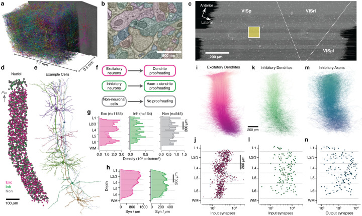
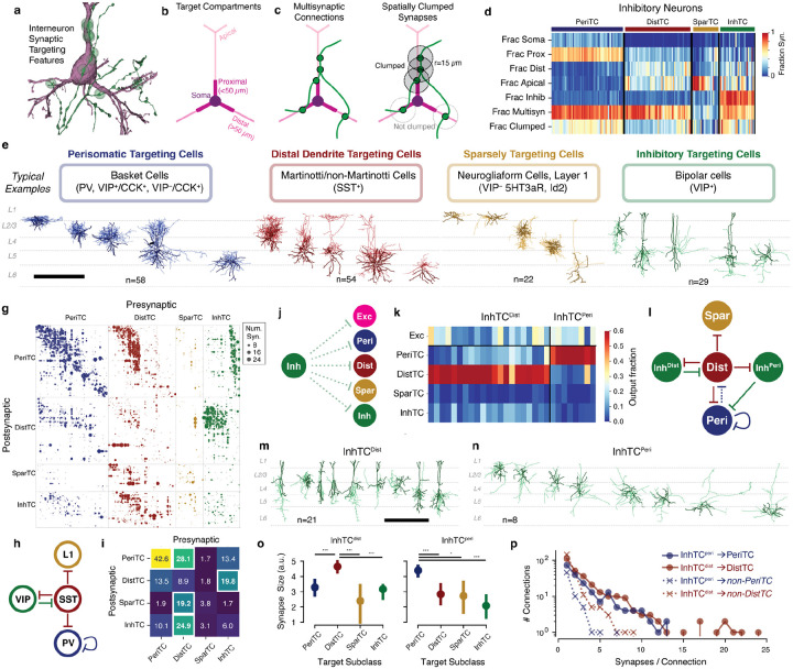
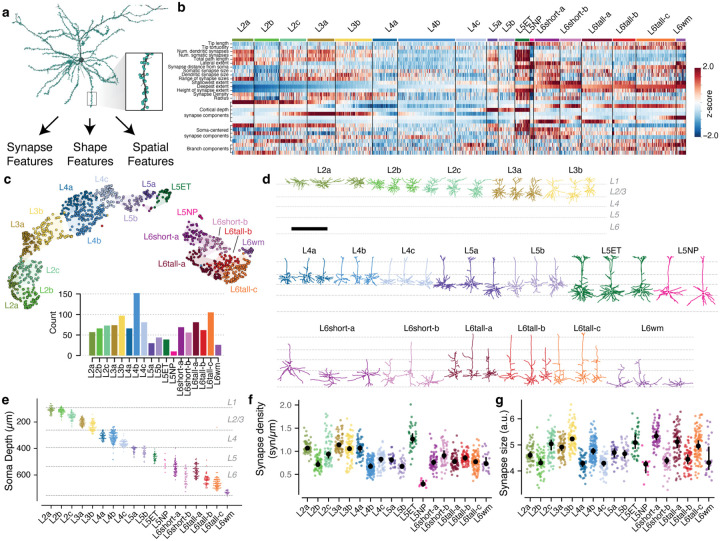
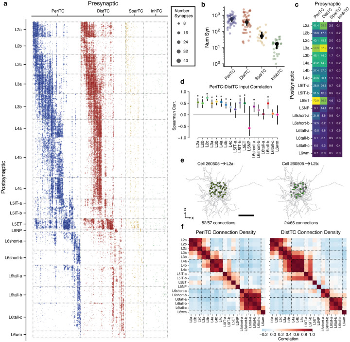
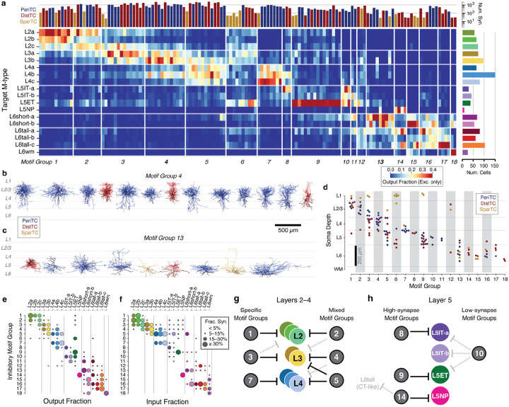
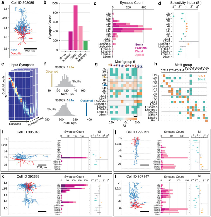
Mammalian cortex features a vast diversity of neuronal cell types, each with characteristic anatomical, molecular and functional properties. Synaptic connectivity powerfully shapes how each cell type participates in the cortical circuit, but mapping connectivity rules at the resolution of distinct cell types remains difficult. Here, we used millimeter-scale volumetric electron microscopy1 to investigate the connectivity of all inhibitory neurons across a densely-segmented neuronal population of 1352 cells spanning all layers of mouse visual cortex, producing a wiring diagram of inhibitory connections with more than 70,000 synapses. Taking a data-driven approach inspired by classical neuroanatomy, we classified inhibitory neurons based on the relative targeting of dendritic compartments and other inhibitory cells and developed a novel classification of excitatory neurons based on the morphological and synaptic input properties. The synaptic connectivity between inhibitory cells revealed a novel class of disinhibitory specialist targeting basket cells, in addition to familiar subclasses. Analysis of the inhibitory connectivity onto excitatory neurons found widespread specificity, with many interneurons exhibiting differential targeting of certain subpopulations spatially intermingled with other potential targets. Inhibitory targeting was organized into "motif groups," diverse sets of cells that collectively target both perisomatic and dendritic compartments of the same excitatory targets. Collectively, our analysis identified new organizing principles for cortical inhibition and will serve as a foundation for linking modern multimodal neuronal atlases with the cortical wiring diagram.


Figures

Figure 1.
Figure 1.
A columnar reconstruction of mouse visual cortex. a) The millimeter-scale EM volume is large enough to capture complete dendrites of cells across all layers. Neurons shown are a random subset of the volume, with a single example at right for clarity. b) The autosegmented EM data reveals ultrastructural features such as membranes, synapses, and mitochondria. c) Top view of EM data with approximate regional boundaries indicated. The yellow box indicates the 100 μm × 100 μm column of interest. d) All nuclei within the column colored by cell class. e) Example neurons from along the column. Note that anatomical continuity required adding a slant in deeper layers. f) Proofreading workflow by cell class. g) Cell density for column cells along cortical depth by cell class. h) Input synapse count per μm of depth across all excitatory (purple) and inhibitory (green) column cells along cortical depth by target neuronal cell class. i) All excitatory dendrites, with arbors of cells with deeper somata colored darker. Same orientation as in d. j) Number of input synapses for each excitatory neuron as a function of soma depth. k) As in j, but for inhibitory neurons. l) As in k, but for inhibitory neurons. m) As in j, but for the proofread axons of inhibitory neurons. n) As in k, but for number of synaptic outputs on inhibitory neuron axons.
Figure 2.
Figure 2.
Data-driven characterization of inhibitory cell subclasses and their connectivity with one another. a) For determining inhibitory cell subclasses, connectivity properties were used such as an axon (green) making synapses (green dots) the perisomatic region of a target pyramidal cell (purple). b) Dendritic compartment definitions for excitatory neurons. c) Cartoon of a multisynaptic connection (left) and the synapses within the multisynaptic connection considered “clumped” along the presynaptic axon (right). d) Targeting features for all inhibitory neurons, measured as fraction of synapses onto column cells (for “Fraction clumped” only, all synapses in multisynaptic connections), sorted by target subclass. e) Relationship between anatomical connectivity categories (top), typical associated classical cell categories (middle), and anatomical examples (bottom) of the four inhibitory subclasses. Dendrite is darker, axon is lighter. Scale bar is 500 μm. g) Inhibition of inhibition connectivity. Each dot represents a connection from a presynaptic to a postsynaptic cell, with dot size proportional to synapse count. Dots are colored by presynaptic subclass and ordered by subclass, connectivity group (see Figure 5), and soma depth. h) Standard connectivity model of inhibition of inhibition based on molecular subclasses. i) Heatmap showing the mean number of synaptic inputs a postsynaptic cell received from all cells of a given presynaptic subclass. Note that the top five connections (highlighted in bold and white outlines) align with those arrows in (h). j) Diagram of potential InhTC connectivity. k) Heatmap showing the fraction of synaptic outputs each InhTC places onto cells of other subclasses. InhTCs are clustered into two subtypes, one that targets DistTCs (InhTCdist) and one that targets PeriTCs (InhTCperis). l) Connectivity diagram for InhTCperis suggested by data. m) Morphology of example InhTCdists. Scale bar is 500 μm. n) Morphology of all InhTCperis. Scale bar same as (m). o) Median synapse size from InhTCdist (left) and InhTCperi (right) onto inhibitory subclasses. Error bars indicate 95% confidence interval. T-test p-values indicated; *: p<0.05, ***: p<0.005 after Holm-Sidak correction. p) Distribution of synapses per connection for InhTCperi and InhTCdist onto their preferred and non-preferred targets.
Figure 3.
Figure 3.
Data-driven characterization of excitatory neuron morphological types (M-types). a) Morphology (black) and synapse (cyan dots) properties were used to extract features for each excitatory neuron, such as this layer 2/3 pyramidal cell shown. b) Heatmap of z-scored feature values for all excitatory neurons, ordered by anatomical cluster (see text) and soma depth. See Methods for detailed feature descriptions. c) UMAP projection of neuron features colored by anatomical cluster. Inset shows number of cells per cluster. d) Example morphologies for each cluster. See (Extended Data Fig. 12) for all excitatory neurons. Scale bar is 500 μm. e) Soma depth of cells in each anatomical cluster. f) Median linear density of input synapses across dendrites by M-type. g) Median synapse size (arbitrary units, see Methods). In f and g, colored dots indicate single cells, black dots and error bars indicate a bootstrapped (n=1000) estimate of the median and 95% confidence interval.
Figure 4.
Figure 4.
Inhibition of excitatory neurons. a) Connectivity from all inhibitory neurons (columns) onto all excitatory neurons, sorted by M-type and soma depth. Dot size indicates net number of synapses observed. b) Net synapses onto column cells for each inhibitory subclass. Black dots indicate median, bars show 5% confidence interval. c) Mean net synapses per target cell from each inhibitory subclass onto each excitatory M-type. d) Spearman correlation of PeriTC and DistTC net input onto individual cells, measured within each M-type. Bars indicate 95% confidence interval based on bootstrapping (n=2000). Stars indicate M-types significantly different from zero with a p-value < 0.05 after Holm-Sidak multiple test correction. e) Example of connectivity density calculation. Connectivity density from a single interneuron (gray) onto all cells within two example M-types (left: L2a right, L2b). Potential target cell body positions shown as dots, filled if synaptically connected and gray otherwise. Scale bar is 100μm. f) Pearson correlation of connectivity density between excitatory M-types, based on PeriTCs (left) and DistTCs (right). Dotted lines indicate groups of cells roughly within a layer.
Figure 5.
Figure 5.
Inhibitory motif groups organize inhibitory connectivity. a) Distribution of synaptic output for all interneurons, clustered into motif groups with common target distributions. Each row is an excitatory target M-type, each column is an interneuron, and color indicates fraction of observed synapses from the interneuron onto the target M-type. Only synapses onto excitatory neurons are used to compute the fraction. Neurons are ordered by motif group and soma depth. Bar plots along top indicate number of synapses onto column cells, with color showing subclass (as in d). Bar plots along right indicate number of cells in target M-type. b) All cells in Group 4. Colors as in d. c) All cells in Group 13. Colors as in d. d) Soma depth and subclass for cells in each motif group. e) Net synaptic output distribution across M-types for each motif group. f) Synaptic input for each M-type from each motif group as a fraction of all within-column inhibition. g) Schematic of motif group connectivity in upper layers. h) Schematic of motif group connectivity in Layer 5.
Figure 6.
Figure 6.
Synaptic selectivity and cell connectivity cards. a) Example inhibitory neuron (Cell ID 303085). Axon in blue, dendrite in red. b) Distribution of synaptic outputs across target compartments for the cell in a. c) Distribution of synaptic outputs across M-types (bar length) and compartments (bar colors) for the cell in a. d) Selectivity index (SI) values for the cell in a, measured as the ratio of observed synapse count to median shuffled synapse count for a null model as described below. Error bars indicate 95 percentile interval. Colored dots (blue: low, orange: high) indicate significant differences (two-sided p<0.05) relative to the shuffle distribution after Holm-Sidak multiple test correction. e) As a baseline synapse distribution for null models, all synaptic inputs onto all cells in the column were binned by compartment, depth, and M-type. See (Extended Data Fig. 17) for more details. f) Shuffled connectivity for the cell in a was computed by sampling from the baseline synapse distribution with the observed depth and compartment bins and counting (N=1,000) and counting synapses onto each M-type. Example shuffle values for L3a (top) and L4a (bottom) M-types vs. observed synapses are shown. g) SI for all cells in Motif group 5. Non-significant values are assigned a value of 1. The cell in a is highlighted by a black box. h) Direction of the median cell’s SI from each motif group onto each M-type. Orange indicates more connected, blue less connected. Connections where the median SI was non-significant are indicated with a dot. i—l) Compact cell connectivity cards encapsulating anatomy (left), M-type target distribution (middle, bar length), compartment targeting (middle, bar colors as in d), and SI (right, as in g) for four example neurons. Full connectivity cards for all cells can be found in Extended Data Cell Atlas.

References

    1. Consortium, M. et al. Functional connectomics spanning multiple areas of mouse visual cortex. bioRxiv, 2021.07.28.454025 (July 29, 2021).
    1. Lorente de Nò R. in Physiology of the Nervous System 288–312 (Oxford University Press, 1949).
    1. Mountcastle V. Modality and topographic properties of single neurons of cat’s somatic sensory cortex. J Neurophysiol 20, 408–434 (July 1957). - PubMed
    1. Hubel D. & Wiesel T. Rec eptive fields, binocular interaction and functional architecture in the cat’s visual cortex. J Physiol 160, 106–154 (1962). - PMC - PubMed
    1. Da Costa N. M. & Martin K. A. C. Whose Cortical Column Would that Be? Frontiers in neuroanatomy 4, 16 (2010). - PMC - PubMed

Publication types