[Preprint]. 2023 Jan 27:rs.3.rs-2499973.
doi: 10.21203/rs.3.rs-2499973/v1.

Extracellular Matrix Composition Alters Endothelial Force Transmission

Affiliations

Extracellular Matrix Composition Alters Endothelial Force Transmission

V A SubramanianBalachandar et al. Res Sq. .

Update in

Abstract

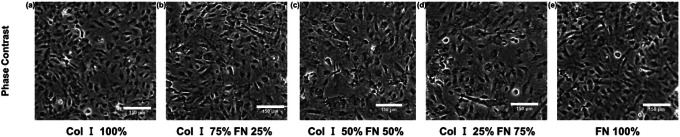
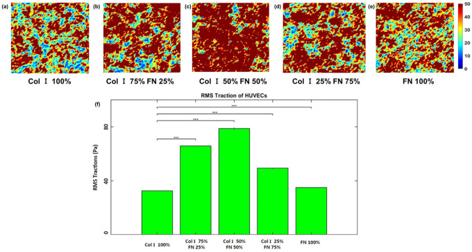
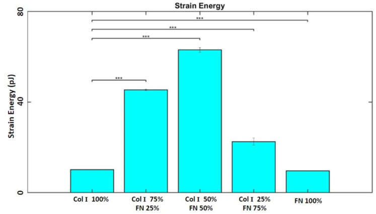
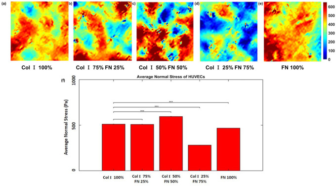
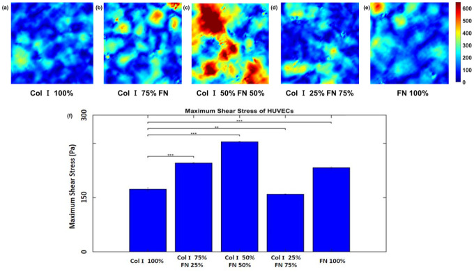
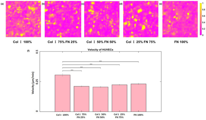
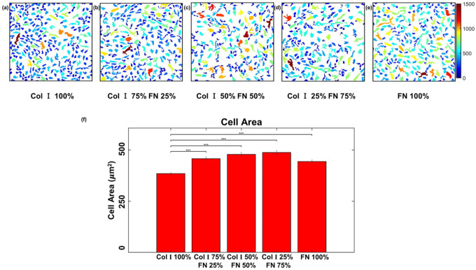
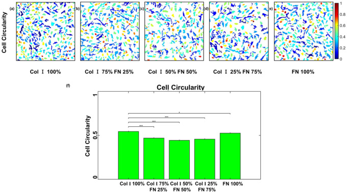
ECM composition is important in a host of pathophysiological processes such as angiogenesis, atherosclerosis, and diabetes, for example and during each of these processes ECM composition has been reported to change over time. However, the impact ECM composition has on the endothelium’s ability to respond mechanically is currently unknown. Therefore, in this study we seeded human umbilical vein endothelial cells (HUVECs) onto soft hydrogels coated with an ECM concentration of 0.1 mg/mL at the following collagen I (Col-I) and fibronectin (FN) ratios: 100%Col-I, 75%Col-I-25%FN, 50%Col-I-50%FN, 25%Col-I-75%FN, and 100%FN. We subsequently measured tractions, intercellular stresses, strain energy, cell morphology, and cell velocity. Our results revealed huvecs seeded on gels coated with 50% Col-I - 50% FN to have the highest intercellular stresses, tractions, strain energies, but the lowest velocities and cell circularity. Huvecs seeded on 100% Col-I had the lowest tractions, cell area while havingthe highest velocities and cell circularity. In addition, cells cultured on 25% Col-I and 75% FN had the lowest intercellular stresses, but the highest cell area. Huvecs cultured on 100% FN yielded the lowest strain energies. We believe these results will be of great importance to the cardiovascular field, biomedical field, and cell mechanics. Summary: Study the influence of different Col-I - FN ECM compositions on endothelial cell mechanics and morphology.


Conflict of interest statement

Conflict of Interest Statement

Ethics approval and consent to participate

Competing interests

The authors declare that they have no competing interests

Figures

Figure 1
Figure 1
Cropped HUVEC monolayer phase contrast images (within 651 × 651 μm2) for different Col-I and FN coating concentration ratios: Col-I 100% (a), Col-I 75% FN 25% (b), Col-I 50% FN 50% (c), Col-I 25% FN 75% (d), FN 100% (e).
Figure 2
Figure 2
RMS traction (within 651 × 651 μm2 cropped section) distributions (Pa) for different Col-I and FN coating concentration ratios: Col-I 100% (a), Col-I 75% FN 25% (b), Col-I 50% FN 50% (c), Col-I 25% FN 75% (d), FN 100% (e) and average RMS tractions (Pa) for different Col-I and FN coating concentrations based on averages from five samples for each ratio (f). * represents statistical significance (* p <= 0.05; ** p <= 1E-2; *** p<= 1E-3, no star p > 0.05).
Figure 3
Figure 3
Average strain energies (pJ) for different Col-I and FN coating concentrations based on averages from five samples for each ratio. * represents statistical significance (* p <= 0.05; ** p <= 1E-2; *** p<= 1E-3, no star p > 0.05).
Figure 4
Figure 4
Average normal stress (within 651 × 651 μm2 cropped section) distributions (Pa) for different Col-I and FN coating concentration ratios: Col-I 100% (a), Col-I 75% FN 25% (b), Col-I 50% FN 50% (c), Col-I 25% FN 75% (d), FN 100% (e) and average normal stress (Pa) for different Col-I and FN coating concentrations based on averages from five samples for each ratio (f). * represents statistical significance (* p <= 0.05; ** p <= 1E-2; *** p<= 1E-3, no star p > 0.05).
Figure 5
Figure 5
Maximum shear stress (within 651 × 651 μm2 cropped section) distributions (Pa) for different Col-I and FN coating concentration ratios: Col-I 100% (a), Col-I 75% FN 25% (b), Col-I 50% FN 50% (c), Col-I 25% FN 75% (d), FN 100% (e) and average maximum shear stress (Pa) for different Col-I and FN coating concentrations based on averages from five samples for each ratio (f). * represents statistical significance (* p <= 0.05; ** p <= 1E-2; *** p<= 1E-3, no star p > 0.05).
Figure 6
Figure 6
RMS velocity (within 651 × 651 μm2 cropped section) distributions (μm/min) for different Col-I and FN coating concentration ratios: Col-I 100% (a), Col-I 75% FN 25% (b), Col-I 50% FN 50% (c), Col-I 25% FN 75% (d), FN 100% (e) and average RMS (Pa) for different Col-I and FN coating concentrations based on averages from five samples for each ratio (f). * represents statistical significance (* p <= 0.05; ** p <= 1E-2; *** p<= 1E-3, no star p > 0.05).
Figure 7
Figure 7
HUVEC area in μm2 (within 651 × 651 μm2 cropped section) for different Col-I and FN coating concentration ratios: Col-I 100% (a), Col-I 75% FN 25% (b), Col-I 50% FN 50% (c), Col-I 25% FN 75% (d), FN 100% (e) and average cell area (μm2) for different Col-I and FN coating concentrations based on averages of 1057 to 1460 cells from five samples for each concentration ratio (f). * represents statistical significance (* p <= 0.05; ** p <= 1E-2; *** p<= 1E-3, no star p > 0.05).
Figure 8
Figure 8
HUVEC circularity computed as (4×Area×pi)/(Perimeter2) with a value of 1 for a perfect circle (within 651 × 651 μm2 cropped section) for different Col-I and FN coating concentration ratios: Col-I 100% (a), Col-I 75% FN 25% (b), Col-I 50% FN 50% (c), Col-I 25% FN 75% (d), FN 100% (e) and average cell area (μm2) for different Col-I and FN coating concentrations based on averages of 1057 to 1460 cells from five samples for each concentration ratio (f). * represents statistical significance (* p <= 0.05; ** p <= 1E-2; *** p<= 1E-3, no star p > 0.05).

References

    1. Gao Y. et al., “The ECM-cell interaction of cartilage extracellular matrix on chondrocytes,” (in eng), BiomedRes Int, vol. 2014, pp. 648459–648459, 2014, doi: 10.1155/2014/648459. - DOI - PMC - PubMed
    1. Wagenseil J. E. and P Mecham R., “Elastin in large artery stiffness and hypertension,” (in eng), J Cardiovasc Transl Res, vol. 5, no. 3, pp. 264–273, 2012, doi: 10.1007/s12265-012-9349-8. - DOI - PMC - PubMed
    1. Chiu J.-J. and Chien S., “Effects of Disturbed Flow on Vascular Endothelium: Pathophysiological Basis and Clinical Perspectives,” Physiological Reviews, vol. 91, no. 1, pp. 327–387, 2011, doi: 10.1152/physrev.00047.2009. - DOI - PMC - PubMed
    1. Chow M.-J., Turcotte R., P Lin C., and Zhang Y., “Arterial extracellular matrix: a mechanobiological study of the contributions and interactions of elastin and collagen,” (in eng), Biophysical journal, vol. 106, no. 12, pp. 2684–2692, 2014, doi: 10.1016/j.bpj.2014.05.014. - DOI - PMC - PubMed
    1. Sonbol H. S., “Extracellular matrix remodeling in human disease,” Journal of microscopy and ultrastructure, vol. 6, no. 3, p. 123, 2018. - PMC - PubMed

Publication types

LinkOut - more resources