[Preprint]. 2023 Jan 27:2023.01.27.525817.
doi: 10.1101/2023.01.27.525817.

Cannabidiol sensitizes TRPV2 channels to activation by 2-APB

Affiliations

Cannabidiol sensitizes TRPV2 channels to activation by 2-APB

Aaron Gochman et al. bioRxiv. .

Update in

Abstract

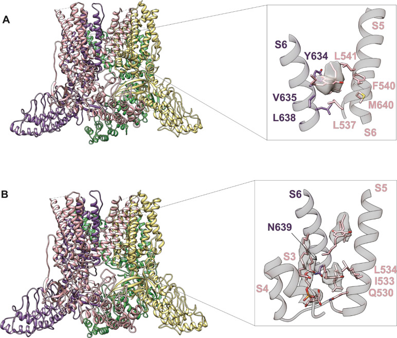
The cation-permeable TRPV2 channel is essential for cardiac and immune cells. Cannabidiol (CBD), a non-psychoactive cannabinoid of clinical relevance, is one of the few molecules known to activate TRPV2. Using the patch-clamp technique we discover that CBD can sensitize current responses of the rat TRPV2 channel to the synthetic agonist 2-aminoethoxydiphenyl borate (2- APB) by over two orders of magnitude, without sensitizing channels to activation by moderate (40 ⁰C) heat. Using cryo-EM we uncover a new small-molecule binding site in the pore domain of rTRPV2 that can be occupied by CBD in addition to a nearby CBD site that had already been reported. The TRPV1 and TRPV3 channels share >40% sequence identity with TRPV2 are also activated by 2-APB and CBD, but we only find a strong sensitizing effect of CBD on the response of mouse TRPV3 to 2-APB. Mutations at non-conserved positions between rTRPV2 and rTRPV1 in either the pore domain or the CBD sites failed to confer strong sensitization by CBD in mutant rTRPV1 channels. Together, our results indicate that CBD-dependent sensitization of TRPV2 channels engages multiple channel regions and possibly involves more than one CBD and 2-APB sites. The remarkably robust effect of CBD on TRPV2 and TRPV3 channels offers a promising new tool to both understand and overcome one of the major roadblocks in the study of these channels - their resilience to activation.


Figures

Figure 1.
Figure 1.. CBD strongly sensitizes rTRPV2 channels to activation by 2-APB in whole-cell and in excised patch recordings.
(A) Representative whole-cell gap-free current recording at −80mV from a cell expressing rTRPV2 channels. The colored horizontal lines denote the duration of exposure to test compounds, and the red dotted line denotes the zero-current level. The insert shows a magnified view of a segment of the recording. (B) Mean current-voltage relations recorded in control solution and in the presence of 0.5 mM 2-APB + 10 μM obtained from un-transfected cells in the whole-cell configuration (n = 8). (C) Representative current families elicited by voltage steps from −100 to +100 mV obtained from a rTRPV2-expressing cell exposed to control solution, 0.5 mM 2-APB or 10 μM CBD. The dotted lines indicate the zero-current level. (D) Mean current-voltage relations obtained from data as in (C) and normalized to the mean value at +100 mV in the presence of 0.5 mM 2-APB. Data are shown as mean ± SEM (n = 5). (E) Dose-response relation for rTRPV2 channel activation by CBD measured at −80 mV in the whole-cell configuration (mean ± SEM; n = 4). The continuous curve is a fit to the Hill equation with parameters: EC50 = 4.3 ± 1.4 μM and Hill coefficient (nH) = 1.7 ± 0.1. (F) Concentration-response relations for rTRPV1 channel activation by 2-APB at −80 mV in the whole-cell configuration measured in the absence (blue symbols) or presence (black symbols) of 10 μM CBD (mean ± SEM; n = 7). Hill equation parameters: no CBD, EC50 = 1.5 ± 0.05 mM, Hill coefficient (nH) = 3.3 ± 0.2; 10 μM CBD, EC50 = 159.9 ± 7.7 μM, Hill coefficient (nH) = 2.9 ± 0.3. (G) Representative whole-cell gap-free recording at −80 mV in a rTRPV2-expressing cell. The bottom-left insert shows group data for the mean current amplitude at each of the three stimulations with 6 mM 2-APB, normalized to the amplitude at the first stimulation with 0.5 mM 2-APB (solid squares – mean ± SEM, n = 5; empty circles – data from individual cells). The bottom-right insert displays group data measured over the different intervals that indicated by circled numbers on the current trace, and normalized to the first stimulation with 0.5 mM 2-APB. The empty circles are data from individual cells. (H) Group data for experiments in the whole-cell configuration as in (A) - first set of bars (n = 5), (G) - second set of bars (n = 5), or from outside-out patches as in Fig. 1 – Fig. Suppl. 3A – third set of bars (n = 6) or inside-out patches as in Fig. 1 – Fig. Suppl. 3B – fourth set of bars (n = 9). Data was normalized to the first stimulation with 0.5 mM 2-APB and shown as mean ± SEM or values from individual cells (empty circles). Figure 1 – source data 1. Excel file with group data from electrophysiological recordings shown in Figure 1.
Figure 2.
Figure 2.. CBD sensitization of rTRPV2 channels involves an increase in open probability.
(A) Data obtained from six rTRPV2-expressing inside-out patches at +80 mV under conditions of low open probability. Each row shows data from a different patch, and columns separate between recordings at different experimental conditions. 50 current sweeps of 500 ms duration are displayed vertically stacked at each experimental condition for each patch. Data points at each sweep are colored by their current amplitude as indicated by the color-bar at the top. (B) Data obtained from nine inside-out patches at +80 mV obtained from un-transfected cells, displayed as in (A). (C) Representative current traces from the same rTRPV2-expressing patch in the presence of 50 μM 2-APB and 10 μM CBD showing two distinct open current amplitudes (S1 and O1) and simultaneous opening of two channels (O2). Data points in each trace are colored as in (A). The red dotted line indicates the zero-current amplitude measured when all channels are closed. (D) All-points histograms for data in (A). Each vertical lane is a histogram, with the single-channel current amplitude bins along the y-axis and the number of points per bin shown as a log-scale color heat-map (see scale bar on the left) and normalized to the peak centered at 0 pA (black dotted line). Histograms from each patch are displayed in the same order as in (A). Asterisks at peak values of 8.5 or 10.5 pA indicate the patches where these single-channel amplitudes were observed.
Figure 3.
Figure 3.. Identifying CBD binding sites in TRPV2.
(A) Overall structure of the rTRPV2 channel in lipid nanodiscs in conformation A with one CBD molecule bound to each monomer (pdb ID 8FX8). Magnified view of the single CBD binding site was shown in the right panel. Both CBD and interacting residues ae presented in stick with the cryo-EM density (EMD-29526) corresponding to CBD shown as a white surface. (B) Overall structure of the rTRPV2 channel in lipid nanodiscs in conformation B with two CBD molecules bound to each monomer (pdb ID 8FXG). Magnified view of the two CBD binding sites are shown in the right panel. CBD with interacting residues and lipid were presented in stick and cryo-EM density (EMD-29532) corresponding to CBD and lipid are shown as a white surface.
Figure 4.
Figure 4.. CBD sensitizes rTRPV1 channels weakly and mTRPV3 channels strongly to activation by 2-APB.
(A) Representative whole-cell current families obtained from rTRPV1 expressing cells in control or in the presence of 10 μM CBD. Currents were elicited by voltage steps from −100 to +100 mV. The red dotted line denotes the zero-current level. (B) Steady-state current-voltage relations obtained from data as in (A) and normalized to the steady-state current magnitude at + 100 mV in CBD. Data is shown as mean ± SEM (n = 6). (C) Representative whole-cell current families from mTRPV3 expressing cells obtained as in (A). (D) Steady-state mTRPV3 channel current-voltage relations obtained from data as in (C) (mean ± SEM, n = 7). (E) Representative whole-cell recording at −80 mV obtained from a rTRPV1-expressing cell. (F) Group data from experiments as in (E) obtained using 10 or 40 μM CBD and normalized to the current magnitude measured at steady state during the first stimulation with 50 μM 2-APB. Data are shown as mean ± SEM (10 μM CBD, n = 6; 40 μM CBD, n = 5) or as individual cells (empty circles). (G) Representative whole-cell gap-free recording at −60 mV obtained from a cell expressing mTRPV3 channels stimulated with 2-APB and CBD. The first exposure to 2-APB alone (10 s duration) was used for normalization. (H) Representative experiment showing repeated stimulation with 2-APB. The first exposure to 2-APB (10 s duration) was used for normalization. Red curves are fits to a double-exponential function of time, with time-constants shown on (K) in magenta and yellow as mean ± SEM (filled symbols) or individual fit values (open symbols). (I, J) Representative experiments for sensitized responses to (I) 2-APB and CBD or (J) 2-APB alone measured after three exposures to 3 mM 2-APB applied at −10 mV to preserve patch integrity (dotted black lines). The first exposure to 2-APB (10 s duration) was used for normalization. The insert shows current amplitudes at each stimulation with 3 mM 2-APB (filled squares, mean ± SEM; empty circles, data from individual cells, n = 5). (K) Mean time-courses for mTRPV3 channel activation by 60 μM 2-APB in the absence (blue symbols) or presence of 10 μM CBD (black symbols), measured in experiments as in (G) and (H), as well as sensitized responses to 2-APB (light blue symbols) or 2-APB + CBD (grey symbols) measured as in (I) and (J). The time-course for the sensitized response to 2-APB alone does not start at t = 0 to account for the sensitizing exposures to 3 mM 2-APB. Data are shown as mean ± SEM (n = 5 for each condition). Data were fit to a mono-exponential function of time, with time constants shown as mean ± SEM (bars) or individual data points. The magenta curve is a fit to a mono-exponential function. Figure 4 – source data 1. Excel file with group data from electrophysiological recordings shown in Figure 4B and D. Figure 4 – source data 2. Excel file with group data from electrophysiological recordings shown in Figure 4F. Figure 4 – source data 3. Excel file with group data from electrophysiological recordings show in Figure 4I–K.
Figure 5.
Figure 5.. CBD binding site in TRPV2 channels and its conservation in TRPV1 channels.
(A) Structure of full-length rTRPV2 in nanodiscs with CBD bound in conformation A (with one CBD per subunit; pdb ID 8FX8). The S6 helix from one subunit is shown in orange, and in the adjacent subunit the S5 and S6 helices, the selectivity filter (S.F.), the pore-helix (P.H.), and the S4-S5 linker helix are shown in grey. Residues near the CBD site are shown in stick representation, and the bound CBD molecule in green. Residues that are similar in rTRPV2 and mTRPV3 channels but different in rTRPV1 channels are shown in purple. (B) Structure of apo full-length rTRPV2 in nanodiscs (pdb ID 6U84) (Pumroy et al., 2019). Same color coding as in A. (C) Structure of apo rTRPV1 (pdb ID 5IRZ) (Gao et al., 2016b) depicting the CBD binding region from rTRPV2. Same color coding as in A and B.
Figure 6.
Figure 6.. Role of non-conserved residues at the CBD site in determining rTRPV1 and rTRPV2 channel sensitivity to CBD and 2-APB.
(A-C) Representative gap-free whole-cell recordings at −80 mV obtained from cells expressing WT or mutant rTRPV2 channels. Horizontal bars denote the time of exposure to 2-APB (blue bars, mM concentrations) or CBD (green bars, μM concentrations). The red dotted line denotes the zero-current level. (D-F) Representative gap-free whole-cell recordings at −80 mV obtained from cells expressing WT or mutant rTRPV1 channels. The inserts show magnified views of the current traces boxed by the dotted black lines. (G, squares) Group data for WT and mutant rTRPV1 (empty symbols) and rTRPV2 (filled symbols) channels obtained from experiments as in (A-F) showing the relation between the normalized sensitized response to 2-APB (ICBD+2APB/I2APB, left axis) and the maximal response to 2-APB (I2APB,max/I2APB, bottom axis). The blue line denotes ICBD+2APB/I2APB = 1 where the response to 2-APB in the presence of CBD is the same as in its absence (i.e. no sensitization). Data are shown as mean ± SEM (WT rTRPV2, n = 4; L538C, n = 4; L541M, n = 4; L538C+L541M, n = 4; WT rTRPV1, n = 5; M572V, n = 5; C578L, n = 5; M581L, n = 5; C578L+M581L, n = 5; M572V+C578L+M581L, n = 6). (G, circles) Relation between the EC50 for channel activation by 2-APB (right axis) and the concentration of 2-APB used in experiments as in (A-F) to probe sensitization by CBD (top axis) in WT and mutant rTRPV1 (empty symbols) and rTRPV2 (filled symbols) channels. The EC50 values for WT rTRPV1 and rTRPV2 channels are highlighted by grey dotted and solid lines, respectively. EC50 data obtained from fits to the Hill equation and shown as mean ± SEM (WT rTRPV2, n = 5; L538C, n = 4; L541M, n = 4; L538C+L541M, n = 3; WT rTRPV1, n = 5; M572V, n = 5; C578L, n = 5; M581L, n = 5; C578L+M581L, n = 4; M572V+C578L+M581L, n = 4; dose-response relations shown in Fig. 6 – Fig. Suppl. 1E and H). (H) Mean sensitized response of WT and rTRPV2 channel mutants to 2-APB (ICBD+2APB) relative to the maximal activation by 2-APB (I2APB,max) as a function of the concentration of CBD that was used to sensitize channels, obtained from experiments as in (A-C). Data for WT rTRPV1 is also shown as reference. (I) Mean sensitized response to 2-APB (ICBD+2APB) relative to the non-sensitized response (I2APB) shown as a function of the concentration of CBD that was used to sensitize channels, obtained from experiments as in (D-F). Figure 6 – source data 1. Excel file with group data from electrophysiological recordings of CBD-dependent sensitization shown in Figure 6G-H, obtained from experiments as in (A)-(F). These data are also displayed on Figure 6 – Supplement 1F–G. Figure 6 – source data 2. Excel file with 2-APB dose-response relation data for rTRPV1 and rTRPV2 mutants shown in Figure 6G. These data are also displayed in Figure 6 – Fig. Supplement 1E, H and I.
Figure 7.
Figure 7.. Cumulative substitutions at non-conserved positions in the rTRPV1 pore do not enhance sensitization.
Representative time courses for (A) rTRPV1-12M channels (E570Q + M572V + C578L + M581L + S592A + T593V + V595L + T597S + S632D + T633A + T650Q + I660L) or (B) rTRPV1-14M channty54rels (E570Q + M572V + C578L + M581L + S592A + T593V + V595L + T597S + S632D + T633A + Y653L + D654R + T650Q + I660L) displaying sensitization by CBD of the response to 12 mM 2-APB. The red dotted line indicates the zero-current level. The insert in (B) is a magnification of the region within the dotted box. Concentrations of 60 or 100 μM CBD were tested in separate experiments for rTRPV1-12M and in the same experiment for rTRPV1-14M. (C) Group data for the experiments in (A) and (B), showing leak-subtracted current responses to each of the stimuli, normalized to the current magnitude in the presence of 12 mM 2-APB. Data points from individual experiments are shown as open symbols. The triangles are not normalized capsaicin response in the experiments for rTRPV1-12M and 100 μM CBD. Data are shown as mean ± SEM (n = 4 for rTRPV1-12M and 60 or 100 μM CBD; n = 5 for rTRPV1-14M). Figure 7 – source data 1. Excel file with group data from electrophysiological recordings of CBD-dependent sensitization shown in Figure 7.

References

    1. Adams P. D.; Afonine P. V.; Bunkóczi G.; Chen V. B.; Davis I. W.; Echols N.; Headd J. J.; Hung L.-W.; Kapral G. J.; Grosse-Kunstleve R. W.; et al. PHENIX: A Comprehensive Python-Based System for Macromolecular Structure Solution. Acta Crystallogr. D Biol. Crystallogr. 2010, 66, 213–221. - PMC - PubMed
    1. Bai D.; del Corsso C.; Srinivas M.; Spray D. C. Block of Specific Gap Junction Channel Subtypes by 2-Aminoethoxydiphenyl Borate (2-APB). J. Pharmacol. Exp. Ther. 2006, 319, 1452–1458. - PubMed
    1. Bang S.; Kim K. Y.; Yoo S.; Lee S.-H.; Hwang S. W. Transient Receptor Potential V2 Expressed in Sensory Neurons Is Activated by Probenecid. Neurosci. Lett. 2007, 425, 120–125. - PubMed
    1. Belkacemi A.; Trost C. F.; Tinschert R.; Flormann D.; Malihpour M.; Wagner C.; Meyer M. R.; Beck A.; Flockerzi V. The TRPV2 Channel Mediates Ca2+ Influx and the Δ9-THC-Dependent Decrease in Osmotic Fragility in Red Blood Cells. Haematologica 2021, 106, 2246–2250. - PMC - PubMed
    1. Bisogno T.; Hanus L.; De Petrocellis L.; Tchilibon S.; Ponde D. E.; Brandi I.; Moriello A. S.; Davis J. B.; Mechoulam R.; Di Marzo V. Molecular Targets for Cannabidiol and Its Synthetic Analogues: Effect on Vanilloid VR1 Receptors and on the Cellular Uptake and Enzymatic Hydrolysis of Anandamide. Br. J. Pharmacol. 2001, 134, 845–852. - PMC - PubMed

Publication types

LinkOut - more resources