[Preprint]. 2023 Jan 26:rs.3.rs-2402606.
doi: 10.21203/rs.3.rs-2402606/v1.

Targeting Immune-Fibroblast Crosstalk in Myocardial Infarction and Cardiac Fibrosis

Affiliations

Targeting Immune-Fibroblast Crosstalk in Myocardial Infarction and Cardiac Fibrosis

Junedh M Amrute et al. Res Sq. .

Update in

  • Targeting immune-fibroblast cell communication in heart failure.
    Amrute JM, Luo X, Penna V, Yang S, Yamawaki T, Hayat S, Bredemeyer A, Jung IH, Kadyrov FF, Heo GS, Venkatesan R, Shi SY, Parvathaneni A, Koenig AL, Kuppe C, Baker C, Luehmann H, Jones C, Kopecky B, Zeng X, Bleckwehl T, Ma P, Lee P, Terada Y, Fu A, Furtado M, Kreisel D, Kovacs A, Stitziel NO, Jackson S, Li CM, Liu Y, Rosenthal NA, Kramann R, Ason B, Lavine KJ. Amrute JM, et al. Nature. 2024 Nov;635(8038):423-433. doi: 10.1038/s41586-024-08008-5. Epub 2024 Oct 23. Nature. 2024. PMID: 39443792 Free PMC article.

Abstract

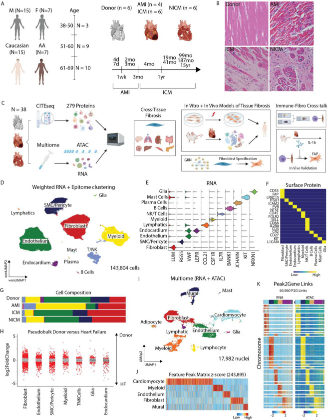
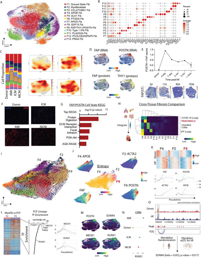
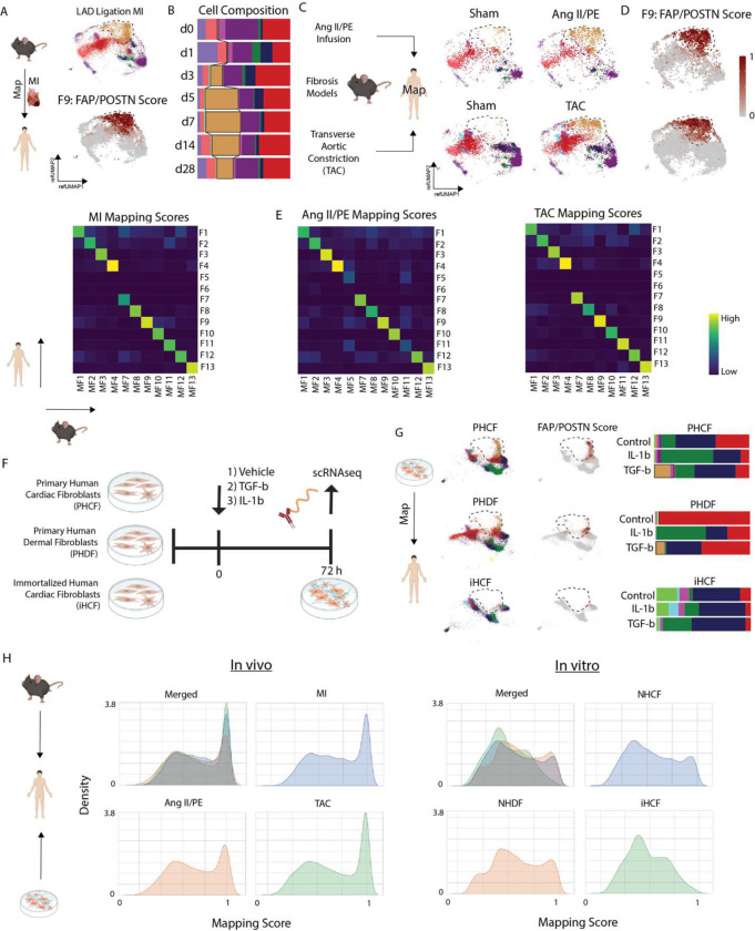
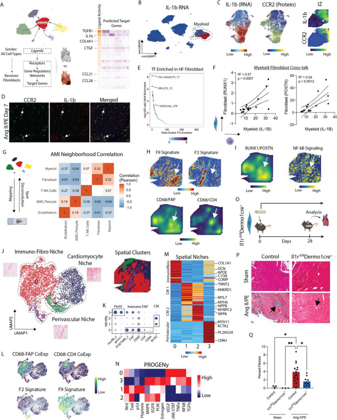
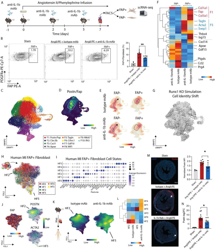
Inflammation and tissue fibrosis co-exist and are causally linked to organ dysfunction. However, the molecular mechanisms driving immune-fibroblast crosstalk in human cardiac disease remains unexplored and there are currently no therapeutics to target fibrosis. Here, we performed multi-omic single-cell gene expression, epitope mapping, and chromatin accessibility profiling in 38 donors, acutely infarcted, and chronically failing human hearts. We identified a disease-associated fibroblast trajectory marked by cell surface expression of fibroblast activator protein (FAP), which diverged into distinct myofibroblasts and pro-fibrotic fibroblast populations, the latter resembling matrifibrocytes. Pro-fibrotic fibroblasts were transcriptionally similar to cancer associated fibroblasts and expressed high levels of collagens and periostin (POSTN), thymocyte differentiation antigen 1 (THY-1), and endothelin receptor A (EDNRA) predicted to be driven by a RUNX1 gene regulatory network. We assessed the applicability of experimental systems to model tissue fibrosis and demonstrated that 3 different in vivo mouse models of cardiac injury were superior compared to cultured human heart and dermal fibroblasts in recapitulating the human disease phenotype. Ligand-receptor analysis and spatial transcriptomics predicted that interactions between C-C chemokine receptor type 2 (CCR2) macrophages and fibroblasts mediated by interleukin 1 beta (IL-1β) signaling drove the emergence of pro-fibrotic fibroblasts within spatially defined niches. This concept was validated through in silico transcription factor perturbation and in vivo inhibition of IL-1β signaling in fibroblasts where we observed reduced pro-fibrotic fibroblasts, preferential differentiation of fibroblasts towards myofibroblasts, and reduced cardiac fibrosis. Herein, we show a subset of macrophages signal to fibroblasts via IL-1β and rewire their gene regulatory network and differentiation trajectory towards a pro-fibrotic fibroblast phenotype. These findings highlight the broader therapeutic potential of targeting inflammation to treat tissue fibrosis and restore organ function.

Keywords: C-C chemokine receptor 2; fibroblast activator protein; fibrosis; heart failure; interleukin 1 beta; macrophages.


Conflict of interest statement

Competing Interests XL, TY, SS, AF, MF, C-ML and BA are or were employed by Amgen.

Figures

Figure 1.
Figure 1.
Study design and integrated multi-omic characterization of human MI and HF. (A) Multiethnic diverse patient demographics. (B) Representative H&E sections from donor, acute MI, ischemic cardiomyopathy, and non-ischemic cardiomyopathy. (C) Study design from multi-omic human sequencing derived hypothesis, cross-tissue fibrosis analysis, in vitro and in vivo experimental characterization, immune-fibroblast crosstalk, and in vivo validation studies. (D) UMAP embedding plot with weighted nearest neighbor clustering (RNA and protein) of CITE-seq data from 22 patients and 143,804 cells. (E) Violin plot for canonical marker genes for cell types. (F) Heatmap of marker proteins for cell types. (G) Cell cluster composition across conditions. (H) Pseudobulk differentially expressed genes between donor and HF samples in major cell types (bottom). Red dots indicate statistically significant genes (log2FC > 0.58 and adjusted p-value < 0.05). (I) UMAP embedding plot of RNA from Multiome (paired RNA and ATAC) specimens from 9 patients and 17,982 nuclei. (J) Marker peaks for the major cell types annotated in (I) with FDR < 0.1 and log2FC > 0.5. (K) Peak to Gene linkage heatmap using paired RNA and ATAC information from the same nucleus.
Figure 2.
Figure 2.
FAP fibroblasts expand post-MI into a PF lineage with a MEOX1/RUNX1 orchestrated gene regulation network. (A) UMAP embedding of CITE-seq fibroblast cell states. (B) Dot Plot of marker genes for fibroblast cell states. (C) Fibroblast cell state composition across four groups (left) and Gaussian kernel density estimation of cells across four groups (right). (D) FAP RNA (top left), POSTN RNA (top right), FAP protein (bottom left), and THY1 protein (bottom right). (E) FAP/POSTN z-score in donor, acute MI, and ICM split by time from MI with spatial transcriptomics samples showing FAP expression at different time points post-MI in human LV specimens. (F) Immunofluorescence of FAP in donor, acute MI (4 days post-MI), ICM, and NICM left ventricle myocardium. (G) Top KEGG pathways for FAP/POSTN cell state. (H) Cross-tissue fibrosis Pearson correlation coefficient between human heart fibroblast cell states and those from other disease contexts. (I) scvelo analysis in force directed layout embedding and (J) Palantir derived entropy with terminal cell states noted and marker genes for terminal states in addition to FAP plotted in FDL embedding highlighting FAP derived fibroblasts diverge into myofibroblats and PFs. (K) Heatmap of pseudobulk differentially expressed genes between donor and HF and terminal state marker genes over pseudotime. (L) Heatmap of differentially expressed genes between F2 and F9 fibroblast cell states (left), TF enrichment analysis for F9 lineage using enrichR (TF-Gene Co-occurrence database) shows key epigenetic regulators of the F9 lineage (middle), and RUNX1/MEOX1 gene expression across pseudotime for 3 lineages showing increased expression along F9 lineage (right). (M) Gene expression density plot in UMAP embedding for POSTN, RUNX1, EDNRA, and MEOX1 (markers distinguishing F2 and F9 lineages). (N) CellOracle gene regulatory network betweenness centrality score for RUNX1 by HF etiology – a higher score indicates that the TF has a greater influence on informational flow in the GRN. (O) ATAC-seq pseudobulk tracts from multiome fibroblasts with peak2gene linkages showing EDNRA locus split by donor and HF; Mendelian Randomization shows that there was a significant positive association between genetically predicted EDNRA gene expression levels and HF in fibroblasts (beta = 0.052, se = 0.022, and p-value = 0.017).
Figure 3.
Figure 3.
Comparison of in vivo and in vitro models to study cardiac tissue fibrosis. (A) Reference mapped mouse MI fibroblasts onto human CITE-seq space with label transfer (top) and prediction score for cells annotated in POSTN/FAP cluster. (B) Cell composition from label-transferred cell states post-MI. (C) Reference mapped mouse fibrosis model fibroblasts from Ang II/PE and TAC split by sham and injury. (D) Prediction score for cells annotated in POSTN/FAP cluster from fibrosis models. (E) Heatmap of average cell prediction score of human fibroblast cell states (y-axis) in annotated mouse fibroblasts (x-axis) in MI, Ang II/PE, and TAC. (F) Experimental set-up of in vitro model. (G) Reference mapped in vitro cell lines post-stimuli (control, TGF-b, and IL-1b) onto human cardiac fibroblasts and corresponding cell composition. (H) Density plot of maximum prediction score for each cell from in vivo and in vitro systems merged and split by in vivo or in vitro condition.
Figure 4.
Figure 4.. CCR2 macrophages co-localize with and signal to fibroblasts via IL-1b in MI and HF.
(A) NicheNet derived predictions of enriched signaling from other cell types to fibroblasts in HF (acute MI, ICM, NICM) relative to donor. (B) IL-1b RNA expression in global CITE-seq UMAP embedding. (C) IL-1b RNA expression in myeloid subsets (left) with corresponding CCR2 protein expression (middle), and IL-1b expression density and imputed CCR2 protein expression in an acute MI IZ spatial transcriptomic sample. (D) Immunofluorescence of IL-1b (red) in CCR2 GFP (green) mice at day 7 post Ang II/PE mini-pump implantation. (E) Rank sorted TF motifs enriched in HF fibroblasts relative to donor fibroblasts from Multiome data show enriched inflammatory signaling. (F) Average expression of IL-1B in macrophages per HF patient versus RUNX1 (left) and POSTN (right) in fibroblasts from the same patient. R2 indicates the regression coefficient and the p-value tests whether the slope is significantly non-zero. (G) SPOTlight derived cell-cell proportion correlation in acute MI patient. (H) z-score for marker genes in F9 (POSTN, COMP, FAP, COL1A1, THBS4, COL3A1) and F2 (ACTA2, TAGN) (top) and CD68/CD4 and CD68/FAP joint density embedding plot (bottom). (I) RUNX1/POSTN co-localization density plot (left) and PROGENy imputed NF-kB pathway score (right). (J) UMAP embedding plot of spatial clusters with three distinct niches and corresponding spatial location of clusters. (K) Dot plot of marker genes and gene set scores for spatial niches. (L) CD68/CD4, CD68/FAP (top) and F2 and F9 fibroblast gene signature (bottom) joint density embedding plot in UMAP embedding. (M) Heatmap of top differentially expressed genes across spatial niches. (N) PROGENy pathway analysis for spatial clusters in (J). (O) Study design for IL-1 receptor deletion in fibroblasts in mice with Ang II/PE minipump implantation, (P) representative images of trichrome staining, and (Q) quantification of fibrosis at day 28.
Figure 5.
Figure 5.
(A) Experimental workflow for cardiac injury experiment with treatment of an IL-1b neutralizing antibody or isotype control. (B) Flow cytometry gating of PDPN vs FAP in a representative sham, Ang II/PE + Isotype, and Ang II/PE + anti-IL-1b mAb mouse heart (left) and FAP+ fibroblasts/total fibroblasts quantified (right). Unpaired t-test and **P = 0.0031 (isotype vs anti-IL-1b mAb). (C) Integrated UMAP for isotype and anti-IL-1b mAb treated mice FAP+/FAP− fibroblasts with annotated clusters. (D) Density plot of Fap/Postn co-localization with area of maximal expression highlighted. (E) Gaussian kernel density plots of 4 conditions in integrated UMAP embedding. (F) Heatmap of key fibroblast cell state genes grouped by four conditions with rows clustered by similarity. (G) CellOracle Runx1 in silico knockout simulated cell identify shift shows a shift away from Fap/Postn fibroblasts. (H) scVelo of human FAP+ fibroblasts in a FDL embedding space colored by re-clustered data. (I) Top marker genes for FAP+ human CITE-seq re-clustered data from (F). (J) FAP, POSTN, and ACTA2 expression in human FAP+ fibroblasts in FDL embedding. (K) Mapping mouse differentially expressed signature between FAP+ isotype and anti-IL-1b mAb treated mice. (L) Heatmap of gene set signature for HF2 (THBS4, POSTN, TNC, APOE, and PENK), isotype treated, HF5 (ACTA2 and TAGLN), and anti-IL-1b mAb treated grouped by FAP+ human fibroblast cell state. (M) IF of POSTN in a representative sham, Ang II/PE + Isotype, and Ang II/PE + anti-IL-1b mAb heart (left) with quantification (right). Unpaired t-test and *P = 0.0243. (N) RT-PCR of Postn in mouse hearts split by 3 conditions. Unpaired t-test and *P = 0.0392.

References

    1. Gutstein D. E. & Fuster V. Pathophysiology and clinical significance of atherosclerotic plaque rupture. Cardiovasc. Res. 41, 323–333 (1999). - PubMed
    1. Thygesen K. et al. Third universal definition of myocardial infarction. Circulation 126, 2020–2035 (2012). - PubMed
    1. Roger V. L. Epidemiology of Heart Failure. Circ. Res. 113, 646 (2013). - PMC - PubMed
    1. Gómez A. M., Guatimosim S., Dilly K. W., Vassort G. & Lederer W. j. Heart Failure After Myocardial Infarction. Circulation 104, 688–693 (2001). - PubMed
    1. Nowbar A. N., Gitto M., Howard J. P., Francis D. P. & Al-Lamee R. Mortality from ischemic heart disease: Analysis of data from the world health organization and coronary artery disease risk factors from NCD risk factor collaboration. Circ. Cardiovasc. Qual. Outcomes 12, (2019). - PMC - PubMed

Publication types

LinkOut - more resources