

Pseudogout of the lumbar spine
- PMID: 36747909
- PMCID: PMC9898284
- DOI: 10.1016/j.radcr.2022.10.057
Pseudogout of the lumbar spine
Abstract
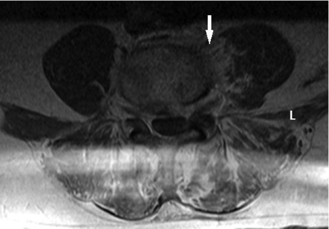
Calcium pyrophosphate deposition involves deposition of calcium pyrophosphate dihydrate crystals in various joints throughout the body. The term "pseudogout" refers to an acute attack of calcium pyrophosphate crystal-induced arthritis. Though clinical presentation and joint involvement vary, involvement of the lumbar spine is rare. We present the case of a 61-year-old male who presented with 3 days of worsening lower back pain. He had elevated inflammatory markers, leukocytosis, and spinal tenderness on exam. Magnetic resonance imaging of the lumbar spine showed likely L4-L5 osteomyelitis; however, biopsy of the disk space revealed extensive calcium pyrophosphate crystal deposition. The patient was treated with prednisone taper with alleviation of symptoms. Though pseudogout of the spine is rare, our report supports literature urging clinicians to consider pseudogout when assessing elderly patients with back pain for prompt and appropriate treatment.
© 2022 The Authors. Published by Elsevier Inc. on behalf of University of Washington.
Figures

References
-
- Sivera F, Andres M, Pascual E. Calcium pyrophosphate crystal deposition. Int J Clin Rheumtol. 2011;6:677–688. doi: 10.2217/ijr.11.60. - DOI
Publication types
LinkOut - more resources
Full Text Sources
