

Testosterone Propionate Promotes Proliferation and Viability of Bone Marrow Mesenchymal Stem Cells while Preserving Their Characteristics and Inducing Their Anti-Cancer Efficacy
- PMID: 36748249
- PMCID: PMC9998833
- DOI: 10.4274/balkanmedj.galenos.2022.2022-10-21
Testosterone Propionate Promotes Proliferation and Viability of Bone Marrow Mesenchymal Stem Cells while Preserving Their Characteristics and Inducing Their Anti-Cancer Efficacy
Abstract
Background: Various studies have reported the effects of testosterone on different cell types, yet bone marrow-derived mesenchymal stem cells’ cellular responses to testosterone remain unknown.
Aims: To investigate the effects of testosterone propionate, an oil-soluble short-acting form of testosterone, on human bone marrow-derived mesenchymal stem cells’ proliferation and viability after 24 hours of incubation. We also investigated the impact of testosterone propionate on bone marrow-derived mesenchymal stem cell’s polarization and cytotoxicity on K562 leukemia cell line.
Study design: In vitro study.
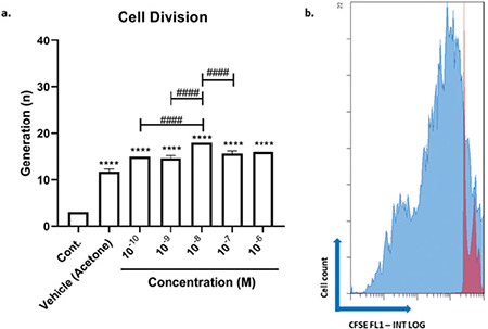
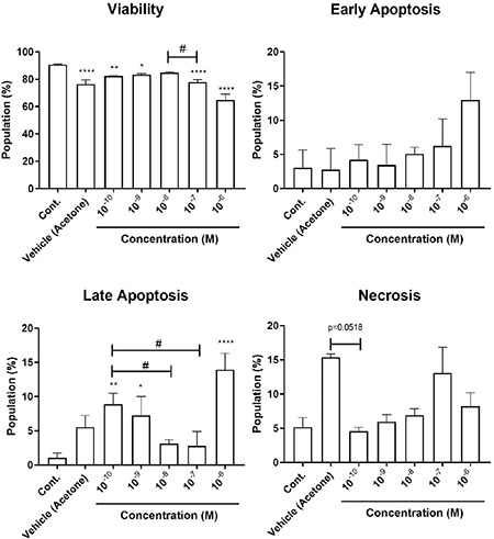
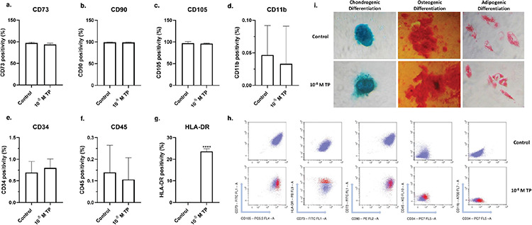
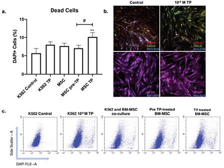
Methods: We expanded commercially available bone marrow derived mesenchymal stem cells in vitro and treated them with testosterone propionate at concentrations ranging from 10-6-10-10 M for 24 hours. Ideal concentration was determined by evaluating cellular viability and proliferation with Annexin V/Propidium Iodide assay and carboxyfluorescein succinimidyl ester staining. The characteristic features of bone marrow-derived mesenchymal stem cells were evaluated by immunophenotyping and investigating their differentiation capacities. Bone marrow-derived mesenchymal stem cells’ cytotoxic properties upon testosterone propionate treatment were determined by co-culturing the cells with K562 cells and with confocal imaging investigating polarization.
Results: Testosterone propionate promoted proliferation and maintained the viability of bone marrow-derived mesenchymal stem at 10-8 M concentration. Further evaluations were conducted with the determined dose. The results showed that, apart from promoting mesenchymal stem cells’ polarization and increasing their cytotoxicity on K562 cells, testosterone propionate did not alter differentiation capacities of bone marrow-derived mesenchymal stem cells and certain cell surface markers, but led to a significant increase in HLA-DR expression.
Conclusion: The findings reveal that testosterone propionate promotes the proliferation and survival of bone marrow-derived mesenchymal stem cells in a dose-dependent manner without hampering their differentiation capacities, induces their polarization to the pro-inflammatory phenotype, and increases their cytotoxicity on the K562 cell line.
Conflict of interest statement
Figures
Similar articles
-
Alteration of cellular and immune-related properties of bone marrow mesenchymal stem cells and macrophages by K562 chronic myeloid leukemia cell derived exosomes.J Cell Physiol. 2019 Apr;234(4):3697-3710. doi: 10.1002/jcp.27142. Epub 2018 Oct 14. J Cell Physiol. 2019. PMID: 30317554
-
Inhibitory effect of alcohol on osteogenic differentiation in human bone marrow-derived mesenchymal stem cells.Alcohol Clin Exp Res. 2004 Mar;28(3):468-79. doi: 10.1097/01.alc.0000118315.58404.c1. Alcohol Clin Exp Res. 2004. PMID: 15084905
-
Testosterone propionate can promote effects of acellular nerve allograft-seeded bone marrow mesenchymal stem cells on repairing canine sciatic nerve.J Tissue Eng Regen Med. 2019 Sep;13(9):1685-1701. doi: 10.1002/term.2922. Epub 2019 Jul 18. J Tissue Eng Regen Med. 2019. PMID: 31267700
-
Resistance of human primary mesenchymal stem cells to cytotoxic effects of nutlin-3 in vitro.J Cell Biochem. 2020 Jan;121(1):788-796. doi: 10.1002/jcb.29324. Epub 2019 Aug 26. J Cell Biochem. 2020. PMID: 31452266
-
Glycyrrhizin Enhances the Proliferation of Diabetic Bone Marrow-derived Mesenchymal Stem Cells: A Potential Therapeutic Agent in Endodontic Surgery.J Contemp Dent Pract. 2023 Jul 1;24(7):494-499. doi: 10.5005/jp-journals-10024-3536. J Contemp Dent Pract. 2023. PMID: 37622629
Cited by
-
Mesenchymal stem cell transplantation plays a role in relieving cancer pain.Front Pharmacol. 2024 Nov 29;15:1483716. doi: 10.3389/fphar.2024.1483716. eCollection 2024. Front Pharmacol. 2024. PMID: 39679363 Free PMC article. Review.
-
Effects of different ratio of sweet sorghum silage to corn silage diets on serum metabolome of lactating dairy cows.Trop Anim Health Prod. 2025 May 24;57(5):229. doi: 10.1007/s11250-025-04483-8. Trop Anim Health Prod. 2025. PMID: 40411653
References
-
- Friedenstein AJ, Petrakova KV, Kurolesova AI, Frolova GP. Heterotopic of bone marrow. Analysis of precursor cells for osteogenic and hematopoietic tissues. Transplantation. 1968;6:230–247. - PubMed
-
- Dominici M, Le Blanc K, Mueller I, et al. Minimal criteria for defining multipotent mesenchymal stromal cells. The International Society for Cellular Therapy position statement. Cytotherapy. 2006;8:315–317. - PubMed
-
- Crisostomo PR, Wang Y, Markel TA, et al. Human mesenchymal stem cells stimulated by TNF-alpha, LPS, or hypoxia produce growth factors by an NF kappa B- but not JNK-dependent mechanism. Am J Physiol Cell Physiol. 2008;294:C675–C682. - PubMed
-
- Caplan AI, Dennis JE. Mesenchymal stem cells as trophic mediators. J Cell Biochem. 2006;98:1076–1084. - PubMed
MeSH terms
Substances
LinkOut - more resources
Full Text Sources
Medical
Research Materials