. 2023 Mar;10(8):e2207301.
doi: 10.1002/advs.202207301. Epub 2023 Feb 7.

A Monoclonal Human Alveolar Epithelial Cell Line ("Arlo") with Pronounced Barrier Function for Studying Drug Permeability and Viral Infections

Affiliations

A Monoclonal Human Alveolar Epithelial Cell Line ("Arlo") with Pronounced Barrier Function for Studying Drug Permeability and Viral Infections

Patrick Carius et al. Adv Sci (Weinh). 2023 Mar.

Abstract

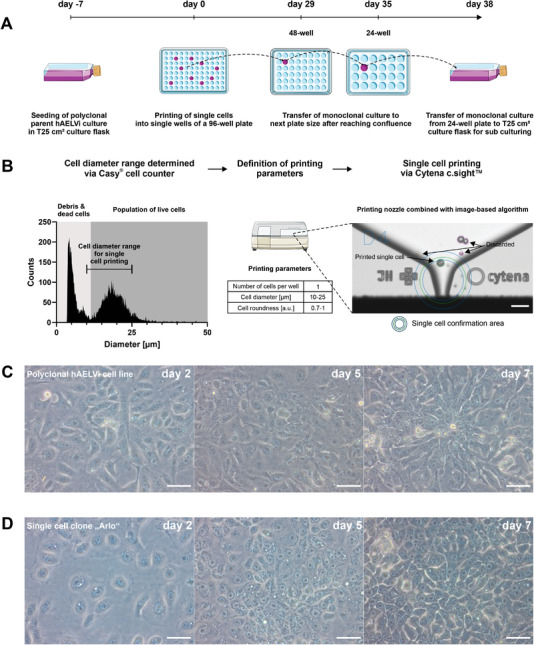
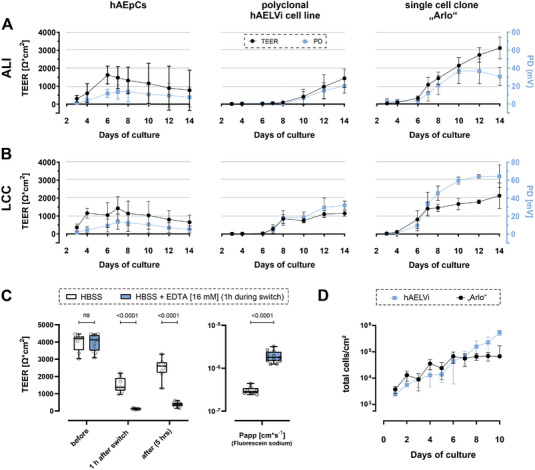
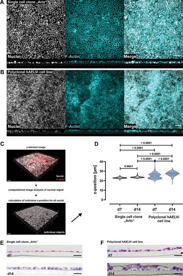
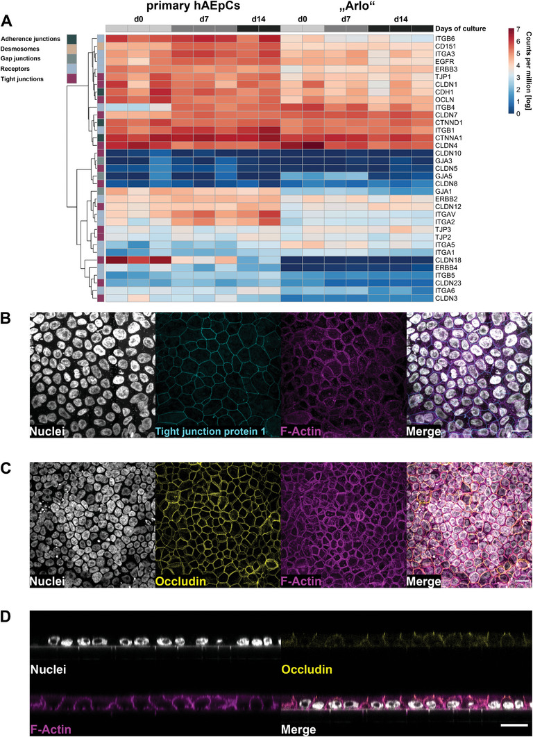
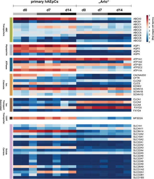
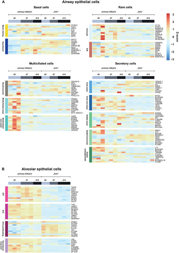
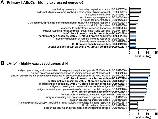
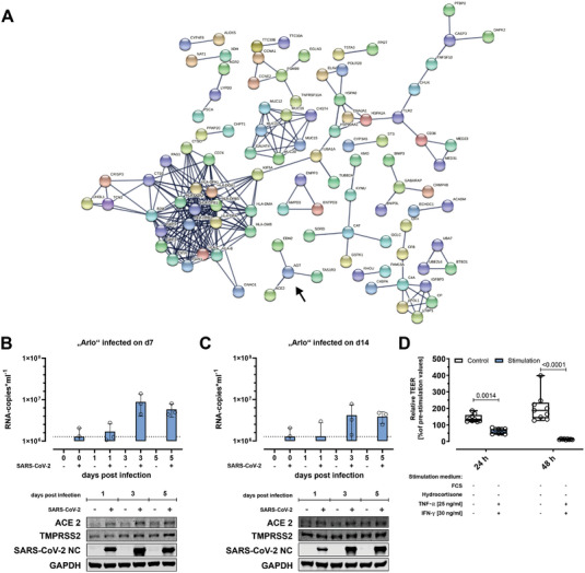
In the development of orally inhaled drug products preclinical animal models regularly fail to predict pharmacological as well as toxicological responses in humans. Models based on human cells and tissues are potential alternatives to animal experimentation allowing for the isolation of essential processes of human biology and making them accessible in vitro. Here, the generation of a novel monoclonal cell line "Arlo," derived from the polyclonal human alveolar epithelium lentivirus immortalized cell line hAELVi via single-cell printing, and its characterization as a model for the human alveolar epithelium as well as a building block for future complex in vitro models is described. "Arlo" is systematically compared in vitro to primary human alveolar epithelial cells (hAEpCs) as well as to the polyclonal hAELVi cell line. "Arlo" cells show enhanced barrier properties with high transepithelial electrical resistance (TEER) of ≈3000 Ω cm2 and a potential difference (PD) of ≈30 mV under air-liquid interface (ALI) conditions, that can be modulated. The cells grow in a polarized monolayer and express genes relevant to barrier integrity as well as homeostasis as is observed in hAEpCs. Successful productive infection with severe acute respiratory syndrome coronavirus 2 (SARS-CoV-2) in a proof-of-principle study offers an additional, attractive application of "Arlo" beyond biopharmaceutical experimentation.

Keywords: Transwell inserts; drug transport; lung; pulmonary drug delivery; tight junctions.


Conflict of interest statement

C.‐M.L., N.S.‐D., and P.C. are the creators of the cell line “Arlo”. A manufacture and distribution license for the cell line “Arlo” was granted to InSCREENeX GmbH, Germany by the Helmholtz Centre for Infection Research (Helmholtz‐Zentrum für Infektionsforschung GmbH) (HZI), Germany. The authors declare that the research was conducted in the absence of any commercial or financial relationships that could be construed as a potential conflict of interest.

Figures

Figure 1
Figure 1
Generation of the single cell clone “Arlo.” A) Schematic depicting the single‐cell printing procedure (detailed in B) and subsequent passaging strategy for the single‐cell clone “Arlo” originating from a polyclonal hAELVi suspension that demonstrated TEER values >1000 Ω cm2 in the previous passage. B) Before single‐cell printing, the cell diameter of the polyclonal hAELVi cell suspension was determined via a Casy cell counter to define the printing parameters. Single cells within the printing parameters (bordered green) were confirmed by an image‐based algorithm and then deposited into a single well of a 96‐well plate. Cells that did not meet the printing criteria were discarded via vacuum aspiration after ejection from the printing nozzle (bordered purple or red). C,D) Light microscopic images showing morphological differences between C) the polyclonal hAELVi cell line and D) the single cell clone “Arlo” when cultured in T25 cm2 culture flasks for 7 d. Scale: 50 µm for all images displayed. Panel A) was partly generated using Servier Medical Art, provided by Servier, licensed under a Creative Commons Attribution 3.0 unported license.
Figure 2
Figure 2
Electrophysiological and functional characterization of barrier properties of the single cell clone “Arlo.” A,B) TEER values (Ω cm2, black curve) as well as epithelial potential difference (PD) (mV, blue curve) of hAEpCs, the polyclonal hAELVi cell line and the single cell clone “Arlo” grown on Transwell inserts either under A) ALI or B) LCC conditions for 14 d. Data represent mean ± S.D. from 12 TWs and three independent biological replicates (hAEpCs day 4: 8 TWs; 2 bio. replicates). C) Apparent permeability of Fluorescein sodium (1 mg mL−1 in HBSS) transported over “Arlo” monolayers after 14 d of culture under LCC. TEER values were measured with cells cultured in the medium before the experiment (before), 1 h after the incubation in transport buffer (1 h after switch), as well as after the transport study (after). Transport buffer (HBSS) was supplemented with EDTA (16 × 10−3 m) during the 1 h incubation to disrupt tight junction complexes in one group. (TEER) 2‐way ANOVA was performed not assuming sphericity and with a Šídák´s multiple comparisons test. (P app) Unpaired t‐test was performed with Welch's Correction; Data represent mean ± S.D.; HBSS: n = 9, HBSS+EDTA: n = 12 from 3 independent biological replicates. D) Growth curve comparing the polyclonal hAELVi cell line with the single cell clone “Arlo”. Data represent mean ± S.D. from at least 2 technical replicates of 3 biological replicates (“Arlo”d10: single biological replicate).
Figure 3
Figure 3
Different tissue morphology between the polyclonal hAELVi cell line and the single cell clone “Arlo.” A,B) Maximum projections from immunofluorescence staining showing cellular distribution as top view (upper panel) and the tissue morphology as orthogonal projections from z‐stacked images (lower panel) for A) the single cell clone “Arlo” as well as B) the polyclonal hAELVi cell line. Nuclei stained with DAPI (gray) as well as F‐Actin stained with phalloidin (cyan). Images are representative for 3 independent biological replicates. Scale bar: 50 µm. C) Schematic depicting the generation of individual digital objects for each cell, based on its nuclear signal by computational image analysis, computed from the information contained in z‐stacked confocal images. D) Computational image analysis of cellular z‐position computed from nuclear signal within different z‐stacks as a quantitative measure of the vertical cellular distribution within each in vitro tissue. 2‐way ANOVA was performed with a Tukey's multiple comparisons test. E,F) Histological tissue sections from the single cell clone “Arlo” as well as from the polyclonal hAELVi cell line stained with hematoxylin and eosin. Cells were grown under ALI conditions for 7 or 14 d. Representative for at least 2 biological replicates. Scale bar: 20 µm. A,B) For details concerning the deviating apparent scale bar please refer to the Experimental section.
Figure 4
Figure 4
Similar expression of barrier relevant genes by “Arlo” in comparison to hAEpCs. A) Expression of 35 genes associated with regulating lung barrier integrity or barrier homeostasis by hAEpCs and the single cell clone “Arlo” cultured under ALI conditions was determined via bulk RNA‐Sequencing. Genes were assigned the following classes: adherence junctions, desmosomes, gap junctions, receptors and tight junctions based on the function of their respective gene products. Data represent at least 2 biological replicates. B‐C) Representative confocal maximum projections from “Arlo” cells on day 14 of culture under ALI conditions, demonstrating homogenously connected networks of the barrier related proteins tight junction protein 1 (B, TJP1, cyan) and Occludin (C, yellow) stained by immunofluorescence. D) Orthogonal projection from confocal microscopy indicating an apically located Occludin signal. Nuclei stained with DAPI (gray) as well as F‐Actin stained with phalloidin (magenta) were included as structural controls in all micrographs. Scale bar: 20 µm.
Figure 5
Figure 5
Expression of genes related to molecular and ion transport by “Arlo." Genes were assigned to the following classes: ABC transporters, aquaporins, ATPases, ion channels, ion transport associated, lipid transporter and solute carriers based on the function of their respective gene products. Genes showed expression in the human lung and were selected from literature.[ 56 , 59 , 60 , 61 , 62 ] Data represent at least 2 biological replicates of cells cultured under ALI conditions and were derived from bulk RNA‐sequencing.
Figure 6
Figure 6
Cell type specific gene signatures in the samples of hAEpCs and “Arlo.” Cell type specific gene signatures of genes whose expression is representative for specific epithelial cell types within the human lung were defined by and derived from the integrated Human Lung Cell Atlas consortium.[ 59 ] A) Airway epithelial cell types are subdivided into basal cells, multiciliated cells, rare cells and secretory cells. Individual cell types are displayed on each heatmap (left, vertically). B) Alveolar epithelial cells are categorized into individual cell types without further sub‐division. Data represent at least 2 biological replicates of cells cultured under ALI conditions and were derived from bulk RNA‐Sequencing.
Figure 7
Figure 7
Gene ontology analysis reveals expression of MHC II surface antigens by hAEpCs and “Arlo.” Gene ontology (GO) analysis of the top 15 highly expressed genes in hAEpCs on A) day 0 after isolation and in “Arlo” on B) day 14 of culture under ALI conditions.[ 69 , 70 , 71 , 72 , 73 ] Shared gene ontology terms are marked in blue. Data represent at least 2 biological replicates of cells cultured under ALI conditions and were derived from bulk RNA‐sequencing.
Figure 8
Figure 8
SARS‐CoV‐2/FFM7 infection as use‐case for using “Arlo” for viral infection studies. A) Cluster analysis by the STRING resource to obtain potential protein‐protein interactions present in samples of the single‐cell clone “Arlo.” Arrow marks a cluster around the gene AGT that indicates an interaction with the gene ACE2. Analysis was derived from bulk RNA‐Sequencing data of cells cultured until day 14 under ALI conditions. Data represent at least 2 biological replicates. B,C) Infection studies performed with “Arlo” either on A) day 7 or B) day 14 of culture under ALI conditions with SARS‐CoV‐2/FFM7 (MOI of 1) or mock (PBS). Upper panels show SARS‐CoV‐2/FFM7 RNA copy numbers (RNA‐copies mL−1) derived from qRT‐PCR of the RNA‐dependent RNA polymerase (RdRp) gene copies present in single apical washes (30 min, PBS) on the given days post‐infection. Lower panels show western blots indicating the cellular presence of angiotensin I converting enzyme 2 (ACE2), transmembrane serine protease 2 (TMPRSS2) and SARS‐CoV‐2/FFM7 nucleocapsid protein (SARS‐CoV‐2 NC) in samples infected with SARS‐CoV‐2/FFM7 (MOI of 1) or mock (PBS). Glyceraldehyde‐3‐phosphate dehydrogenase (GAPDH) was used as a control protein. Data represent single independent experiments. D) Reduction of barrier properties by stimulating “Arlo” monolayers synergistically with TNF‐α [25 ng mL−1] and INF‐γ [30 ng mL−1] for 48 h. 24 h before the experiment, cells were incubated in stimulation medium without FCS and hydrocortisone as an adaptation of a previous protocol.[ 75 ] “Arlo” monolayers were grown under LCC for 10 d before the switch to stimulation medium. TEER values were normalized to the values before stimulation. Data represent mean ± S.D.; n = 9 for each group from 3 independent biological replicates.

References

    1. Artzy‐Schnirman A., Lehr C.‐M., Sznitman J., Expert Opin. Drug Delivery 2020, 17, 621. - PMC - PubMed
    1. Fröhlich E., Pharmaceutics 2019, 11, 316. - PMC - PubMed
    1. Ingber D. E., Adv. Sci. 2020, 7, 2002030. - PMC - PubMed
    1. Seok J., Warren H. S., Cuenca A. G., Mindrinos M. N., Baker H. V., Xu W., Richards D. R., McDonald‐Smith G. P., Gao H., Hennessy L., Finnerty C. C., López C. M., Honari S., Moore E. E., Minei J. P., Cuschieri J., Bankey P. E., Johnson J. L., Sperry J., Nathens A. B., Billiar T. R., West M. A., Jeschke M. G., Klein M. B., Gamelli R. L., Gibran N. S., Brownstein B. H., Miller‐Graziano C., Calvano S. E., Mason P. H., et al., Proc. Natl. Acad. Sci. USA 2013, 110, 3507. - PMC - PubMed
    1. Carius P., Horstmann J. C., Carvalho‐Wodarz S., de C., Lehr C.‐M., Handb. Exp. Pharmacol. 2021, 265, 157. - PubMed

Publication types