. 2023 Feb;16(2):e009768.
doi: 10.1161/CIRCHEARTFAILURE.122.009768. Epub 2023 Feb 7.

Right Ventricular Architectural Remodeling and Functional Adaptation in Pulmonary Hypertension

Affiliations

Right Ventricular Architectural Remodeling and Functional Adaptation in Pulmonary Hypertension

Emilio A Mendiola et al. Circ Heart Fail. 2023 Feb.

Abstract

Background: Global indices of right ventricle (RV) function provide limited insights into mechanisms underlying RV remodeling in pulmonary hypertension (PH). While RV myocardial architectural remodeling has been observed in PH, its effect on RV adaptation is poorly understood.

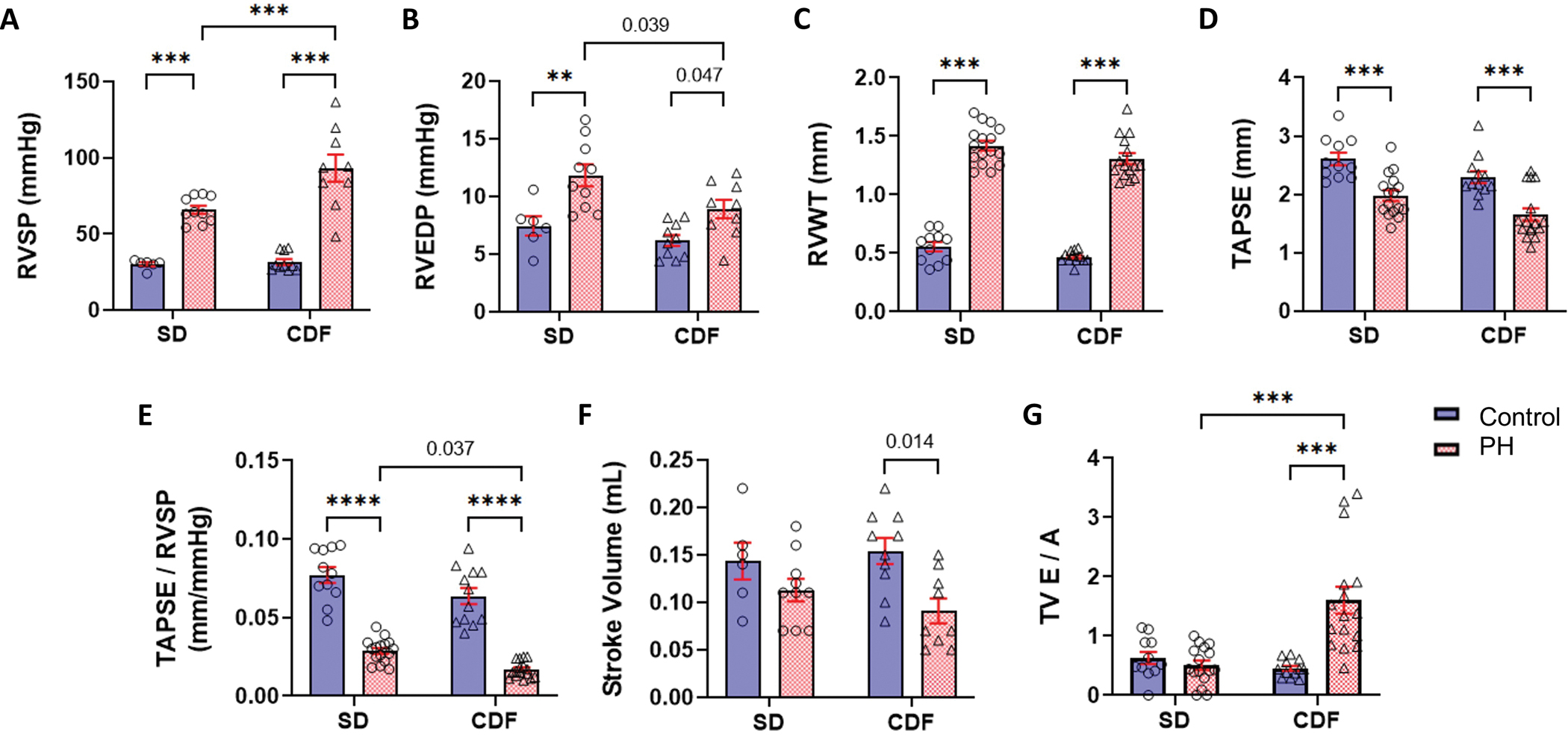
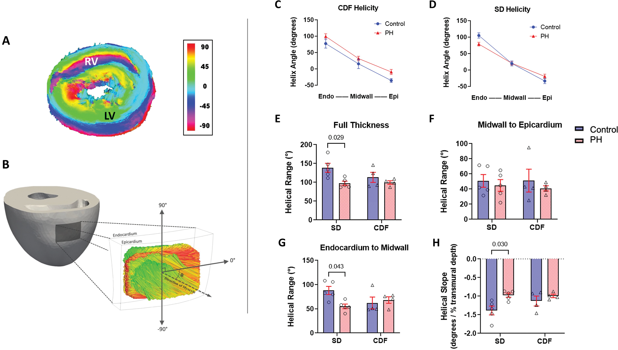
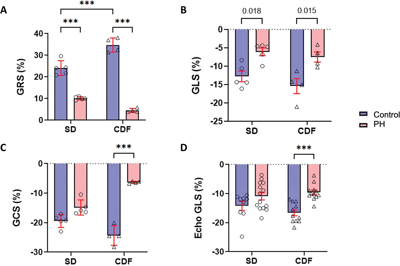
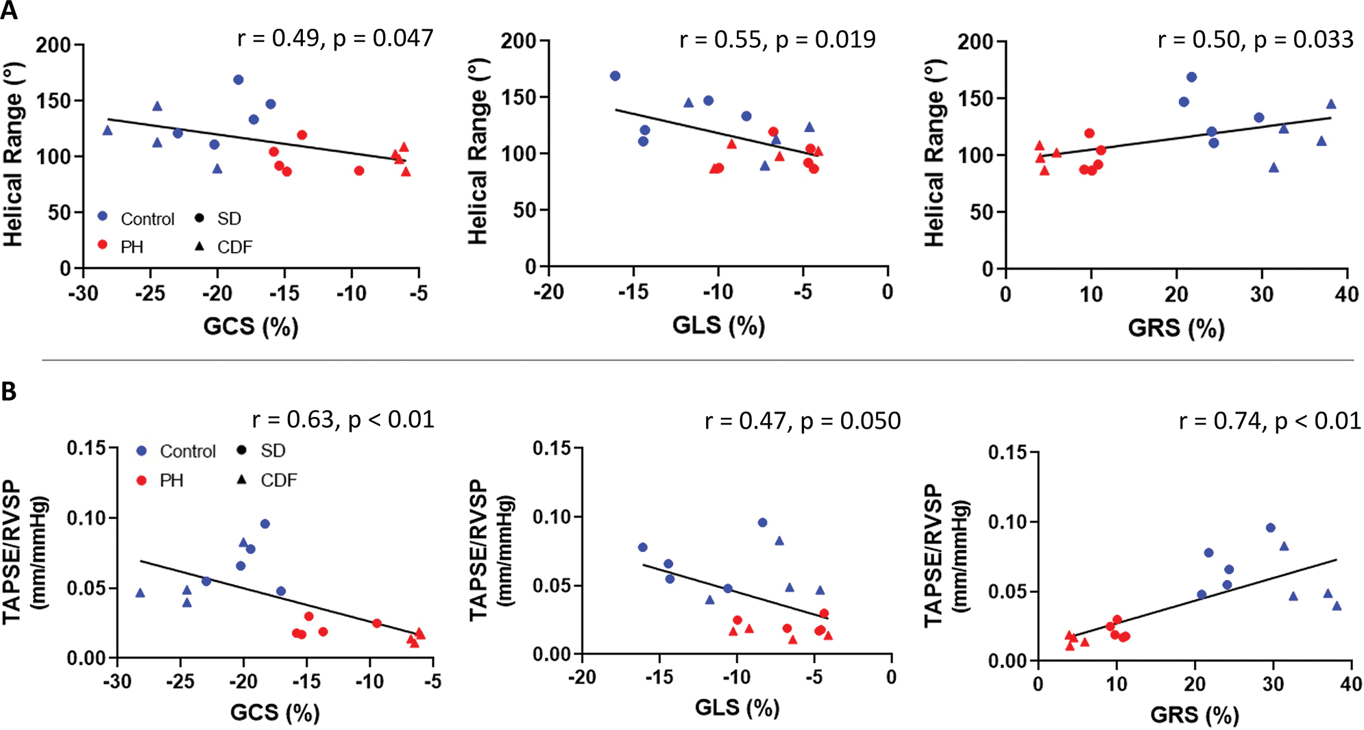
Methods: Hemodynamic assessments were performed in 2 rodent models of PH. RV free wall myoarchitecture was quantified using generalized Q-space imaging and tractography analyses. Computational models were developed to predict RV wall strains. Data from animal studies were analyzed to determine the correlations between hemodynamic measurements, RV strains, and structural measures.

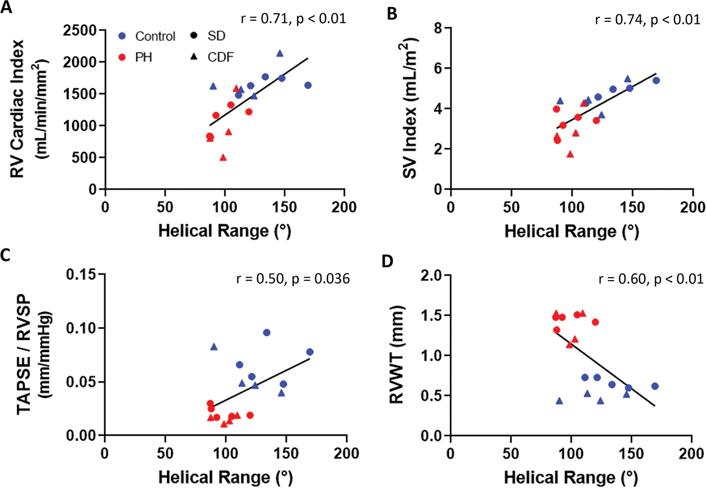
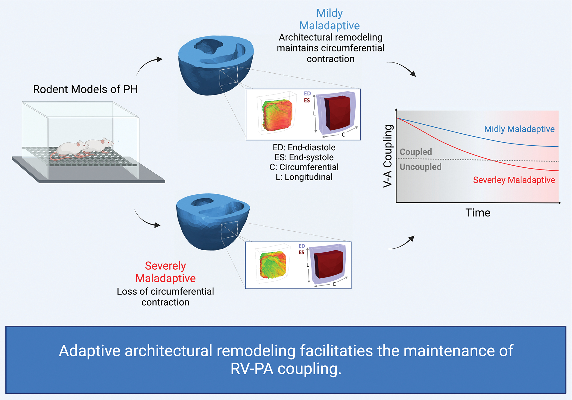
Results: In contrast to the PH rats with severe RV maladaptation, PH rats with mild RV maladaptation showed a decrease in helical range of fiber orientation in the RV free wall (139º versus 97º; P=0.029), preserved global circumferential strain, and exhibited less reduction in right ventricular-pulmonary arterial coupling (0.029 versus 0.017 mm/mm Hg; P=0.037). Helical range correlated positively with coupling (P=0.036) and stroke volume index (P<0.01). Coupling correlated with global circumferential strain (P<0.01) and global radial strain (P<0.01) but not global longitudinal strain.

Conclusions: Data analysis suggests that adaptive RV architectural remodeling could improve RV function in PH. Our findings suggest the need to assess RV architecture within routine screenings of PH patients to improve our understanding of its prognostic and therapeutic significance in PH.

Keywords: physiological adaptation; pulmonary hypertension; right ventricle; strain; ventricle remodeling.


Conflict of interest statement

Disclosures

Authors declare that they have no conflicts of interest.

Figures

Fig. 1.
Fig. 1.. Characterization of PH Models.
PH was confirmed in both models by hemodynamic (A-B) and echocardiographic measurements, as PH rats showed higher RV wall thickness (C), and RV systolic function measured by TAPSE (D) and TAPSE/RVSP (E) were significantly reduced in PH rats. Both PH models showed reductions in stroke volume (F) which were significant only for CFD-PH rats. In addition, the CDF-PH model had lower TAPSE/RVSP (E) and more severe RV dysfunction (G) in comparison to SD-PH rats. CDF: Fischer rat model; E/A: ratio of the early (E) to late (A) RV filling velocities; PH: pulmonary hypertension; RV: right ventricle; RVEDP: right ventricle end-diastolic pressure; RVSP: right ventricle systolic pressure; RVWT: right ventricle wall thickness; SD: Sprague-Dawley rat model; TAPSE: tricuspid annular plane systolic excursion; TAPSE/RVSP: measure of RV-pulmonary artery coupling; TV: tricuspid valve. **p<0.01; ***p<0.001. SD: n=6–11 Control, 10–16 PH; CDF: n=10–12 Control, 9–16 PH. Two-way ANOVA followed by Tukey post hoc test.
Fig 2:
Fig 2:. Architectural Remodeling in Right Ventricle.
(A) DWI visualization example of a SD-control whole heart. Schematic showing the region of the heart from which DWI fiber data was gathered and the convention to calculate helical angle. (C-D) Myocardial fiber helix angles are shown at the endocardium, midwall, and epicardium. (E-G) Mean helical range were calculated across the full and partial transmural RVFW thickness. (H) Mean helical slope. CDF: Fischer rat model; DWI: Diffusion-weighted Imaging; LV: left ventricle; PH: pulmonary hypertension; RV: right ventricle; RVFW: right ventricle free wall; SD: Sprague-Dawley rat model. SD: n=5 Control, 5 PH; CDF: n=4 Control, 4 PH. Two-way ANOVA followed by Tukey post hoc test.
Fig 3:
Fig 3:. Right Ventricular Strains in Pulmonary Hypertension.
(A-C) Mean and SD of simulated global circumferential (GCS), longitudinal (GLS), and radial (GRS) RVFW strains from finite-element modeling. (D) Mean GLS calculated from echocardiography. CDF: Fischer rat model; PH: pulmonary hypertension; RVFW: right ventricle free wall; SD: Sprague-Dawley rat model. ***p<0.001. SD: n=5–9 Control, 5–14 PH; CDF: n=4–11 Control, 4–12 PH. Two-way ANOVA followed by Tukey post hoc test.
Fig 4:
Fig 4:. Correlation Between Strains, Myoarchitecture, and RV-PA Coupling.
(A) RV global circumferential (GCS), longitudinal (GLS), and radial (GRS) strains were associated with helical range. (B) RV circumferential and radial strains were associated with RV-PA coupling, measured by TAPSE/RVSP, while longitudinal strain was not. CDF: Fischer rat model; PA: pulmonary artery; PH: pulmonary hypertension; RV: right ventricle; RVSP: right ventricle systolic pressure; SD: Sprague-Dawley rat model; TAPSE: tricuspid annular plane systolic excursion; TAPSE/RVSP: measure of RV-PA coupling. SD: n=5 Control, 5 PH; CDF: n=4 Control, 4 PH. Linear regression model.
Fig 5:
Fig 5:. Correlation Between Myoarchitecture and RV Function.
Helical range was associated with key RV functional metrics (A-B) and coupling (C), but not with RV wall thickness (D). CDF: Fischer rat model; PH: pulmonary hypertension; RV: right ventricle; RVSP: right ventricle systolic pressure; RVWT: right ventricle wall thickness; SD: Sprague-Dawley rat model; SV: stroke volume; TAPSE: tricuspid annular valve plane systolic excursion; TAPSE/RVSP: measure of RV-pulmonary artery coupling. SD: n=5 Control, 5 PH; CDF: n=4 Control, 4 PH. Linear regression model.
Fig 6:
Fig 6:. Architectural remodeling influences RV-PA coupling.
Adaptive architectural remodeling contributes to preserving transverse (circumferential) contraction that enhances active elastance of the RV and facilitates the maintenance of RV-PA coupling. Theoretically, architectural remodeling can improve RV contractility independent of changes in intrinsic active properties of myofibers. PA: pulmonary artery; PH: pulmonary hypertension; RV: right ventricle; V-A: ventricle-arterial.

Comment in

References

    1. Vonk-Noordegraaf A, Haddad F, Chin KM, Forfia PR, Kawut SM, Lumens J, Naeije R, Newman J, Oudiz RJ, Provencher S. Right heart adaptation to pulmonary arterial hypertension: physiology and pathobiology. Journal of the American College of Cardiology. 2013;62:D22–D33. - PubMed
    1. Bogaard HJ, Abe K, Noordegraaf AV, Voelkel NF. The right ventricle under pressure: cellular and molecular mechanisms of right-heart failure in pulmonary hypertension. Chest. 2009;135:794–804. - PubMed
    1. Dong Y, Pan Z, Wang D, Lv J, Fang J, Xu R, Ding J, Cui X, Xie X, Wang X. Prognostic Value of Cardiac Magnetic Resonance–Derived Right Ventricular Remodeling Parameters in Pulmonary Hypertension: A Systematic Review and Meta-Analysis. Circulation: Cardiovascular Imaging. 2020;13:e010568. - PubMed
    1. Vélez-Rendón D, Zhang X, Gerringer J, Valdez-Jasso D. Compensated right ventricular function of the onset of pulmonary hypertension in a rat model depends on chamber remodeling and contractile augmentation. Pulmonary circulation. 2018;8:2045894018800439. - PMC - PubMed
    1. Tello K, Dalmer A, Vanderpool R, Ghofrani HA, Naeije R, Roller F, Seeger W, Wilhelm J, Gall H, Richter MJ. Cardiac magnetic resonance imaging-based right ventricular strain analysis for assessment of coupling and diastolic function in pulmonary hypertension. JACC: Cardiovascular Imaging. 2019;12:2155–2164. - PubMed

Publication types