Fracture lines and comminution zones of traumatic sacral fractures
- PMID: 36748773
- PMCID: PMC10198334
- DOI: 10.14744/tjtes.2022.15163
Fracture lines and comminution zones of traumatic sacral fractures
Abstract
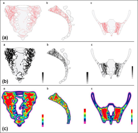
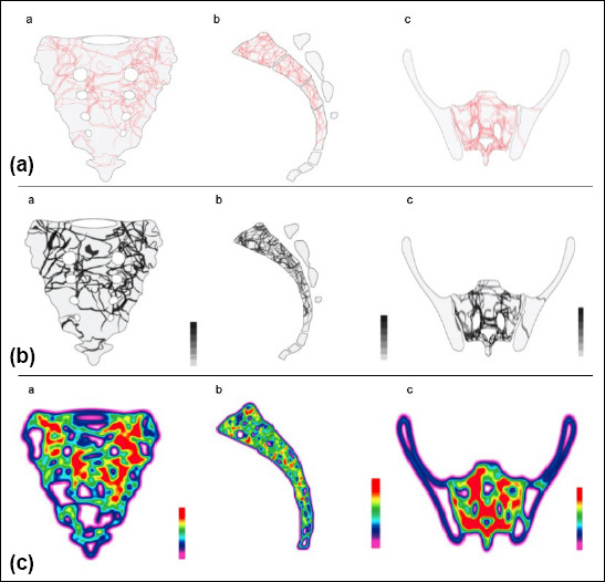
Background: Sacral fractures are uncommon and understanding three-dimensional morphology is needed to obtain proper treatment. The purpose of this study was to identify the repeatable fracture patterns and comminution zones for traumatic sacral fractures and create fracture maps.
Methods: Computerized tomography images of 72 patients with traumatic sacral fracture were included in the study. For each fracture, fracture lines were identified and digitally reduced. All fractures were superimposed over a template and fracture maps; comminution zones and heatmaps were created for each zone.
Results: There were 40 males and 32 females with a mean age of 46.5±19.9. Fifty-three (73.6%) patients sustained major trauma, and 19 (26.4%) had minor trauma. There were 37 (51.4%) Zone 1, 22 (30.6%) Zone 2, and 13 (18.1%) Zone 3 fractures. Each Denis zone showed certain fracture patterns. In Zone 1 fractures, most of the fracture lines were vertical and oblique (up to 45°) orientation on both sides. In Zone 2 fractures, fracture lines were concentrated on the S1 and S2 levels. Anterolateral and posterolateral parts of the sacrum were less affected in right-side fractures. In Zone 3 fractures, fractures were concentrated in S1, S2, and S3 levels around the sacral canal. The median sacral crest and midline remained mostly unaffected.
Conclusion: Sacral fractures showed specific repeatable patterns for each zone. These findings may be helpful for pre-operative planning, placement of fixation material, design of new implants, and modification of current fracture-classification systems.
Conflict of interest statement
Figures

References
-
- Beckmann NM, Chinapuvvula NR. Sacral fractures:Classification and management. Emerg Radiol. 2017;24:605–17. - PubMed
-
- Gutierrez-Gomez S, Wahl L, Blecher R, Olewnik Ł, Iwanaga J, Maulucci CM, et al. Sacral fractures:An updated and comprehensive review. Injury. 2021;52:366–75. - PubMed
-
- Lehman RA, Jr, Kang DG, Bellabarba C. A new classification for complex lumbosacral injuries. Spine J. 2012;12:612–28. - PubMed
-
- Pascal-Moussellard H, Hirsch C, Bonaccorsi R. Osteosynthesis in sacral fracture and lumbosacral dislocation. Orthop Traumatol Surg Res. 2016;102(1 Suppl):S45–57. - PubMed
MeSH terms
LinkOut - more resources
Full Text Sources
Medical