Review
. 2023 Jun;57(6):1621-1640.
doi: 10.1002/jmri.28624. Epub 2023 Feb 7.

QSM Throughout the Body

Affiliations
Review

QSM Throughout the Body

Alexey V Dimov et al. J Magn Reson Imaging. 2023 Jun.

Abstract

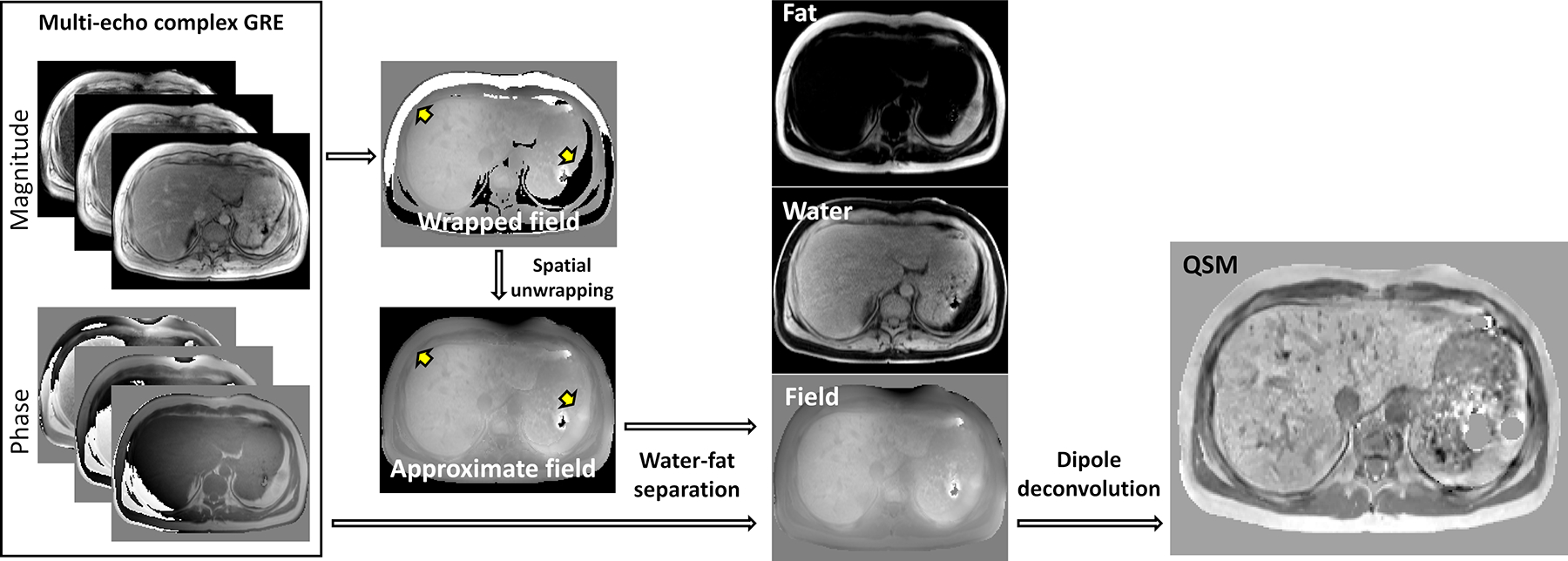
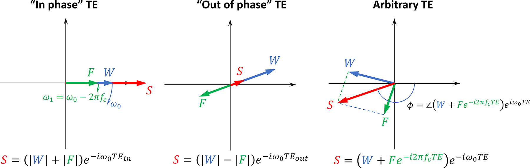
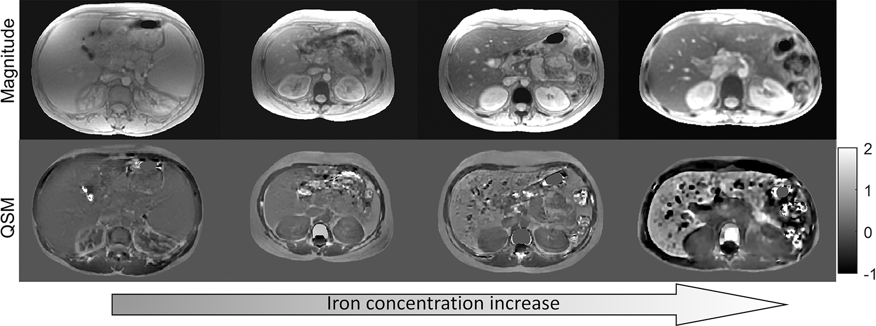
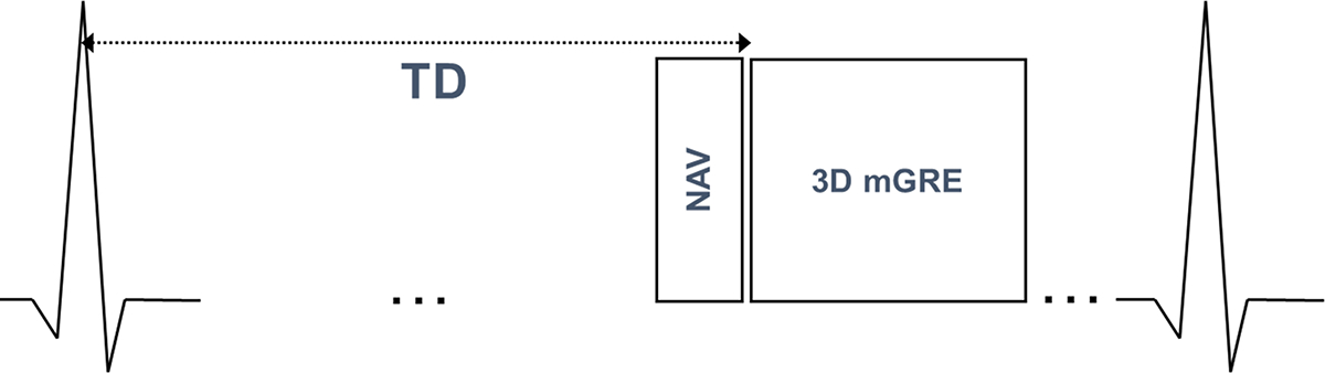
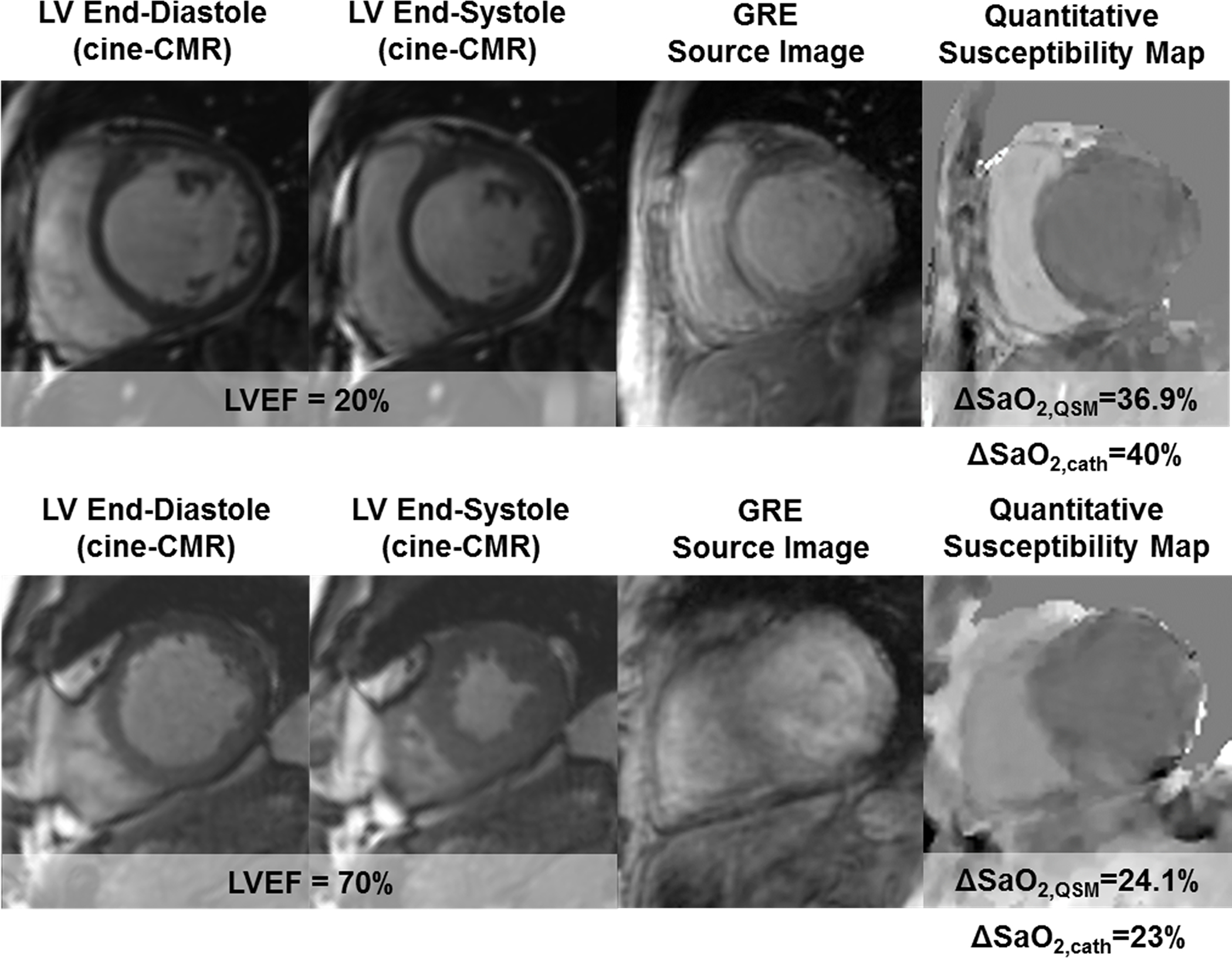
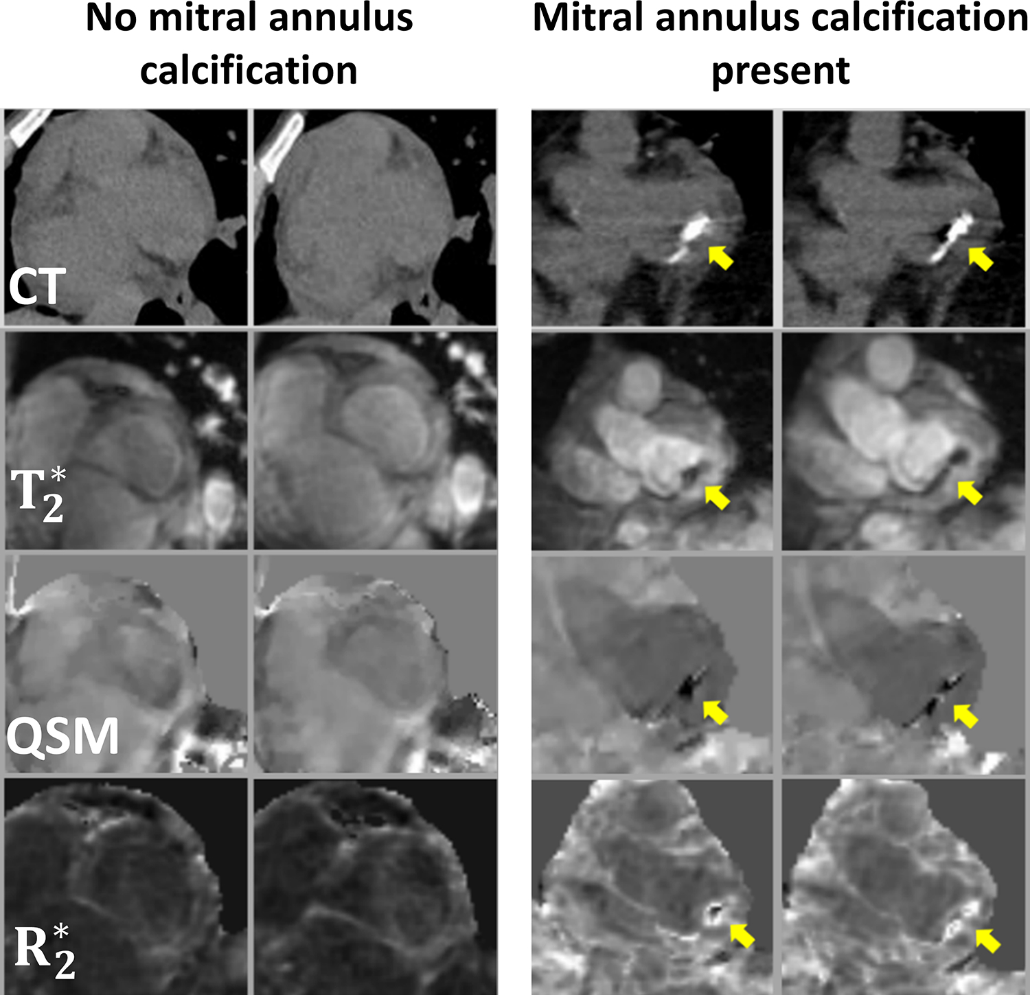
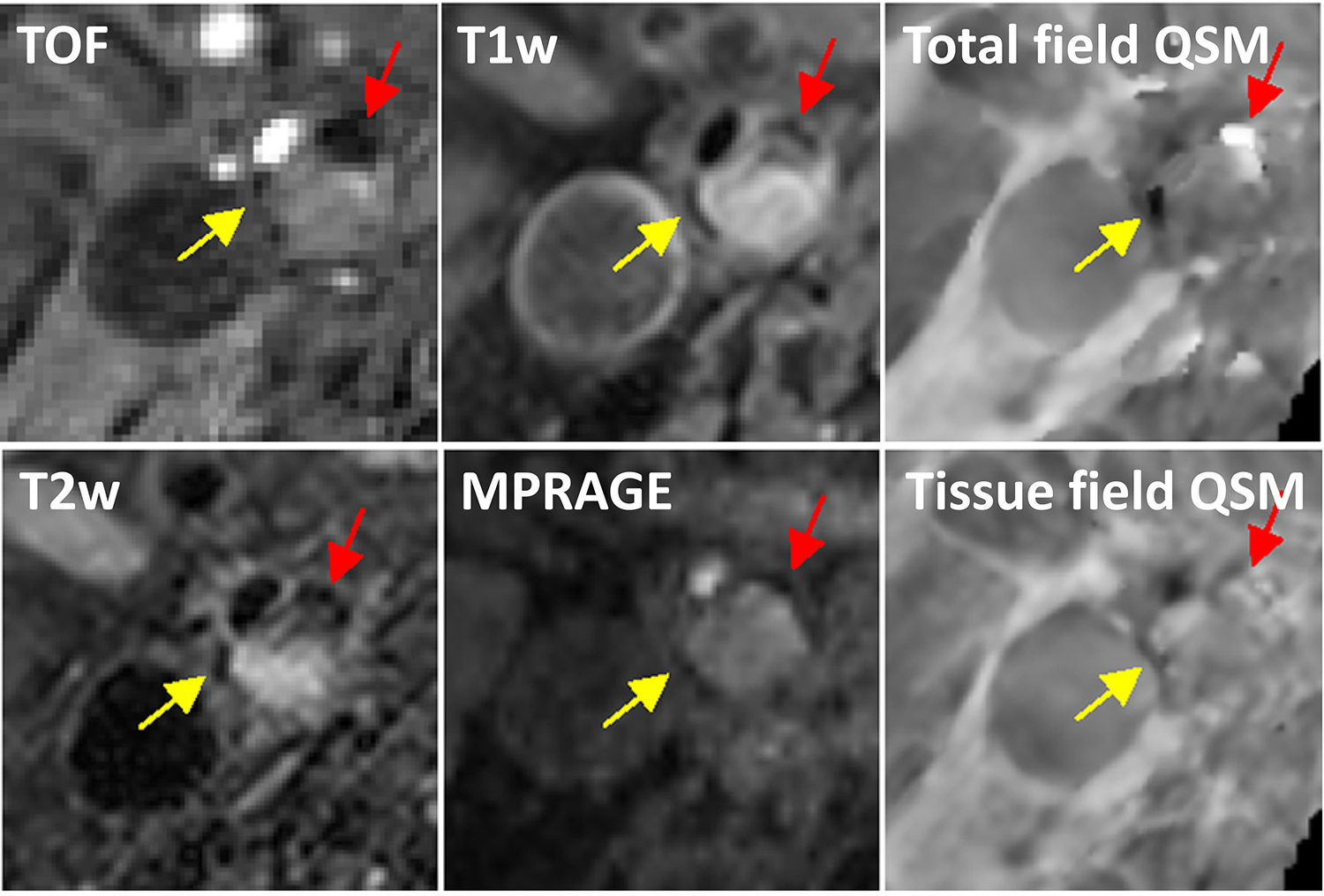
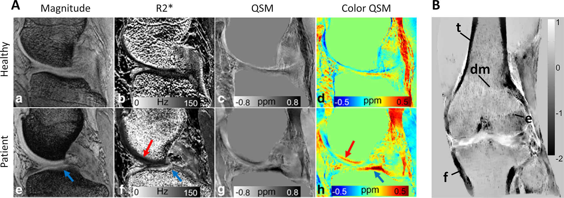
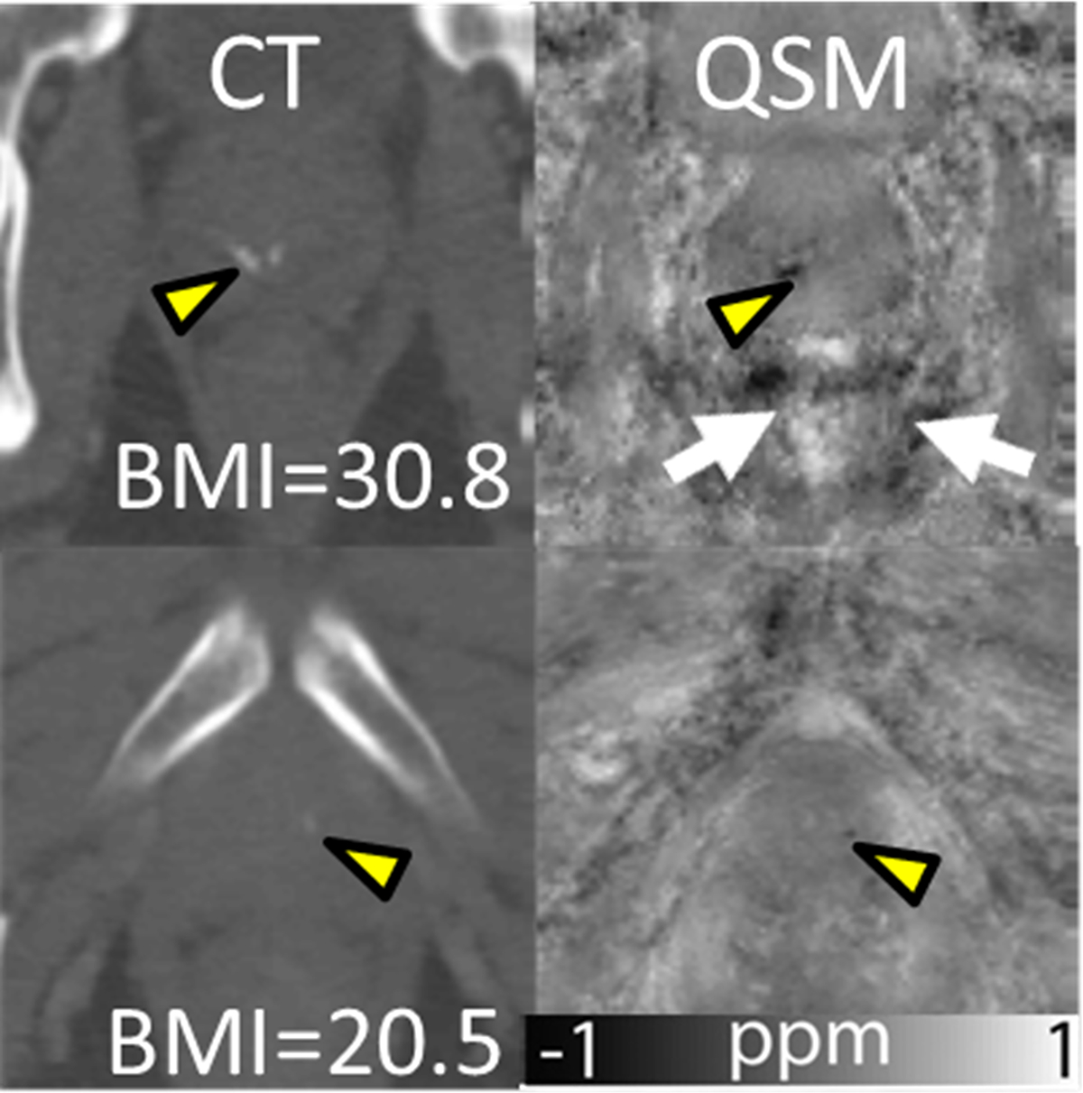
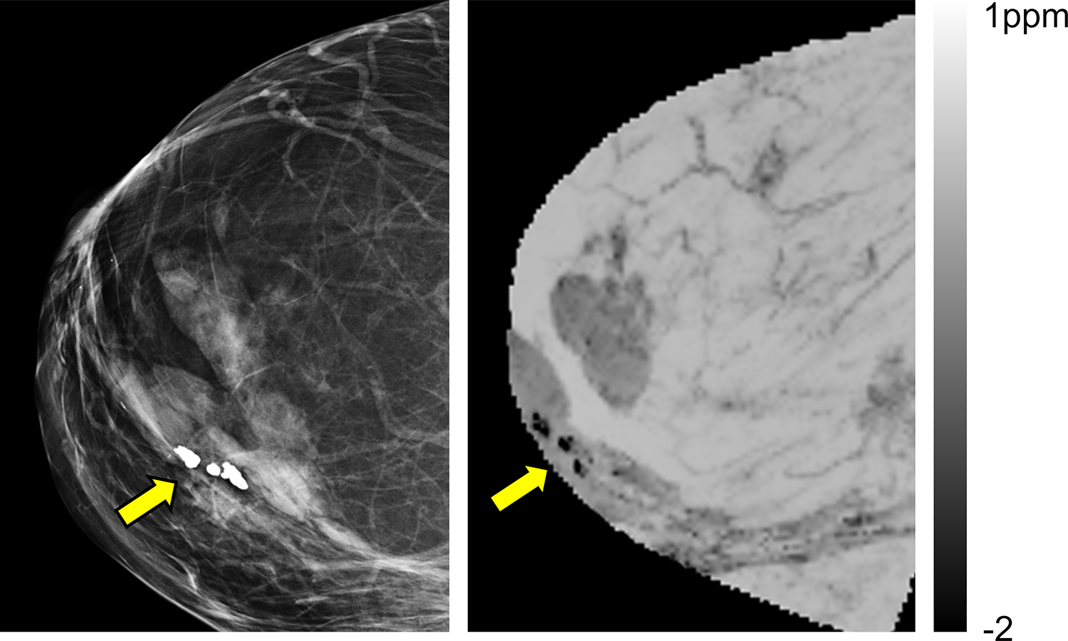
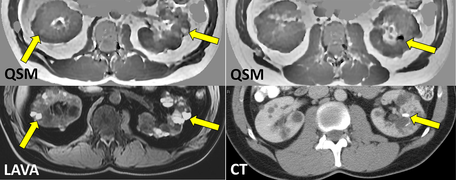
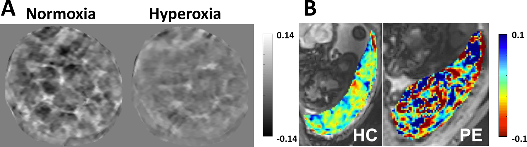
Magnetic materials in tissue, such as iron, calcium, or collagen, can be studied using quantitative susceptibility mapping (QSM). To date, QSM has been overwhelmingly applied in the brain, but is increasingly utilized outside the brain. QSM relies on the effect of tissue magnetic susceptibility sources on the MR signal phase obtained with gradient echo sequence. However, in the body, the chemical shift of fat present within the region of interest contributes to the MR signal phase as well. Therefore, correcting for the chemical shift effect by means of water-fat separation is essential for body QSM. By employing techniques to compensate for cardiac and respiratory motion artifacts, body QSM has been applied to study liver iron and fibrosis, heart chamber blood and placenta oxygenation, myocardial hemorrhage, atherosclerotic plaque, cartilage, bone, prostate, breast calcification, and kidney stone.

Keywords: body imaging; quantitative imaging; quantitative susceptibility mapping.


Figures

Figure 1.
Figure 1.
Flowchart of QSM reconstruction in body applications. Susceptibility mapping requires acquisition and preservation of both magnitude and phase of standard complex gradient echo sequence. Water-fat separation is performed to generate a high-fidelity magnetic field map. Dipole deconvolution to solve the inverse field-to-source problem is performed as the final step
Figure 2.
Figure 2.
Effects of chemical shift in field mapping. Signal vector diagram illustrating GRE phase behavior in presence of fat. Because of the chemical shift, signals of water (W, blue arrow) and fat (F, green arrow) experience different rates of phase accrual, ω0 and ω1; adding and subtracting images S acquired at strategically selected “In phase” and “out of phase” echo times, water and fat images can be generated. At an arbitrary echo time, relationship between the echo time and phase becomes nonlinear, and its dependence on chemical shift value can be exploited for estimation of water, fat images, and field mapping
Figure 3.
Figure 3.
Magnitude and QSM images in four thalassemia major patients. Higher degrees of iron overload manifest in gradual increase of liver parenchymal susceptibility, linearly proportional to iron concentration (adapted from (64))
Figure 4.
Figure 4.
Pulse sequence diagram for cardiac QSM. ECG triggered navigator 3D Cartesian multi-echo gradient echo free breathing acquisition
Figure 5.
Figure 5.
Two representative examples of QSM maps in cardiac patients. In the top patient, who had severely reduced LV function (EF=20%), QSM measured a marked increase in ΔSO2 (36.9%), which agreed well with invasive catheterization (40%). In the bottom patient, who had normal LV function (EF=70%), QSM measured ΔSO2 (24.1%) was within normal limits and was similar to invasive data (23%). Reprinted with permission from (128)
Figure 6.
Figure 6.
QSM of mitral calcification. Representative examples of patients with and without MAC as visualized by CT and T2* weighted magnitude, QSM, and R2* pulse sequences on cardiac MRI. Note that CT evidenced calcium was found to correspond with presence and location of annular susceptibility on cardiac QSM. Reprinted with permission from (133).
Figure 7.
Figure 7.
Comparison between tissue and total field inversion QSM techniques. Two regions at the boundary of a large plaque from a 63-year-old carotid artery stenosis patient appear similarly hypointense on TOF, T1w, T2w, and MPRAGE images. On QSM reconstructed using total field inversion, one region has a strong diamagnetic appearance (yellow arrow), consistent with calcification, while the other region has strongly positive susceptibility indicative of an old hemorrhage with hemosiderin deposition (red arrow). This susceptibility contrast could not be seen well on QSM obtained with a tissue field inversion approach (adapted from (39))
Figure 8.
Figure 8.
QSM in MSK imaging. A) Medial cartilage in a healthy subject (a-d) and a patient with collagen damage (e-h). Patient magnitude and R2* images reveal signal alteration in the tibial plateau compared to the healthy control (arrows). Susceptibility map (h) reflects changes in cartilage composition in the affected regions (adapted from (167)). B) MIP of a whole knee joint QSM in a healthy subject demonstrating delineation of cortical areas of the femur (f) and tibia (t), the depiction of trabeculation, the epiphyseal line (e) and transition from diaphyseal to metaphyseal bone (dm) (adapted from (55))
Figure 9.
Figure 9.
QSM in prostate. Prostatic calcifications (yellow arrowheads) in CT images (first column), and susceptibility maps (second column). Motion/air artifacts and noise can be observed around the prostate (first row), indicated by arrows. Additionally, body mass index (BMI) of patients is provided (adapted from (46)).
Figure 10.
Figure 10.
QSM in breast. Mammogram (left) and QSM (right) of a breast in a female patient with calcified nodules. (adapted from (200)). QSM is able to unambiguously identify of calcifications, which appear hypo-intense due to their diamagnetic susceptibility
Figure 11.
Figure 11.
Kidney QSM in ADPKD. A) Despite hyperintense appearance on 3D T1w LAVA image, only few ADPKD cysts appear as paramagnetic on QSM (yellow arrows), indicating heterogeneity of cyst composition and non-specificity of T1 hyperintensity to presence of hemorrhage and blood products. B) QSM allows detection of calcified kidney stones as confirmed by CT (unpublished data).
Figure 12.
Figure 12.
QSM in placenta. A) Susceptibility maps (ppm) acquired in a healthy pregnancy (gestational age, 29 1/7 weeks) under normoxia and maternal hyperoxia induced by administration of 100% oxygen within the same scan session. Spatial variation of susceptibility was substantially reduced under hyperoxia. B. Susceptibility maps (ppm) acquired in a health pregnancy (left; gestational age, 28 6/7 weeks) and one complicated by preeclampsia (right; gestational age, 35 weeks). Susceptibility of the placenta associated with preeclampsia showed markedly increased spatial variation. Reprinted from (218, 219).

References

    1. Wang Y, Liu T. Quantitative susceptibility mapping (QSM): Decoding MRI data for a tissue magnetic biomarker. Magn Reson Med 2015;73(1):82–101. - PMC - PubMed
    1. Harada T, Kudo K, Fujima N, et al. Quantitative Susceptibility Mapping: Basic Methods and Clinical Applications. Radiographics 2022;42(4):1161–1176. - PubMed
    1. Vinayagamani S, Sheelakumari R, Sabarish S, et al. Quantitative Susceptibility Mapping: Technical Considerations and Clinical Applications in Neuroimaging. J Magn Reson Imaging 2021;53(1):23–37. - PubMed
    1. Haacke EM, Liu S, Buch S, Zheng W, Wu D, Ye Y. Quantitative susceptibility mapping: current status and future directions. Magn Reson Imaging 2015;33(1):1–25. - PubMed
    1. Boyacioglu R, Wang C, Ma D, McGivney DF, Yu X, Griswold MA. 3D magnetic resonance fingerprinting with quadratic RF phase. Magn Reson Med 2021;85(4):2084–2094. - PubMed

Publication types