. 2023 May;68(5):577-590.
doi: 10.1165/rcmb.2022-0362OC.

AKT Phosphorylates FAM13A and Promotes Its Degradation via CUL4A/DDB1/DCAF1 E3 Complex

Affiliations

AKT Phosphorylates FAM13A and Promotes Its Degradation via CUL4A/DDB1/DCAF1 E3 Complex

Lu Gong et al. Am J Respir Cell Mol Biol. 2023 May.

Abstract

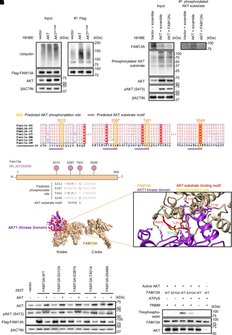
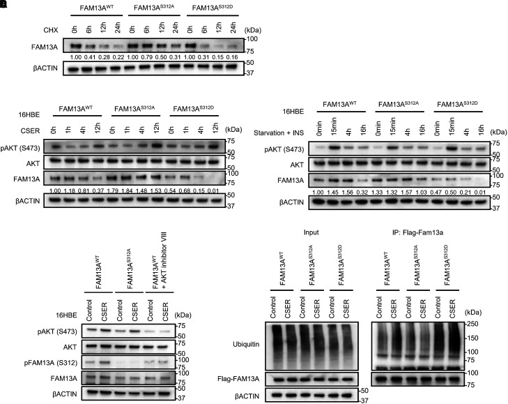
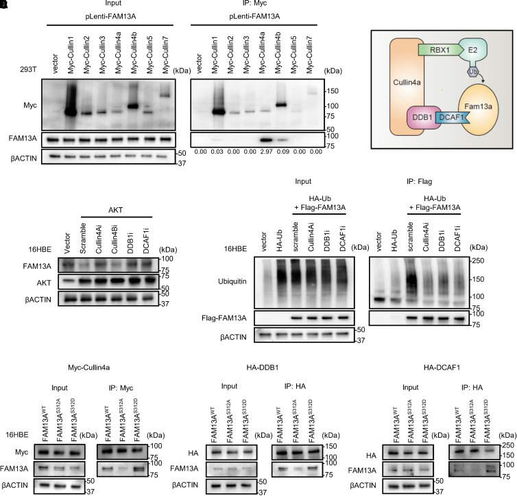
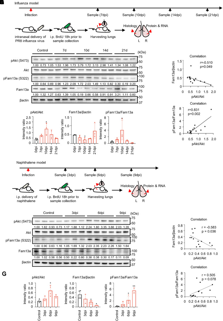
SNPs within FAM13A (family with sequence similarity 13 member A) gene are significantly associated with chronic obstructive pulmonary disease and lung function in genome-wide association studies (GWAS). However, how FAM13A protein is regulated under physiological and pathological conditions remains largely elusive. Herein, we report that FAM13A is phosphorylated at the serine 312 residue by AKT kinase after cigarette smoke extract treatment and thereby recognized by the CULLIN4A/DCAF1 (DDB1 and CUL4 associated factor 1) E3 ligase complex, rendering the ubiquitination-mediated degradation of FAM13A. More broadly, downregulation of FAM13A protein upon AKT activation, as a general cellular response to acute stress, was also detected in influenza- or naphthalene-injured lungs in mice. Functionally, reduced protein levels of FAM13A lead to accelerated epithelial cell proliferation in murine lungs during the recovery phase after injury. In summary, we characterized a novel molecular mechanism that regulates the stability of FAM13A protein, which enables the fine-tuning of lung epithelial repair after injury. These significant findings will expand our molecular understanding of the regulation of protein stability, which may modulate lung epithelial repair implicated in the development of chronic obstructive pulmonary disease and other lung diseases.

Keywords: AKT; E3 ligase; FAM13A; cellular response; protein degradation.


Figures

Figure 1.
Figure 1.
AKT promotes FAM13A (family with sequence similarity 13 member A) degradation. (A) Protein levels of FAM13A were measured in NHBE cells and 16HBE cells after 3% cigarette smoke extract (CSE) treatment for 12 hours with recovery (CSER) or without recovery. Cells without CSE were used as a control. (B) FAM13A protein was detected and quantified in 16HBE cells pretreated with MG132 (10 μM for 3 h), followed by treatment with cycloheximide (CHX; 10 μg/ml) to inhibit new protein synthesis for indicated durations. (C) Normalized FAM13A protein levels from three replicates were fit to the curve of a nonlinear one-phase decay model for calculation of the half-life of FAM13A protein. (D) Protein levels of phosphorylated AKT, total AKT, and FAM13A were measured using IB in 16HBE cells at indicated time points. (E) Quantification of protein bands in D indicates correlation between FAM13A levels and AKT activation. (F) Detection of Flag-tagged FAM13A in 16HBE cells cotransfected with low or high amount of AKT1 or AKT2. (G) Detection of AKT activation and FAM13A in HEK 293T cells cotransfected with Flag-FAM13A and AKT followed by treatment with indicated inhibitors. ALLN = acetylleucyl-leucyl-norleucinal; NHBE = normal human bronchial epithelial; p-AKT = phosphorylated AKT.
Figure 2.
Figure 2.
AKT phosphorylates FAM13A to promote its degradation. (A) Detection of the ubiquitination of Flag-tagged FAM13A in 16HBE cells cotransfected with wild-type (WT) AKT or mutant AKTK179M with defective kinase activity after IP with Flag antibody. Cells were treated with MG132 before IP assay. (B) Detection of FAM13A protein in 16HBE cells cotransfected with siRNA targeting FAM13A or siRNA control and AKT followed by IP using the antibody recognizing phosphorylated AKT substrates. (C) Conserved AKT phosphorylation sites on FAM13A protein across species are indicated inside the orange frame. Highly conserved residues are highlighted in red. (D) Four potential phosphorylation sites on human FAM13A protein contain AKT substrate motif. (E and F) Predicted protein structural model simulating interaction between FAM13A and AKT kinase (E). The area inside the red frame is illustrated in F, with details for predicted interaction interface between FAM13A and AKT kinase domain. (G) Detection of Flag-tagged FAM13A and activated AKT in HEK 293T cells transfected with WT or mutant FAM13A carrying various point mutations at putative phosphorylation sites. (H) AKT-mediated FAM13A phosphorylation was assessed using in vitro kinase assay with purified FAM13A and AKT proteins. PNBM = p-nitrobenzyl mesylate.
Figure 3.
Figure 3.
Phosphorylation of FAM13A at S312/S322 regulates the stability of FAM13A protein. (A) Measurements of the levels of endogenous FAM13A protein in 16HBE clonal lines edited using CRISPR/Cas9 targeting S312 site expressing WT or mutant FAM13A pretreated with MG132 (10 μM for 3 h) and then CHX (10 μg/ml) for indicated durations. (B) Detection of phosphorylated AKT and FAM13A in FAM13AWT, FAM13AS312A, or FAM13AS312D 16HBE clonal lines after CSER treatment. (C) Detection of FAM13A protein in prestarved 16HBE clonal lines (FAM13AWT, FAM13AS312A, or FAM13AS312D), followed by INS (2.5 μg/ml) treatment for indicated durations. (D) S312-phosphorylated FAM13A was assessed using immunoblots in 16HBE clonal lines expressing FAM13AWT or FAM13AS312A under indicated conditions. (E) Ubiquitination of FAM13A was detected using IP in 16HBE cells transfected with Flag-tagged WT FAM13A or various mutants (FAM13AS312A or FAM13AS312D) and treated with CSER. In D and E, cells were pretreated with MG132 to prevent protein degradation. Cas9 = CRISPR associated protein 9; CHX = cycloheximide; INS = insulin.
Figure 4.
Figure 4.
CULLIN4A E3 ligase complex contributes to the degradation of FAM13A. (A) Myc-tagged CULLIN family proteins interact with overexpressed FAM13A as assessed using co-IP assays in HEK 293T cells. Immunoprecipitated FAM13A:CULLIN protein ratios were calculated on the basis of band densitometry as shown. (B) A model diagram showing FAM13A binding to CULLIN4A (CUL4A) E3 ligase complex. (C) Detection of FAM13A in 16HBE cells cotransfected with AKT and siRNA targeting indicated CULLIN4A complex component or scramble siRNA. (D) Ubiquitination of Flag-tagged FAM13A was detected in 16HBE cells cotransfected with HA–tagged ubiquitin, indicated siRNA, and Flag-FAM13A by IP with Flag beads. Cells were pretreated with MG132 (10 μM for 3 h) before IP experiment. (E) Interaction of CULLIN4A complex with endogenous FAM13A in 16HBE clonal lines (FAM13AWT, FAM13AS312A, or FAM13AS312D) detected using IP. DCAF1 = DDB1 and CUL4 associated factor 1; DDB1 = damage specific DNA binding protein 1; HA = hemagglutinin; RBX1 = Ring-Box 1; Ub = ubiquitin.
Figure 5.
Figure 5.
S322-Fam13a was phosphorylated in mouse lung injury models in vivo. (A) Schematic illustration of the influenza-induced lung injury/repair mouse model. (B) Indicated protein levels of Fam13a, S322-phosphorylated Fam13a, and activated Akt were measured in lung tissues from mice after influenza infection. (C) Phosphorylated Akt (S473):total Akt ratio, phosphorylated Fam13a (S322):total Fam13a ratio, and normalized Fam13a protein levels were quantified in B across different time points. (D) Correlations between Akt phosphorylation and Fam13a total protein or Fam13a phosphorylation at S322 were analyzed. (E) Diagram showing the design of the naphthalene-induced airway injury mouse model. (F) Detection of Fam13a protein levels and Akt activation in lungs at different time points. (G) Quantification of Akt activation (phosphorylated Akt [S473]:total Akt ratio) and Fam13a protein in various samples. (H) Correlation between Akt phosphorylation and Fam13a phosphorylation or protein levels. BrdU = bromodeoxyuridine; dpi = days post injury; i.p. = intraperitoneal; L = left; R = right.
Figure 6.
Figure 6.
FAM13A suppresses cell growth in vitro and in vivo. (A) Cell growth curve in 16HBE cells stably infected with lentiviral shRNA targeting FAM13A (FAM13Ash). Cells infected with nontargeting shRNA were used as controls (STDsh). Mean ± SD values are from triplicate wells in one representative repeat from three biological repeats. (B) Cell growth curves in stable 16HBE clonal lines (FAM13AWT, FAM13AS312A, or FAM13AS312D). (C) Immunostaining with club cell 10 kD (CC10) (red), SP-C (surfactant protein C) (green), BrdU (yellow), and DAPI (blue), indicating club cells, alveolar type II (ATII) cells, proliferating cells, and cell nucleus, respectively. Scale bars, 25 μm. (D) Quantification of proliferating ATII cells (BrdU+SP-C+/SP-C+) in Fam13a+/+ and Fam13a−/− mice (n = 4) after influenza infection. Each dot indicates the quantification result of a randomly chosen area (0.5 × 0.4 mm) on the lung specimen. Yellow arrows indicate BrdU+SP-C+ cells. White dashed lines indicate airway boundaries. (E) Immunofluorescence staining in lung specimens from Fam13a+/+ and Fam13a−/− mice after naphthalene treatment with CC10 (red), SP-C (green), BrdU (yellow), and DAPI (blue), indicating club cells, ATII cells, proliferating cells, and cell nucleus, respectively. Yellow arrowheads indicate BrdU+CC10+ cells. White dashed lines indicate airway basal membrane. Scale bars, 25 μm. (F) Quantification on the number of CC10+ cells per airway. (G) Proliferating CC10+ cells (i.e., percentage of BrdU+CC10+ cells/CC10+ in Fam13a+/+ and Fam13a−/− mice) 6 days after naphthalene delivery. Quantification results are from 20 randomly selected areas (0.5 × 0.4 mm) on the lung sections from four mice in each group. (H) Diagram briefly summarizing how proliferative signals generated during tissue repair activate AKT, which in turn phosphorylates FAM13A, which is recognized by the CULLIN4A E3 ligase complex for ubiquitination-mediated degradation. BR = bronchiole.

Comment in

References

    1. Barnes PJ, Burney PGJ, Silverman EK, Celli BR, Vestbo J, Wedzicha JA, et al. Chronic obstructive pulmonary disease. Nat Rev Dis Primers . 2015;1:15076. - PubMed
    1. GBD 2015 Chronic Respiratory Disease Collaborators. Global, regional, and national deaths, prevalence, disability-adjusted life years, and years lived with disability for chronic obstructive pulmonary disease and asthma, 1990-2015: a systematic analysis for the Global Burden of Disease Study 2015. Lancet Respir Med . 2017;5:691–706. - PMC - PubMed
    1. Hindorff LA, Sethupathy P, Junkins HA, Ramos EM, Mehta JP, Collins FS, et al. Potential etiologic and functional implications of genome-wide association loci for human diseases and traits. Proc Natl Acad Sci USA . 2009;106:9362–9367. - PMC - PubMed
    1. Cho MH, Boutaoui N, Klanderman BJ, Sylvia JS, Ziniti JP, Hersh CP, et al. Variants in FAM13A are associated with chronic obstructive pulmonary disease. Nat Genet . 2010;42:200–202. - PMC - PubMed
    1. Hobbs BD, de Jong K, Lamontagne M, Bossé Y, Shrine N, Artigas MS, et al. COPDGene Investigators ECLIPSE Investigators; LifeLines Investigators; SPIROMICS Research Group; International COPD Genetics Network Investigators; UK BiLEVE Investigators; International COPD Genetics Consortium. Genetic loci associated with chronic obstructive pulmonary disease overlap with loci for lung function and pulmonary fibrosis. Nat Genet . 2017;49:426–432. - PMC - PubMed

Publication types

MeSH terms