. 2023 Feb 22;15(7):9726-9739.
doi: 10.1021/acsami.2c20502. Epub 2023 Feb 7.

Interactions of Catalytic Enzymes with n-Type Polymers for High-Performance Metabolite Sensors

Affiliations

Interactions of Catalytic Enzymes with n-Type Polymers for High-Performance Metabolite Sensors

David Ohayon et al. ACS Appl Mater Interfaces. .

Abstract

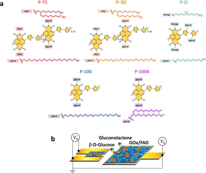
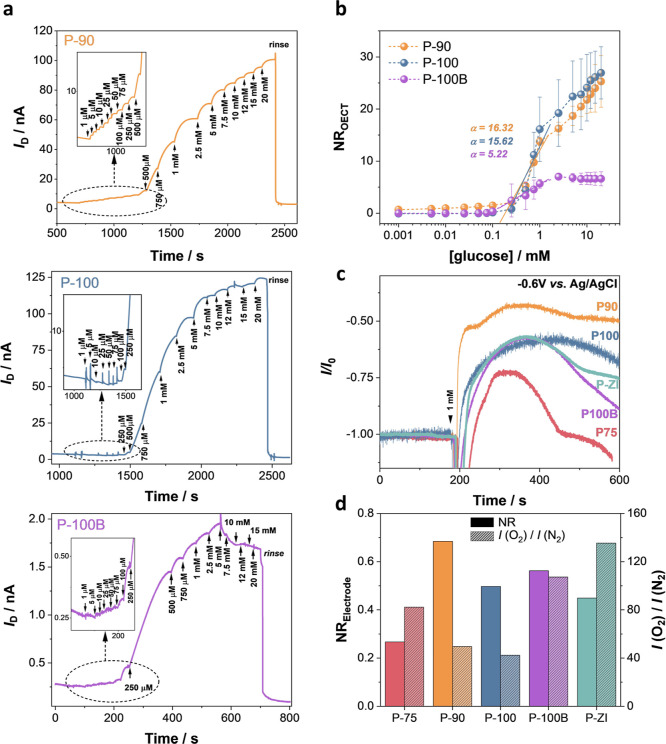
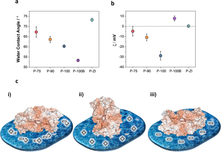
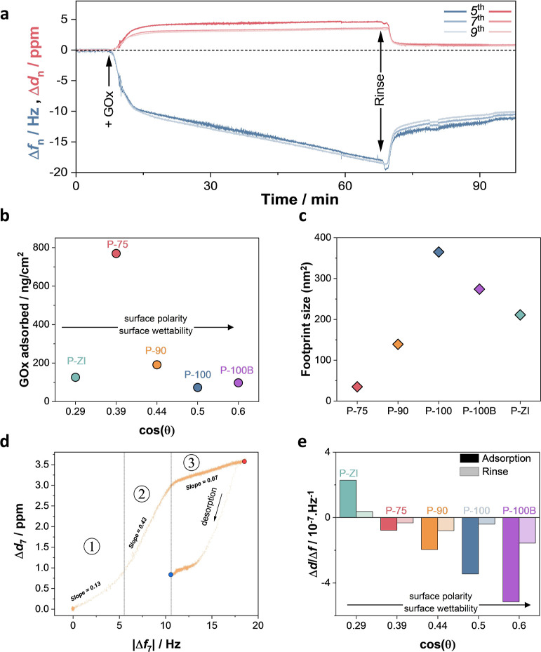
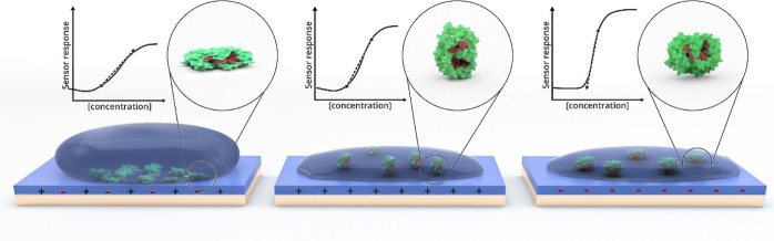
The tight regulation of the glucose concentration in the body is crucial for balanced physiological function. We developed an electrochemical transistor comprising an n-type conjugated polymer film in contact with a catalytic enzyme for sensitive and selective glucose detection in bodily fluids. Despite the promise of these sensors, the property of the polymer that led to such high performance has remained unknown, with charge transport being the only characteristic under focus. Here, we studied the impact of the polymer chemical structure on film surface properties and enzyme adsorption behavior using a combination of physiochemical characterization methods and correlated our findings with the resulting sensor performance. We developed five n-type polymers bearing the same backbone with side chains differing in polarity and charge. We found that the nature of the side chains modulated the film surface properties, dictating the extent of interactions between the enzyme and the polymer film. Quartz crystal microbalance with dissipation monitoring studies showed that hydrophobic surfaces retained more enzymes in a densely packed arrangement, while hydrophilic surfaces captured fewer enzymes in a flattened conformation. X-ray photoelectron spectroscopy analysis of the surfaces revealed strong interactions of the enzyme with the glycolated side chains of the polymers, which improved for linear side chains compared to those for branched ones. We probed the alterations in the enzyme structure upon adsorption using circular dichroism, which suggested protein denaturation on hydrophobic surfaces. Our study concludes that a negatively charged, smooth, and hydrophilic film surface provides the best environment for enzyme adsorption with desired mass and conformation, maximizing the sensor performance. This knowledge will guide synthetic work aiming to establish close interactions between proteins and electronic materials, which is crucial for developing high-performance enzymatic metabolite biosensors and biocatalytic charge-conversion devices.

Keywords: catalytic enzymes; conjugated polymers; electron transporting (n-type) polymers; enzymatic sensors; glucose; organic bioelectronics; organic electrochemical transistor.


Conflict of interest statement

The authors declare no competing financial interest.

Figures

Figure 1
Figure 1
(a) Chemical structure of n-type polymers: the two copolymers with varying EG contents: P-75 and P-90, the fully glycolated (alkyl-free) analogue (P-100), P-100B with a branched EG side chain (branched analogue of P-100), and P-ZI with its zwitterions on the side chain. (b) OECT schematic highlighting the locations of the n-type film with adsorbed GOx (channel and gate). When glucose is present, it gets oxidized to gluconolactone by the active site (FAD) of GOx, following glucose + GOx (FAD) → gluconolactone + GOx (FADH2).
Figure 2
Figure 2
Performance of n-type OECTs and electrodes as glucose sensors. (a) Real-time response of the OECT (source-drain current, ID, as a function of time) as successive amounts of glucose are added to the electrolyte. The gate and drain voltages were kept constant at +0.5 V. The OECTs were operated using a planar gate configuration, where both the channel and gate were functionalized with GOx. Insets represent the real-time response of the devices to glucose concentrations lower than 250 μM. (b) Normalized response of the OECTs (NROECT) to glucose. Error bars represent the standard deviation of at least three different devices. (c) Amperometric response of n-type conjugated polymer electrodes to 1 mM glucose. The working electrode was the n-type film functionalized with GOx, the reference electrode was Ag/AgCl, and the counter electrode was a Pt coil. The arrow represents the time point when 1 mM glucose was added into the electrolyte. (d) NRelectrode to 1 mM glucose (plain bars) and to O2 (patterned bars).
Figure 3
Figure 3
Surface properties of n-type films. (a) Water contact angle and (b) ζ potential of the n-type polymer films. Error bars represent the standard deviation of (a) nine and (b) four different measurements. (c) Orientations of native GOx from A. niger (PDB: 3QVP) as a function of surface charge: (i) “front-lying” orientation on a charge-neutral surface; (ii) “standing” orientation on a positively charged surface; and (iii) “back-lying” orientation on a negatively charged surface. Surface colors on GOx indicate positive and negative electrostatic potentials contoured from 50 kT/e (blue) to −50 kT/e (red). The cofactor is shown in stick representation and highlighted in red.
Figure 4
Figure 4
GOx adsorption on polymer films, analyzed using QCM-D. (a) P-90 QCM-D raw data reporting the change in frequency (Δfn) and dissipation (Δdn) for harmonics fifth, seventh, and ninth. (b) Corresponding mass taken up upon GOx adsorption on each film as a function of surface wettability [represented by cos(θ) where θ is the water contact angle]. The adsorbed mass per area was extracted from the data collected at the end of the rinsing process (see Figure S14). (c) Footprint of the enzyme on each polymer film. The footprint was calculated from the enzyme surface coverage and average dimensions of the crystal structure of a deglycosylated GOx molecule (60 Å × 52 Å × 77 Å), assuming an even distribution of the enzyme on a laterally homogeneous surface. (d) P-90 Δd vs Δf plots (seventh harmonic). The numbers define the linear regions in the plots with different slopes. The red and blue dots demark the end of the adsorption and desorption processes, respectively. (e) Δdf ratio, calculated from the values measured at the end of the adsorption and the rinsing steps, as a function of wettability cos(θ).
Figure 5
Figure 5
GOx adsorption kinetics. (a) dm/dt vs Sauerbrey mass uptake (seventh harmonic) plots for GOx adsorption on P-90 and (b) plot of C1 as a function of ka.
Figure 6
Figure 6
High-resolution N 1s XPS spectra of the polymer films after enzyme adsorption. Yellow circles highlight the chemical bonds involving N atoms in the polymers, including the NDI backbone and side chains and those in the primary amino acids present in GOx. The amino acids displayed in the figure correspond to a selected few in the protein sequence: lysine (LYS), glutamine (GLN), and arginine (ARG).
Figure 7
Figure 7
Influence of n-type polymer film surface properties on GOx adsorption. The surface hydrophilicity and charge govern enzyme adsorption behavior on the film surface. Hydrophobic surfaces tend to retain more enzymes. Surfaces that are too hydrophobic, however, lead to complete denaturation of the enzyme structure. GOx adopts a flattened conformation on hydrophilic/polar surfaces. The surface charge influences GOx orientation. A neutral surface (left) leads to a “front-lying” orientation, where the active site of the enzyme faces downward and is inaccessible to the glucose. A positively charged surface (middle) leads to the enzyme adsorbing in a “standing” orientation, where the active site is accessible. A negatively charged surface (right) leads to a “back-lying” orientation, with the active site facing up and in closer proximity to the sensor surface than in a standing fashion. The latter surface, which is hydrophilic, negatively charged, and homogeneous, is most desirable to build top-performer glucose sensors.

References

    1. Ngandu Mpoyi E.; Cantini M.; Reynolds P. M.; Gadegaard N.; Dalby M. J.; Salmerón-Sánchez M. Protein Adsorption as a Key Mediator in the Nanotopographical Control of Cell Behavior. ACS Nano 2016, 10, 6638–6647. 10.1021/acsnano.6b01649. - DOI - PMC - PubMed
    1. Cho W.; Stahelin R. V. Membrane-Protein Interactions in Cell Signaling and Membrane Trafficking. Annu. Rev. Biophys. Biomol. Struct. 2005, 34, 119–151. 10.1146/annurev.biophys.33.110502.133337. - DOI - PubMed
    1. Xu L.-C.; Bauer J. W.; Siedlecki C. A. Proteins, Platelets, and Blood Coagulation at Biomaterial Interfaces. Colloids Surf., B 2014, 124, 49–68. 10.1016/j.colsurfb.2014.09.040. - DOI - PMC - PubMed
    1. Urbani A.; Sirolli V.; Lupisella S.; Levi-Mortera S.; Pavone B.; Pieroni L.; Amoroso L.; Di Vito R.; Bucci S.; Bernardini S.; Sacchetta P.; Bonomini M. Proteomic Investigations on the Effect of Different Membrane Materials on Blood Protein Adsorption During Haemodialysis. Blood Transfus. 2012, 10 Suppl 2, s101–s112. 10.2450/2012.014S. - DOI - PMC - PubMed
    1. Russo M. J.; Han M.; Desroches P. E.; Manasa C. S.; Dennaoui J.; Quigley A. F.; Kapsa R. M. I.; Moulton S. E.; Guijt R. M.; Greene G. W.; Silva S. M. Antifouling Strategies for Electrochemical Biosensing: Mechanisms and Performance toward Point of Care Based Diagnostic Applications. ACS Sens. 2021, 6, 1482–1507. 10.1021/acssensors.1c00390. - DOI - PubMed