. 2023 May 18;141(20):2417-2429.
doi: 10.1182/blood.2022018380.

The role of CD8+ T-cell clones in immune thrombocytopenia

Affiliations

The role of CD8+ T-cell clones in immune thrombocytopenia

Amna Malik et al. Blood. .

Abstract

Immune thrombocytopenia (ITP) is traditionally considered an antibody-mediated disease. However, a number of features suggest alternative mechanisms of platelet destruction. In this study, we use a multidimensional approach to explore the role of cytotoxic CD8+ T cells in ITP. We characterized patients with ITP and compared them with age-matched controls using immunophenotyping, next-generation sequencing of T-cell receptor (TCR) genes, single-cell RNA sequencing, and functional T-cell and platelet assays. We found that adults with chronic ITP have increased polyfunctional, terminally differentiated effector memory CD8+ T cells (CD45RA+CD62L-) expressing intracellular interferon gamma, tumor necrosis factor α, and granzyme B, defining them as TEMRA cells. These TEMRA cells expand when the platelet count falls and show no evidence of physiological exhaustion. Deep sequencing of the TCR showed expanded T-cell clones in patients with ITP. T-cell clones persisted over many years, were more prominent in patients with refractory disease, and expanded when the platelet count was low. Combined single-cell RNA and TCR sequencing of CD8+ T cells confirmed that the expanded clones are TEMRA cells. Using in vitro model systems, we show that CD8+ T cells from patients with ITP form aggregates with autologous platelets, release interferon gamma, and trigger platelet activation and apoptosis via the TCR-mediated release of cytotoxic granules. These findings of clonally expanded CD8+ T cells causing platelet activation and apoptosis provide an antibody-independent mechanism of platelet destruction, indicating that targeting specific T-cell clones could be a novel therapeutic approach for patients with refractory ITP.


Conflict of interest statement

Conflict-of-interest disclosure: The authors declare no competing financial interests.

Figures

None
Graphical abstract
Figure 1.
Figure 1.
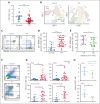
TEMRA cells without features of exhaustion are expanded in patients with ITP and correlate with disease activity. (A) Peripheral CD4+:CD8+ ratio is significantly lower in patients with ITP than in controls, indicating CD8-mediated disease. (B) A t-distributed stochastic neighbor embedding (tSNE) plot of the T-cell CD4+ and CD8+ subsets based on their surface expression of CD45RA and CD62L in a control patient and a patient with ITP. Expanded TEMRA cells from a patient with ITP are shown. (C) An example of a dot plot from flow cytometry analysis of a control vs a patient with ITP, using CD45RA and CD62L. (D) Patients with ITP compared with controls have significantly higher numbers of TEMRA cells. (E) Patients with platelet counts <30 × 109/L have higher numbers of TEMRA cells than those with platelet counts ≥30 × 109/L. (F) An example of a dot plot from flow cytometry analysis of a control vs a patient with ITP, showing expression of IFN-γ and TNFα. (G) CD8+ T cells in patients with ITP have increased IFN-γ, TNFα, and granzyme B; polyfunctional CD8+ T cells (expressing IFN-γ, TNFα, and granzyme B) are also increased in patients with ITP. (H) PD-1 expression has not changed, and Tim-3 expression is reduced in TEMRA cells. ∗P ≤ .05; ∗∗P ≤ .01; ∗∗∗P ≤ .001.
Figure 2.
Figure 2.
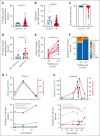
High-throughput TCR sequencing reveals expansion of private clones associated with decreased TCR diversity in patients with ITP vs healthy controls: as clones expand, the platelet count falls. (A) The percentage of space occupied by the expanded clones (defined as clones occupying >5% of the repertoire) is significantly higher in patients with ITP than in healthy controls. (B) Number of productive unique CDR3 sequences (unique clones) per 103 unique clones is significantly lower in patients with ITP than in healthy controls. (C) Simpson diversity index is significantly lower in patients with ITP than in healthy controls. (D) In patients with refractory ITP, the percentage of space occupied by the expanded clones is significantly higher than in patients with nonrefractory ITP. (E) The number of productive unique clones per 103 unique CDR3 sequences (diversity) is reduced in patients with a platelet count of <30 × 109/L and recovers in individual patients as the count increases (platelet count of ≥30 × 109/L). (F) The amount of the T-cell repertoire/space taken up by expanded clones is higher in patients with a platelet count <30 × 109/L and falls in individual patients as the count returns to normal levels, reflecting the changes in T-cell repertoire. (G) In 2 patients with chronic ITP, individual clones were followed-up for over a number of years (point 0 is the first time that TCR is measured) and compared with the overall T-cell diversity and the platelet count. TCR diversity falls with the platelet count and increases as it recovers. Correspondingly, the individual clones expand as the platelet count decreases and contract as the platelet count increases. ∗P ≤ .05; ∗∗P ≤ .01; ∗∗∗P ≤ .001; ∗∗∗∗P ≤ .0001.
Figure 3.
Figure 3.
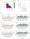
Expanded unique T-cell clones originate in the TEMRA compartment. (A) CMH, ranging from 0 (the samples are entirely different) to 1 (the samples are identical), shows the overlap between clones detected in the overall T cells and clones detected in the TEMRA cells from the same patients. The mean CMH value of the TCR repertoires was >0.4 between T-cell and TEMRA cell compartments. In comparison, there was no overlap between patients. (B) Percentage space occupied by the largest clonotype among T-cell and TEMRA compartment in 3 patients with ITP. (C) 3D-plot visualization of the composite TRBV and TRBJ repertoire of the TEMRA compartment compared with non-TEMRA CD8+ T cells and CD4+ cells combined.
Figure 4.
Figure 4.
Combined single-cell RNA and TCR sequencing defines expanded clones as TEMRA cells. (A) Sorted CD8+ T cells are clustered using their gene expression profile into naïve, TEMRA, central memory (CM), effector memory (EM), and CD161+ high cells. (B) Heatmap of genes in sorted CD8+ T cells differentially expressed in accordance with clusters, including naïve, TEMRA, CM, EM, and CD161+ high cells. (C) Large clones (top 10 clones) are predominantly within the TEMRA subset, shown in red. (D) Patients with a platelet count <30 × 109/L have more TEMRA T cells compared with patients with a platelet count ≥30 × 109/L.
Figure 5.
Figure 5.
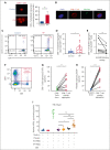
CD8+ T cells interact with platelets causing T-cell activation, IFN-γ release, and CD107a and platelet activation. CD8+ T-cell–platelet aggregates are inhibited with MHC class I blocking on platelets. (A) When CD8+ T cells flow along a chamber coated with platelets, CD8+ T cells from patients with ITP were 4 times more likely to slow down and stop along the platelet coated surface than controls. (B) Confocal imaging of CD8+ T-cell interactions with platelets in a patient with ITP (×20 lens objective). (C) Example of a dot plot from flow cytometry analysis of a control vs patient with ITP when CD8+ T cells are cocultured with platelets, showing CD8+ T-cell–platelet aggregates (CD8+CD41+) from total CD8+ T cells. (D) CD8+ T-cell–platelet aggregates are higher in patients with ITP than in healthy controls and (E) are inhibited when MHC class I HLA-A, -B, and -C receptors on platelets are blocked. (F) In CD8+ T-cell–platelet coculture, (G) CD107a is increased in the CD8+ T-cell–platelet aggregates compared with CD8+ T cells cultured alone, consistent with release of granzyme B; (H) platelets in the CD8+ T-cell–platelet aggregates show increased CD62P, consistent with platelet activation. (I) IFN-γ ELISpot assay of T cells cultured with autologous platelets shows that T cells from patients with ITP have increased secretion of IFN-γ when cultured with platelets (detected by IFN-γ–forming spots). ∗P ≤ .05; ∗∗P ≤ .01; ∗∗∗P ≤ .001. HC, healthy control; SEB, staphylococcal enterotoxin.

Comment in

  • TEMRA: the CD8 subset in chronic ITP?
    Lazarus AH, Semple JW. Lazarus AH, et al. Blood. 2023 May 18;141(20):2409-2410. doi: 10.1182/blood.2023019859. Blood. 2023. PMID: 37200060 No abstract available.

References

    1. Rodeghiero F, Stasi R, Gernsheimer T, et al. Standardization of terminology, definitions and outcome criteria in immune thrombocytopenic purpura of adults and children: report from an international working group. Blood. 2009;113(11):2386–2393. - PubMed
    1. Cooper N, Ghanima W. Immune thrombocytopenia. N Engl J Med. 2019;381(10):945–955. - PubMed
    1. Cooper N, Kruse A, Kruse C, et al. Immune thrombocytopenia (ITP) world impact survey (I-WISh): impact of ITP on health-related quality of life. Am J Hematol. 2021;96(2):199–207. - PMC - PubMed
    1. Terrell DR, Neunert CE, Cooper N, et al. Immune thrombocytopenia (ITP): current limitations in patient management. Medicina (Mex) 2020;56(12):667. - PMC - PubMed
    1. Provan D, Arnold DM, Bussel JB, et al. Updated international consensus report on the investigation and management of primary immune thrombocytopenia. Blood Adv. 2019;3(22):3780–3817. - PMC - PubMed

Publication types

Substances